The hornworts: morphology, evolution and development - PubMed (original) (raw)
Review
. 2021 Jan;229(2):735-754.
doi: 10.1111/nph.16874. Epub 2020 Sep 15.
Affiliations
- PMID: 32790880
- PMCID: PMC7881058
- DOI: 10.1111/nph.16874
Review
The hornworts: morphology, evolution and development
Eftychios Frangedakis et al. New Phytol. 2021 Jan.
Abstract
Extant land plants consist of two deeply divergent groups, tracheophytes and bryophytes, which shared a common ancestor some 500 million years ago. While information about vascular plants and the two of the three lineages of bryophytes, the mosses and liverworts, is steadily accumulating, the biology of hornworts remains poorly explored. Yet, as the sister group to liverworts and mosses, hornworts are critical in understanding the evolution of key land plant traits. Until recently, there was no hornwort model species amenable to systematic experimental investigation, which hampered detailed insight into the molecular biology and genetics of this unique group of land plants. The emerging hornwort model species, Anthoceros agrestis, is instrumental in our efforts to better understand not only hornwort biology but also fundamental questions of land plant evolution. To this end, here we provide an overview of hornwort biology and current research on the model plant A. agrestis to highlight its potential in answering key questions of land plant biology and evolution.
Keywords: Anthoceros; bryophytes; chloroplast; development; evolution; hornworts; symbiosis.
© 2020 The Authors New Phytologist © 2020 New Phytologist Trust.
Figures
Fig. 1:. Life and laboratory cycle of the hornwort A. agrestis.
(a) A. agrestis has two life cycle phases. A dominant haploid phase called gametophyte and a diploid phase called sporophyte. The life cycle of A. agrestis starts with the germination of the haploid spores (1) which develop into an irregularly shaped thallus (2). A. agrestis is monoecious, with both male and female reproductive organs present on the same individual. Male (antheridia) and female (archegonia) reproductive organs are embedded in the thallus and mitotically produce sperm and egg, respectively (3). Biflagellated motile sperm cells swim in water to the archegonium where the egg is fertilised (4). The resulting diploid zygote divides first by a longitudinal division and subsequent divisions to form the embryo, which is initially composed of three tiers. The bottom tier produces the foot. The middle tier gives rise to the basal meristem and the top tier forms the tip of the sporophyte (5). The sporophyte develops within the gametophyte and is nourished through the placenta, the junction between foot and gametophyte cells. Meiosis and sporogenesis occur progressively from the base of the sporophyte upwardly, leading to spore formation: sporogenous tissue at the base of the sporophyte produces spore mother cells that, via meiosis, produce spore tetrads and spores that are released at the tip where the sporophyte separates into two valves. (6). n: haploid, 2n: diploid. (b - c) Laboratory cycle of A. agrestis: (b) Plants can be easily propagated in axenic culture by transferring small thallus fragments (typically 1 mm x 1 mm) onto plates with fresh growth media using sterile scalpels. (c) In laboratory conditions A. agrestis sporophyte induction can be achieved in 1-2 months under axenic conditions using a small thallus fragment as starting material.
Fig. 2:. Key morphological features of A. agrestis
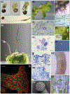
(a) Light micrograph (LM) of germinating spores. Upper three images, successive stages in globose sporeling production. Lowermost: under low light conditions spore germination involves a germ tube, a long single-celled filament that develops a terminate globose sporeling. Scale bars: 50 μm. (b) Surface view of the irregularly shaped thallus. Blue arrow: rhizoids. Orange arrow: wavy thallus edge. Scale bar: 2.0 mm. (c) Top: LM of single-celled rhizoids on the ventral thallus (red arrow). Scale bar: 150 μm. Bottom: LM of single-celled rhizoid tips (yellow arrowhead) Scale bar: 100 μm (d) Sporophytes (blue arrow) growing on the gametophyte (yellow arrow). Scale bar: 3.0 mm. (e) LM of longitudinal section of thallus with mucilage canals (red arrows). Mucilage clefts on the ventral side indicated with red arrowheads. Scale bar: 50 μm. (h) LM of antheridia (red arrow) in an antheridial chamber in longitudinal section. Scale bar: 50 μm. (f) Surface view of antheridial chamber with yellow antheridia embedded in the dorsal thallus. Scale bar: 10 μm (h). (g) Antheridium removed from chamber in (f) showing antheridial body with sperm cells inside and stalk to the lower right. LM of two archegonia embedded in the dorsal thallus in longitudinal section showing from the base up: egg cell, ventral canal cell, neck cells and cover cells. Scale bar: 10 μm. (i) LM of biflagellate sperm. Coiled cell body is on the left and the flagella are on the right (yellow arrow). Scale bar: 5.0 μm. (m) LM longitudinal section of thallus with an open archegonium containing only the egg cell near the apical notch. Dorsal side (red asterisk) and ventral side (orange asterisk). Scale bar: 25 μm. (k) Sporophyte with stomata. Scale bar: 20 μm. (l) Confocal fluorescence microscopy image of transgenic gametophyte showing single plastids in each cell. Green: green fluorescent protein localised in the plasma membrane expressed under the CaMV 35S promoter. Red: chlorophyll autofluorescence. Scale bar 50 μm. (m) (n) Higher magnification LM of a stoma with two guard cells surrounding a pore. Scale bar: 10 μm. (o) Scanning electron micrograph (SEM) of distal side of a spinose spore. Scale bar: 10 μm.
Fig. 3:. Phylogeny of land plants and hornworts
(a-c) Competing hypotheses about the phylogenetic position of hornworts among land plants. (a) Liverworts, mosses and hornworts are successive sister lineages to tracheophytes (Qiu et al. 2006). (b) Hornworts sister to all other land plants with liverworts and mosses monophyletic (Wickett et al., 2014). (c) Monophyletic bryophytes with hornworts sister to Setaphyta that include mosses and liverworts (Li et al., 2020; Renzanglia et al., 2018). (d) Phylogeny of hornworts based on Villarreal & Renner (2012), numbered circles next to the names in the phylogenetic tree correspond to species example images below. Phaeoceros photo credit: John Baker, University of Oxford. Presence and/or absence of stomata and pyrenoid is indicated next to each genera name. * exception: stomata absent in Folioceros incurvus (Renzaglia et al., 2009; Villarreal & Renner, 2012). (e) Phylogeny of the Anthoceros agrestis/Anthoceros punctatus group based on Dawes et al., 2020.
Fig. 4 :. A. agrestis gametophyte apical growth
(a) Surface view of A. agrestis gametophyte. Arrows indicate apical notches. Scale bar: 1.0 mm (b) LM surface view of apical notch (yellow arrowhead) showing row of apical cell and immediate derivatives and single chloroplasts in older cells. Scale bar: 50 μm (c) LM surface section of apical notch covered by mucilage (arrowhead). Scale bar: 50 μm. (d) LM transverse section of thallus showing four rectangular cells that include the apical cell (red arrowhead) and three immediate derivatives in a growing notch covered by mucilage. The more abundant cells on either side are from divisions in the lateral derivatives. Scale bar: 50 μm. (e) Schematic representation of gametophyte apical cell (pink) with four cutting faces and four derivatives (blue).
Fig. 5:. Key developmental genes of land plants
Key genes controlling, gametophyte, embryo, sporophyte, rhizoid and stomata development in bryophytes. Bottom: Phylogenetic relationships of the major lineages of land plants illustrating the monophyly of bryophytes, lycophytes, ferns, gymnosperms and angiosperms (tracheophytes) (Li et al., 2020). CLE, refers to the number of CLE signalling peptide encoding genes. Kn: Klebsormidium nitens, Aa: Anthoceros agrestis, Mp: Marchantia polymorpha, Pp: Physcomitrella patens, Sm: Selaginella moellendorffii, Af: Azolla filiculoides, Pa_: Picea abies_ and At: Arabidopsis thaliana. Numbers in yellow boxes indicate the number of homologs in the corresponding species genome. Purple boxes with “x” indicate the absence of the homolog (Miwa et al., 2009; Bowman et al., 2017; Li et al., 2018; Whitewoods et al., 2018; Zhang et al., 2020; Li et al., 2020; Nystedt et al., 2013) .
Fig. 6:. A. agrestis embryo and sporophyte
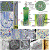
(a) Differential interference contrast image of embryo with first longitudinal division (yellow arrowhead). Scale bar 10 μm. (b) _S_porophyte. Spores mature progressively from the bottom to the top of the sporophyte. Involucre indicated with black arrow. Scale bar: 0.5 mm. (c) Schematic representation of sporophyte. The sporophyte has a foot, a basal meristem, the columella, a spore layer with pseudoelaters (for spore dispersal), a multicellular assimilative layer, and stomata. Stomata successive developmental stages are shown next to the sporophyte. Opening of the pore occurs near the base of the sporophyte then guard cells wall thickens, guard cells collapse toward the upper part of the sporophyte, allowing dehydration and dehiscence into two valves. Involucre indicated with black arrow. Numbered circles indicate the relative position on the sporophyte that corresponds to the cartoons on the right of panel: 1. Schematic representation of longitudinal section directly above the foot showing the basal meristem and differentiating sporogenous tissue. 2. Schematic representation of transverse section showing from centre to outside, columella, spores, pseudoelaters, assimilative tissue, epidermis and stomata with substomatal cavities. (d) LM longitudinal section of bulbous foot and basal meristem surrounded by the involucre. The placenta consists of elongated haustorial foot cells adjacent to small gametophyte cells. Scale bar: 50 μm. (e) Enlargement of placental cells in (d) showing smooth walled haustorial cells (red arrowhead) intermixed with gametophyte cells (orange arrowhead) with conspicuous cell wall ingrowths (orange arrowhead) Scale bar: 10 μm (f) LM longitudinal section of archesporial tissue (orange arrowhead) surrounding columella (red arrowhead) directly above the basal meristem. Scale bar: 20 μm (g) Transmission electron microscopy (TEM) image of columella in cross section showing 4 by 4 arrangement of the 16 living cells that contain dense cytosol and chloroplast (yellow arrowhead). Scale bar: 4.0 μm (h) LM section showing spore mother cells (red asterisks) with three of four large starch-filled plastids in four poles and central nucleus preparing for meiosis. Scale bar: 20 μm (i) TEM of dying and collapsing stoma (brown in (c)) showing thickened walls, inner and outer ledges of guard cells, and substomatal cavity. This section is on the polar end of the guard cells, away from the pore. Scale bar: 5 μm (j) SEM of a proximal surface of spore with a defined trilete mark. Scale bar: 10 μm. (k) TEM of spore in a tetrad still surrounded by the spore mother cell wall showing three-layered wall with ornamentation. The aperture on the proximal wall where spores in a tetrad meet each other has a thick intine and includes the trilete mark (red arrow). Scale bar 4.0 μm. (l) TEM of mature spore wall composed of outer exine (orange arrow), thick inner exine with compressed globular sporopollenin (green arrow) and thin intine that is much like a primary cell wall (red arrow). Protein bodies fill the spore (pink arrow). Scale bar 0.5 μm. (m) LM of dissected columella with elongated multicellular pseudoelaters attached. Scale bars: 25 μm.
Fig. 7:. A. agrestis chloroplast
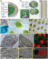
(a) Schematic representation of C. reinhardtii CCM model. The carbonic anhydrase (CAH2) converts CO2 into HCO3− (dicarboxylate (DIC)) in the periplasmic space. DIC is pumped across membranes via DIC transporters localised in the plasma membrane (low-CO2 inducible 1 (LCI1) and high light activated 3 (HLA3)), the chloroplast envelope (low-CO2 inducible A (LCIA)) and the thylakoid membrane (low-CO2 inducible 11 (LCI11), Cre16.g662600 and Cre16.g663400). The carbonic anhydrase 3 (CAH3) in the thylakoid lumen converts HCO3− into CO2. supplied to RuBiSCo. EPYC1 is acting as glue between RuBiSCo units in the pyrenoid. The low-CO2 inducible B and C (LCIB/C) proteins are thought to form a molecular “ring” around the pyrenoid that acts as a barrier to CO2 leakage transferring CO2 back to the thylakoid via the DIC pumps. (b) Top left boxes: Genes of prokaryotic origin (in blue) and genes of land plant origin (in green) involved in chloroplast division in A. thaliana. Genes absent in A. agrestis genome are in grey. Top right and bottom: Schematic representation of the key elements of chloroplast division machinery in A. thaliana (Chen et al., 2018). FtsZ1 and FtsZ2 self-assemble and then recruit ARC6, PARC6, PDV1, PDV2 and DRP5B forming a ring around the chloroplast that mediates its division. IE: inner envelope, OE: outer envelope. (c) Surface view of sporophyte showing chloroplasts. Scale bar: 100 μm. (d) LM section of spore mother cell with plastids (three out of four visible) at poles indicated with asterisks. Scale bar: 15 μm (e) LM of spore mother cell stained with DAPI showing DNA in central nucleus and plastids (orange arrowhead). Scale bar: 15 μm (f) Cells of young gametophyte tissue with a single chloroplast and central pyrenoid surrounded by starch grains. Scale bar: 10 μm (g) Cells of mature gametophyte tissue with single chloroplasts that have several protrusions that are likely stromules (orange arrowhead). Scale bar: 50 μm. (h-k) TEMs of chloroplasts of A. agrestis. (h) Chloroplast in an assimilative cell of sporophyte near intercellular space, with pyrenoid (red arrowhead) traversed by thylakoids and small grana (orange arrowhead). Starch granules indicated by yellow arrowhead and thylakoids enlarged in (i) at yellow arrowhead. Scale bar: 2.0 μm. (i) Details of thylakoids and grana showing absence of end membranes. Scale bar: 0.5 μm. (j&k) Sporophyte chloroplast in an assimilative cell near intercellular space traversed by channel thylakoids and grana stacks (yellow arrowhead). Starch grains indicated by orange arrowhead. Pyrenoid is not in this non-median section. Scale bar: 500 nm. (k) Higher magnification (scale bar: 0.5 μm) of region indicated in (j) showing channel thylakoids (red arrowhead). Scale bar: 2.0 μm. (l) Confocal microscopy image of A. agrestis transgenic gametophyte. Green: green fluorescent protein localised in the plasma membrane expressed under the CaMV 35S promoter. Red: chlorophyll autofluorescence. Orange arrowhead indicates chloroplast protrusions likely to be stromules. Scale bar: 10 μm (m) Confocal microscopy image of a germinating spore highlighting the wavy 3D structure of the chloroplast. Red: chlorophyll autofluorescence. Scale bar: 20 μm.
Fig. 8:. Hornwort symbiotic relationships
(a) Surface view of A. puncatus thallus colonised by cyanobacteria (yellow arrowheads). Scale bar: 450 μm. (b-c) Hand sections of A. punctatus thallus showing ellipsoidal cavities colonised by cyanobacteria. In (c) cyanobacteria indicated with red arrowhead Scale bars: 100 μm (b) and 10 μm (c). (d) LM section of Nostoc colony showing algal cells (red arrowhead) with intermingling gametophyte cells in A. agrestis. Scale bar: 10 μm (e) LM surface view of ventral mucilage cleft. Scale bar: 15 μm. (f) Longitudinal section of a mucilage cleft (red arrowhead) leading to small intercellular space near apical notch of A. agrestis. Scale bar: 50 μm (g) LM transverse section of sporophyte showing guard cell in epidermis that lead to substomatal cavities. Guard cells are larger than epidermal cells and have differentially thickened cell walls with inner and outer ledges and are different from mucilage cleft cells in (f) that have evenly thickened walls. Scale bar: 50 μm. (h) LM showing surface view of a mucilage cleft and attracted cyanobacteria just entered the cleft in Phaeoceros carolinianus. Scale bar:20 μm (i, j) Hornwort symbiotic relationship with arbuscular mycorrhizal fungi. Hand section LM of P. carolinianus thallus cells with fungal hyphae (red arrowhead). Scale bar: 10 μm. (j) LM section of gametophyte cells containing vesicles (circles) and arbuscules (masses of hyphae in cells). Scale bar: 20 μm
Similar articles
- An optimized transformation protocol for Anthoceros agrestis and three more hornwort species.
Waller M, Frangedakis E, Marron AO, Sauret-Güeto S, Rever J, Sabbagh CRR, Hibberd JM, Haseloff J, Renzaglia KS, Szövényi P. Waller M, et al. Plant J. 2023 May;114(3):699-718. doi: 10.1111/tpj.16161. Epub 2023 Apr 11. Plant J. 2023. PMID: 36811359 Free PMC article. - Major transitions in the evolution of early land plants: a bryological perspective.
Ligrone R, Duckett JG, Renzaglia KS. Ligrone R, et al. Ann Bot. 2012 Apr;109(5):851-71. doi: 10.1093/aob/mcs017. Epub 2012 Feb 22. Ann Bot. 2012. PMID: 22356739 Free PMC article. Review. - Anthoceros genomes illuminate the origin of land plants and the unique biology of hornworts.
Li FW, Nishiyama T, Waller M, Frangedakis E, Keller J, Li Z, Fernandez-Pozo N, Barker MS, Bennett T, Blázquez MA, Cheng S, Cuming AC, de Vries J, de Vries S, Delaux PM, Diop IS, Harrison CJ, Hauser D, Hernández-García J, Kirbis A, Meeks JC, Monte I, Mutte SK, Neubauer A, Quandt D, Robison T, Shimamura M, Rensing SA, Villarreal JC, Weijers D, Wicke S, Wong GK, Sakakibara K, Szövényi P. Li FW, et al. Nat Plants. 2020 Mar;6(3):259-272. doi: 10.1038/s41477-020-0618-2. Epub 2020 Mar 13. Nat Plants. 2020. PMID: 32170292 Free PMC article. - The hornwort genome and early land plant evolution.
Zhang J, Fu XX, Li RQ, Zhao X, Liu Y, Li MH, Zwaenepoel A, Ma H, Goffinet B, Guan YL, Xue JY, Liao YY, Wang QF, Wang QH, Wang JY, Zhang GQ, Wang ZW, Jia Y, Wang MZ, Dong SS, Yang JF, Jiao YN, Guo YL, Kong HZ, Lu AM, Yang HM, Zhang SZ, Van de Peer Y, Liu ZJ, Chen ZD. Zhang J, et al. Nat Plants. 2020 Feb;6(2):107-118. doi: 10.1038/s41477-019-0588-4. Epub 2020 Feb 10. Nat Plants. 2020. PMID: 32042158 Free PMC article. - The cell wall of hornworts and liverworts: innovations in early land plant evolution?
Pfeifer L, Mueller KK, Classen B. Pfeifer L, et al. J Exp Bot. 2022 Jul 16;73(13):4454-4472. doi: 10.1093/jxb/erac157. J Exp Bot. 2022. PMID: 35470398 Review.
Cited by
- Cell wall polymers in the Phaeoceros placenta reflect developmental and functional differences across generations.
Henry JS, Ligrone R, Vaughn KC, Lopez RA, Renzaglia KS. Henry JS, et al. Bryophyt Divers Evol. 2021 Jun 30;43(1):265-283. doi: 10.11646/bde.43.1.19. Bryophyt Divers Evol. 2021. PMID: 34532591 Free PMC article. - Accelerating gametophytic growth in the model hornwort Anthoceros agrestis.
Gunadi A, Li FW, Van Eck J. Gunadi A, et al. Appl Plant Sci. 2022 Mar 6;10(2):e11460. doi: 10.1002/aps3.11460. eCollection 2022 Mar-Apr. Appl Plant Sci. 2022. PMID: 35495194 Free PMC article. - The bryophytes Physcomitrium patens and Marchantia polymorpha as model systems for studying evolutionary cell and developmental biology in plants.
Naramoto S, Hata Y, Fujita T, Kyozuka J. Naramoto S, et al. Plant Cell. 2022 Jan 20;34(1):228-246. doi: 10.1093/plcell/koab218. Plant Cell. 2022. PMID: 34459922 Free PMC article. Review. - Symbiosis between cyanobacteria and plants: from molecular studies to agronomic applications.
Álvarez C, Jiménez-Ríos L, Iniesta-Pallarés M, Jurado-Flores A, Molina-Heredia FP, Ng CKY, Mariscal V. Álvarez C, et al. J Exp Bot. 2023 Oct 13;74(19):6145-6157. doi: 10.1093/jxb/erad261. J Exp Bot. 2023. PMID: 37422707 Free PMC article. Review. - Newly found and rediscovered hornworts (Anthocerotophyta) in Poland: Indicators of climate change impact in Central Europe.
Plášek V, Číhal L, Müller F, Pöltl M, Wierzgoń M, Ochyra R. Plášek V, et al. PhytoKeys. 2024 Oct 31;248:237-261. doi: 10.3897/phytokeys.248.134729. eCollection 2024. PhytoKeys. 2024. PMID: 39525526 Free PMC article.
References
- Adams DG. 2002. Cyanobacteria in symbiosis with hornworts and liverworts In: Rai AN, Bergman B, Rasmussen U, eds. Cyanobacteria in Symbiosis. Springer, Dordrecht, 117–135.
- Adams DG, Duggan PS. 2008. Cyanobacteria-bryophyte symbioses. Journal of experimental botany 59: 1047–1058. - PubMed
- Albert VA. 1999. Shoot apical meristems and floral patterning: an evolutionary perspective. Trends in Plant Science 4: 84–86.
- Barkan A, Small I. 2014. Pentatricopeptide repeat proteins in plants. Annual Review of Plant Biology 65: 415–442. - PubMed
Publication types
MeSH terms
LinkOut - more resources
Full Text Sources


