Effects of Methanolic Extract Based-Gel From Saudi Pomegranate Peels With Enhanced Healing Potential on Excision Wounds in Diabetic Rats - PubMed (original) (raw)
Effects of Methanolic Extract Based-Gel From Saudi Pomegranate Peels With Enhanced Healing Potential on Excision Wounds in Diabetic Rats
Shahid Karim et al. Front Pharmacol. 2021.
Abstract
Introduction: Current study was designed to evaluate the wound healing activity of a Saudi pomegranate peel extract on excision wound healing in experimentally induced diabetes in rats. Methodology: Animals were divided into three groups: diabetic excision wound with no treatment, diabetic excision wound with gel alone and diabetic excision wound with Saudi pomegranate peel extract in gel. Animals were monitored for clinical signs, weekly body weight, morbidity and mortality during entire study period. The efficacy parameters evaluated were percent wound contraction, Hydroxyproline content, estimation of Transforming Growth Factor ß1 (TGF-ß1), Vascular Endothelial Growth Factor (VEGF), and Epidermal Growth Factor (EGF) in wound lysates by ELISA, mRNA expression of TGF-ß1, VEGF, and EGF in wound lysates by qPCR, Estimation of nitric oxide (NO) and NO synthase (NOS) in Wound Lysates and histopathology of skin for reepithelization, neovascularization, and inflammation. Results: The Saudi pomegranate peel extract in gel (5.0 g extract per 100 g gel) showed significant wound healing activity when compared to the vehicle control [p < 0.05] following 21 days of treatment. Animals in the control and treatment groups were apparently normal through the study with no significant differences in body weights between groups. Expression of mRNA of TGFβ1, EGF and VEGF in wounds was the highest on day 14 post treatment 4.3, 3.5 and 0.9 fold higher respectively in the treatment group when compared to vehicle control, and on day 21, the values were 0.12, 0.3 and 0.83, respectively. No statistically significant differences were observed in TGF-ß1 levels in wounds on days 4, 7, 14 and 21 post treatment when compared to the vehicle control (_p_ > 0.05). Significantly higher levels of VEGF were observed in treatment group on day 7 and 21 when compared to vehicle control (p < 0.05). Significantly higher levels of EGF were observed in treatment group on day 7 and 21 when compared to vehicle control (p < 0.05). Mean hydroxyproline levels were higher in treatment group on days 4 and 7 when compared to vehicle control. NO levels in treatment group were significantly lower on days 7, 14 and 21 when compared to vehicle control (p < 0.05). NOS activity in treatment group were significantly lower on days 4 and 7 when compared to vehicle control (p < 0.05). Histopathological changes in skin wound in the treatment group were consistent with wound healing when compared to the vehicle group. Conclusion: This study's findings suggest that topical application of SPPE gel effectively enhanced wound healing in experimentally induced diabetic conditions.
Keywords: EGF; TGF beta 1; VEGF; diabetes; nitric oxide; pomegranate peel extract; topical gel; wound healing.
Copyright © 2021 Karim, Alkreathy, Ahmad and Khan.
Conflict of interest statement
The authors declare that the research was conducted in the absence of any commercial or financial relationships that could be construed as a potential conflict of interest.
Figures
FIGURE 1
(A) Calibration curve of standard gallic acid for determination of total phenolic content in SPPE. (B) Calibration curve of standard quercetin for determination of total flavonoid content in SPPE.
FIGURE 2
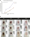
Effect of SPPE gel on wound contraction in diabetic rats. (A) The percentage wound contraction was measured at 4, 7, 14, and 21 days after wound creation. Data expressed as mean ± SEM (n = 6). (B) Representative pictures of wound contraction at 4, 7, 14, and 21 days after wound creation.
FIGURE 3
Effect of treatments with Diabetic control (G1), Vehicle (G2), and SPPE gel (G3) on hydroxyproline contents in wound tissues of rats at day 4, 7, 14 and 21 post wounding. Values are represented as mean ± SEM (n = 6).
FIGURE 4
Effect of SPPE gel on expression of TGF-β1 (A), VEGF (B), and EGF (C) in wound tissues of rats at day 4, 7, 14, and 21 post treatment Diabetic control (G1), Vehicle (G2), SPPE gel (G3). Values are represented as mean ± SEM (n = 6).
FIGURE 5
Nitric Oxide (NO) levels (A) and Nitric Oxide synthase (NOS) activity (B) in skin homogenate following treatments with diabetic control (G1), Vehicle (G2), SPPE gel (G3) Values are represented as mean ± SEM (n = 6).
FIGURE 6
(A) G1, Day 0; Skin with normal epidermis (EP) and dermis (H&E X 100×). (B) G1, day 4: Skin with inflammatory cells (IN), collagen formation (C), neovascularization (arrowhead) and ulceration (U) (H&E X 100×). (C) G2, Day 4: Skin with inflammatory cells (IN) and collagen formation (C) (H&E X 100×) (D) G3, Day 4: Skin with inflammatory cells (IN), collagen formation (C) and fibroblast proliferation (F) and neovascularization (arrowhead) (H&E X 100×). (E) G1, Day 7: Skin with inflammatory cells (IN), collagen formation (C), fibroblast proliferation (F) and neovascularization (arrowhead) (H&E X 100×). (F) G2, day 7: Skin with inflammatory cells (IN), granulation tissue (G), fibroblast proliferation (arrow), neovascularization (arrowhead) and immature epidermis (IE) (H&E X 100×). (G) G3, Day 7: Skin with inflammatory cells (IN), collagen formation (C), granulation tissue (G), fibroblast proliferation (arrow), neovascularization (arrowhead) and immature epidermis (IE) (H&E X 100×). (H) G1, Day 14: Skin with inflammatory cells (IN), collagen formation (C), neovascularization (arrowhead) and immature epidermis (IE) (H&E X 100×). (I) G2, Day 14: Skin with inflammatory cells (IN) and collagen formation (C) (H&E X 100×). (J) G3, Day 14: Skin with inflammatory cells (IN), collagen formation (C), granulation tissue (G), fibroblast proliferation (arrow), neovascularization (arrowhead) and immature epidermis (IE) (H&E X 100×). (K) G1, Day 21: Skin with inflammatory cells (IN), collagen formation (C), granulation tissue (G), fibroblast proliferation (arrow), neovascularization (arrowhead) and immature epidermis (IE) (H&E X 100×). (L) G2_,_ Day 21: Skin with inflammatory cells (IN), collagen formation (C), granulation tissue (G), fibroblast proliferation (arrow), neovascularization (arrowhead) and immature epidermis (IE) (H&E X 100×). (M) G3, Day 21: Skin with inflammatory cells (IN), granulation tissue (G), neovascularization (arrowhead) and immature epidermis (IE) (H&E X 100×).
FIGURE 7
Box plots of histopathology scores for skin parameters through treatment. Horizontal lines in boxes represent median and error bars represent minimum and maximum. *Significantly different from G2 [p < 0.05, Kruskal-Wallis test]. 0 = None, 1 = Rare or Minimal, 2 = Moderate, 3 = Abundant, and 4 = Severe or Marked.
Similar articles
- Effect of pomegranate peel polyphenol gel on cutaneous wound healing in alloxan-induced diabetic rats.
Yan H, Peng KJ, Wang QL, Gu ZY, Lu YQ, Zhao J, Xu F, Liu YL, Tang Y, Deng FM, Zhou P, Jin JG, Wang XC. Yan H, et al. Chin Med J (Engl). 2013;126(9):1700-6. Chin Med J (Engl). 2013. PMID: 23652054 - [Effects and mechanism of rat epidermal stem cells treated with exogenous vascular endothelial growth factor on healing of deep partial-thickness burn wounds in rats].
Shi Y, Tu LX, Deng Q, Zhang YP, Hu YH, Liu DW. Shi Y, et al. Zhonghua Shao Shang Za Zhi. 2020 Mar 20;36(3):195-203. doi: 10.3760/cma.j.cn501120-20191125-00441. Zhonghua Shao Shang Za Zhi. 2020. PMID: 32241045 Chinese. - Study on wound healing activity of Punica granatum peel.
Murthy KN, Reddy VK, Veigas JM, Murthy UD. Murthy KN, et al. J Med Food. 2004 Summer;7(2):256-9. doi: 10.1089/1096620041224111. J Med Food. 2004. PMID: 15298776 - [The modern approach to wound treatment].
Komarcević A. Komarcević A. Med Pregl. 2000 Jul-Aug;53(7-8):363-8. Med Pregl. 2000. PMID: 11214479 Review. Croatian. - EGF and TGF-alpha in wound healing and repair.
Schultz G, Rotatori DS, Clark W. Schultz G, et al. J Cell Biochem. 1991 Apr;45(4):346-52. doi: 10.1002/jcb.240450407. J Cell Biochem. 1991. PMID: 2045428 Review.
Cited by
- Environmental and Economic Benefits of Using Pomegranate Peel Waste for Insulation Bricks.
Ragab A, Zouli N, Abutaleb A, Maafa IM, Ahmed MM, Yousef A. Ragab A, et al. Materials (Basel). 2023 Jul 31;16(15):5372. doi: 10.3390/ma16155372. Materials (Basel). 2023. PMID: 37570075 Free PMC article. - Effects of Pomegranate on Wound Repair and Regeneration.
Bahadoram M, Hassanzadeh S, Bahadoram S, Mowla K. Bahadoram M, et al. World J Plast Surg. 2022 Mar;11(1):157-159. doi: 10.52547/wjps.11.1.157. World J Plast Surg. 2022. PMID: 35592232 Free PMC article. No abstract available. - Pomegranate Peel Phytochemistry, Pharmacological Properties, Methods of Extraction, and Its Application: A Comprehensive Review.
Singh J, Kaur HP, Verma A, Chahal AS, Jajoria K, Rasane P, Kaur S, Kaur J, Gunjal M, Ercisli S, Choudhary R, Bozhuyuk MR, Sakar E, Karatas N, Durul MS. Singh J, et al. ACS Omega. 2023 Sep 19;8(39):35452-35469. doi: 10.1021/acsomega.3c02586. eCollection 2023 Oct 3. ACS Omega. 2023. PMID: 37810640 Free PMC article. Review. - Cordycepin- melittin nanoconjugate intensifies wound healing efficacy in diabetic rats.
Shaik RA, Alotaibi MF, Nasrullah MZ, Alrabia MW, Asfour HZ, Abdel-Naim AB. Shaik RA, et al. Saudi Pharm J. 2023 May;31(5):736-745. doi: 10.1016/j.jsps.2023.03.014. Epub 2023 Mar 27. Saudi Pharm J. 2023. PMID: 37181143 Free PMC article. - Horsetail (Equisetum hyemale) Extract Accelerates Wound Healing in Diabetic Rats by Modulating IL-10 and MCP-1 Release and Collagen Synthesis.
Aguayo-Morales H, Sierra-Rivera CA, Claudio-Rizo JA, Cobos-Puc LE. Aguayo-Morales H, et al. Pharmaceuticals (Basel). 2023 Mar 30;16(4):514. doi: 10.3390/ph16040514. Pharmaceuticals (Basel). 2023. PMID: 37111271 Free PMC article.
References
- Andrade M. A., Lima V., Sanches Silva A., Vilarinho F., Castilho M. C., Khwaldia K., et al. (2019). Pomegranate and Grape By-Products and Their Active Compounds: Are They a Valuable Source for Food Applications?. Trends Food Sci. Tech. 86, 68–84. 10.1016/j.tifs.2019.02.010 - DOI
LinkOut - more resources
Full Text Sources


