Investigating Tumor Heterogeneity in Mouse Models - PubMed (original) (raw)
Investigating Tumor Heterogeneity in Mouse Models
Tuomas Tammela et al. Annu Rev Cancer Biol. 2020 Mar.
Abstract
Cancer arises from a single cell through a series of acquired mutations and epigenetic alterations. Tumors gradually develop into a complex tissue comprised of phenotypically heterogeneous cancer cell populations, as well as noncancer cells that make up the tumor microenvironment. The phenotype, or state, of each cancer and stromal cell is influenced by a plethora of cell-intrinsic and cell-extrinsic factors. The diversity of these cellular states promotes tumor progression, enables metastasis, and poses a challenge for effective cancer treatments. Thus, the identification of strategies for the therapeutic manipulation of tumor heterogeneity would have significant clinical implications. A major barrier in the field is the difficulty in functionally investigating heterogeneity in tumors in cancer patients. Here we review how mouse models of human cancer can be leveraged to interrogate tumor heterogeneity and to help design better therapeutic strategies.
Keywords: cancer stem cells; epigenetics; genetics; mouse models; tumor heterogeneity; tumor microenvironment.
Figures
Figure 1
Heterogeneity in cancer. Tumors have several levels of heterogeneity. (a) During tumorigenesis, mutations in the DNA and genetic heterogeneity (represented by different colors) can arise from normal cell division, from exposure to mutagens, or in response to treatment in the clinic. This may lead to the appearance (and disappearance) of subclones. (b) Even when cancer cells are identical or very similar at the genetic level, tumors are often organized in a hierarchical manner in which cancer stem cells can self-renew and give rise to daughter cells with different transcriptional programs (represented by different shapes). (c) Cancer cells in tumors are intimately connected with several noncancer cells; heterogeneity can also be found within each subtype of noncancer cells in the microenvironment (e.g., fibroblasts, endothelial cells, or T cells).
Figure 2
Lineage-tracing strategies for the prospective interrogation of cell fate determination in cancer. (Top) Classic lineage tracing is based on the introduction of a heritable genetic mark, e.g., a fluorescent protein (green). Cancer cells with stem-like properties marked in this way will clonally expand upon tumor growth. scRNA-seq can be used in such an experiment to determine whether the progeny of a stem-like cell can give rise to all cancer cell subtypes. (Middle) CRISPR genome recording can be used to connect a cell’s transcriptional state at the time of analysis to its clonal ancestry. This method can be used to reveal dominant clones. (Bottom) CRISPR recording can also be used to record the cumulative activity of a pathway of interest. A history of pathway activity associated with increased fitness or stemness will be detected in most cancer cells. Conversely, the bulk tumor cell population will display few recorded events of a pathway that suppresses tumor progression and clonal expansion. Abbreviations: scRNA-seq, single-cell RNA sequencing; t-SNE dim n, _n_th dimension of t-distributed stochastic neighbor embedding.
Figure 3
Cell-extrinsic signals that can generate epigenetic heterogeneity in cancer cells. (a) The epigenetic state of cancer cells is under the control of multiple factors in the microenvironment. (b) Representative images of heterogeneity in mouse models of small cell lung carcinoma (SCLC) and lung adenocarcinoma (LUAD). (Left) Immunohistochemistry (brown) for Hes1 marks the nucleus of Notch-active SCLC cells in a p53/Rb/p130_-mutant mouse. The counterstain is hematoxylin (purple). (Right) Immunostaining for green fluorescent protein (GFP; green) and porcupine (red) in a subcutaneous transplant of primary Kras_G12D/+;p53_Δ/Δ;Lgr5_GFP-CreER/+ mouse LUAD cells three weeks after transplantation. DNA is stained in blue. Note the juxtaposition between the green GFP+ Lgr5+ cells and the red porcupine+ cells. SCLC image courtesy of the Sage lab; LUAD image reproduced with permission from Tammela et al. (2017). (c) In SCLC, some of the cancer stem cells change their fate upon activation of Notch signaling: The newly formed non-neuroendocrine cancer cells (Hes1+) serve as a supportive niche that produces growth and survival factors. Blocking Notch activation may be a therapeutic strategy to prevent the generation of the supportive niche population. (d) In LUAD, cancer stem cells rely on Wnt signaling for long-term expansion. Some of these stem-like cancer cells (Lgr5+) can differentiate into a supportive niche population that secretes active Wnt ligands (porcupine+), thereby allowing the maintenance of the reciprocal interaction. Blocking the generation of active Wnt ligands or Wnt signaling may be a therapeutic strategy to prevent the long-term expansion of LUAD stem cells.
Figure 4
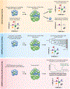
Strategies to target heterogeneity in cancer. (Top) Tumors contain heterogeneous subpopulations of cancer cells that are intrinsically resistant to therapies, enabling the tumor to acutely evade therapy and develop adaptive resistance. (Bottom) Targeting mechanisms that drive heterogeneity can either push cancer cells into states that are responsive to conventional therapies (phenotype switching) or eliminate specific subpopulations through the identification of critical druggable dependencies before or during treatment of the tumor with conventional therapy.
Similar articles
- Cell-of-Origin and Genetic, Epigenetic, and Microenvironmental Factors Contribute to the Intra-Tumoral Heterogeneity of Pediatric Intracranial Ependymoma.
Servidei T, Lucchetti D, Navarra P, Sgambato A, Riccardi R, Ruggiero A. Servidei T, et al. Cancers (Basel). 2021 Dec 3;13(23):6100. doi: 10.3390/cancers13236100. Cancers (Basel). 2021. PMID: 34885210 Free PMC article. Review. - Roles of phenotypic heterogeneity and microenvironment feedback in early tumor development.
Smart M, Goyal S, Zilman A. Smart M, et al. Phys Rev E. 2021 Mar;103(3-1):032407. doi: 10.1103/PhysRevE.103.032407. Phys Rev E. 2021. PMID: 33862830 - The role of tumor microenvironment in drug resistance: emerging technologies to unravel breast cancer heterogeneity.
Salemme V, Centonze G, Avalle L, Natalini D, Piccolantonio A, Arina P, Morellato A, Ala U, Taverna D, Turco E, Defilippi P. Salemme V, et al. Front Oncol. 2023 May 17;13:1170264. doi: 10.3389/fonc.2023.1170264. eCollection 2023. Front Oncol. 2023. PMID: 37265795 Free PMC article. Review. - Heterogeneity of Colon Cancer Stem Cells.
Hirata A, Hatano Y, Niwa M, Hara A, Tomita H. Hirata A, et al. Adv Exp Med Biol. 2019;1139:115-126. doi: 10.1007/978-3-030-14366-4_7. Adv Exp Med Biol. 2019. PMID: 31134498 Review. - Cancer stem cells: a major culprit of intra-tumor heterogeneity.
Naz F, Shi M, Sajid S, Yang Z, Yu C. Naz F, et al. Am J Cancer Res. 2021 Dec 15;11(12):5782-5811. eCollection 2021. Am J Cancer Res. 2021. PMID: 35018226 Free PMC article. Review.
Cited by
- Lymphatic-specific methyltransferase-like 3-mediated m6A modification drives vascular patterning through prostaglandin metabolism reprogramming.
Shi L, Lu S, Han X, Ye F, Li X, Zhang Z, Jiang Q, Yan B. Shi L, et al. MedComm (2020). 2024 Oct 4;5(10):e728. doi: 10.1002/mco2.728. eCollection 2024 Oct. MedComm (2020). 2024. PMID: 39372388 Free PMC article. - Robotic data acquisition with deep learning enables cell image-based prediction of transcriptomic phenotypes.
Jin J, Ogawa T, Hojo N, Kryukov K, Shimizu K, Ikawa T, Imanishi T, Okazaki T, Shiroguchi K. Jin J, et al. Proc Natl Acad Sci U S A. 2023 Jan 3;120(1):e2210283120. doi: 10.1073/pnas.2210283120. Epub 2022 Dec 28. Proc Natl Acad Sci U S A. 2023. PMID: 36577074 Free PMC article. - Microfluidic harvesting of breast cancer tumor spheroid-derived extracellular vesicles from immobilized microgels for single-vesicle analysis.
Rima XY, Zhang J, Nguyen LTH, Rajasuriyar A, Yoon MJ, Chiang CL, Walters N, Kwak KJ, Lee LJ, Reátegui E. Rima XY, et al. Lab Chip. 2022 Jun 28;22(13):2502-2518. doi: 10.1039/d1lc01053k. Lab Chip. 2022. PMID: 35579189 Free PMC article. - Rodent Models to Analyze the Glioma Microenvironment.
Hetze S, Sure U, Schedlowski M, Hadamitzky M, Barthel L. Hetze S, et al. ASN Neuro. 2021 Jan-Dec;13:17590914211005074. doi: 10.1177/17590914211005074. ASN Neuro. 2021. PMID: 33874781 Free PMC article. Review. - DLL3 regulates Notch signaling in small cell lung cancer.
Kim JW, Ko JH, Sage J. Kim JW, et al. iScience. 2022 Nov 16;25(12):105603. doi: 10.1016/j.isci.2022.105603. eCollection 2022 Dec 22. iScience. 2022. PMID: 36483011 Free PMC article.
References
- Assenov Y, Brocks D, Gerhauser C. 2018. Intratumor heterogeneity in epigenetic patterns. Semin. Cancer Biol 51:12–21 - PubMed
- Balkwill FR, Capasso M, Hagemann T. 2012. The tumor microenvironment at a glance. J. Cell Sci 125:5591–96 - PubMed
- Barker N, van Es JH, Kuipers J, Kujala P, van den Born M, et al. 2007. Identification of stem cells in small intestine and colon by marker gene Lgr5. Nature 449:1003–7 - PubMed
Grants and funding
- U54 CA217450/CA/NCI NIH HHS/United States
- U01 CA231851/CA/NCI NIH HHS/United States
- K99 CA187317/CA/NCI NIH HHS/United States
- R01 CA201513/CA/NCI NIH HHS/United States
- R35 CA231997/CA/NCI NIH HHS/United States
- R01 CA206540/CA/NCI NIH HHS/United States
- U01 CA213273/CA/NCI NIH HHS/United States
- R00 CA187317/CA/NCI NIH HHS/United States
LinkOut - more resources
Full Text Sources


