Retinal inter-eye difference and atrophy progression in multiple sclerosis diagnostics - PubMed (original) (raw)
Retinal inter-eye difference and atrophy progression in multiple sclerosis diagnostics
Jenny Nij Bijvank et al. J Neurol Neurosurg Psychiatry. 2022 Feb.
Abstract
Background: The visual system could be included in the diagnostic criteria for multiple sclerosis (MS) to demonstrate dissemination in space (DIS) and dissemination in time (DIT).
Objective: To investigate the diagnostic value of retinal asymmetry in MS.
Methods: A prospective, longitudinal study in individuals with MS (n=151) and healthy controls (n=27). Optical coherence tomography (OCT) was performed at 0, 2 and 4 years. Macular ganglion cell and inner plexiform layer (mGCIPL) thickness was determined as well as measures for retinal asymmetry: the inter-eye percentage difference (IEPD) and inter-eye absolute difference (IEAD). Receiver operator characteristics curves were plotted and the area under the curve (AUC) was calculated for group comparisons of the mGCIPL, IEPD, IEAD and atrophy rates.
Results: The diagnostic accuracy of both the IEPD and IEAD for differentiating bilateral and unilateral MS optic neuritis was high and stable over time (AUCs 0.88-0.93). The IEPD slightly outperformed the IEAD. Atrophy rates showed low discriminatory abilities for differentiating MS from controls (AUC 0.49-0.58).
Conclusion: The inter-eye differences of the mGCIPL have value for demonstration of DIS but in individuals with longstanding MS not for DIT. This may be considered as a test to detect DIS in future diagnostic criteria. Validation in a large prospective study in people presenting with symptoms suggestive of MS is required.
Keywords: multiple sclerosis; neuroophthalmology.
© Author(s) (or their employer(s)) 2022. Re-use permitted under CC BY-NC. No commercial re-use. See rights and permissions. Published by BMJ.
Conflict of interest statement
Competing interests: JNB is supported by the Dutch MS Research Foundation, grant nr. 18-1027. BMJU has received consultancy fees from Biogen Idec, Genzyme, Merck Serono, Novartis, Roche and Teva. AP reports personal fees from Novartis, Heidelberg Engineering, Zeiss, grants from Novartis, outside the submitted work; and AP is part of the steering committee of the OCTiMS study which is sponsored by Novartis. AP is part of the steering committee of Angio-OCT which is sponsored by Zeiss. He does not receive honorary as part of these activities. The NIHR BRC at Moorfields Eye Hospital supported AP.
Figures
Figure 1
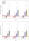
Box-and-Whisker plots of (A) the inter-eye percentage differences (IEPD) and (B) the inter-eye absolute difference (IEAD) at baseline, year 2 and year 4. The red line indicates the 5% cut-off for the IEPD and the 4 µm cut-off of the IEAD. BON, bilateral MS associated optic neuritis; HC, healthy controls; MS, multiple sclerosis; NON, no MS associated optic neuritis; ON, unilateral MS associated optic neuritis. The median (bold horizontal line), 25–75 percentiles (box), 5–95 percentiles (whiskers), mean (symbol in the box) and outliers (symbols outside the box) are shown.
Similar articles
- Diagnostic accuracy of optical coherence tomography inter-eye percentage difference for optic neuritis in multiple sclerosis.
Coric D, Balk LJ, Uitdehaag BMJ, Petzold A. Coric D, et al. Eur J Neurol. 2017 Dec;24(12):1479-1484. doi: 10.1111/ene.13443. Epub 2017 Oct 9. Eur J Neurol. 2017. PMID: 28887838 - Retinal asymmetry in multiple sclerosis.
Petzold A, Chua SYL, Khawaja AP, Keane PA, Khaw PT, Reisman C, Dhillon B, Strouthidis NG, Foster PJ, Patel PJ; UK Biobank Eye and Vision Consortium. Petzold A, et al. Brain. 2021 Feb 12;144(1):224-235. doi: 10.1093/brain/awaa361. Brain. 2021. PMID: 33253371 Free PMC article. - Diagnostic value of intereye difference metrics for optic neuritis in aquaporin-4 antibody seropositive neuromyelitis optica spectrum disorders.
Oertel FC, Zimmermann HG, Motamedi S, Chien C, Aktas O, Albrecht P, Ringelstein M, Dcunha A, Pandit L, Martinez-Lapiscina EH, Sanchez-Dalmau B, Villoslada P, Palace J, Roca-Fernández A, Leite MI, Sharma SM, Leocani L, Pisa M, Radaelli M, Lana-Peixoto MA, Fontenelle MA, Havla J, Ashtari F, Kafieh R, Dehghani A, Pourazizi M, Marignier R, Cobo-Calvo A, Asgari N, Jacob A, Huda S, Mao-Draayer Y, Green AJ, Kenney R, Yeaman MR, Smith TJ, Cook L, Brandt AU, Paul F, Petzold A. Oertel FC, et al. J Neurol Neurosurg Psychiatry. 2023 Jul;94(7):560-566. doi: 10.1136/jnnp-2022-330608. Epub 2023 Feb 21. J Neurol Neurosurg Psychiatry. 2023. PMID: 36810323 Free PMC article. - Optical coherence tomography in optic neuritis and multiple sclerosis: a review.
Kallenbach K, Frederiksen J. Kallenbach K, et al. Eur J Neurol. 2007 Aug;14(8):841-9. doi: 10.1111/j.1468-1331.2007.01736.x. Eur J Neurol. 2007. PMID: 17662003 Review. - Retinal layer segmentation in multiple sclerosis: a systematic review and meta-analysis.
Petzold A, Balcer LJ, Calabresi PA, Costello F, Frohman TC, Frohman EM, Martinez-Lapiscina EH, Green AJ, Kardon R, Outteryck O, Paul F, Schippling S, Vermersch P, Villoslada P, Balk LJ; ERN-EYE IMSVISUAL. Petzold A, et al. Lancet Neurol. 2017 Oct;16(10):797-812. doi: 10.1016/S1474-4422(17)30278-8. Epub 2017 Sep 12. Lancet Neurol. 2017. PMID: 28920886 Review.
Cited by
- Diagnosis of multiple sclerosis using optical coherence tomography supported by explainable artificial intelligence.
Dongil-Moreno FJ, Ortiz M, Pueyo A, Boquete L, Sánchez-Morla EM, Jimeno-Huete D, Miguel JM, Barea R, Vilades E, Garcia-Martin E. Dongil-Moreno FJ, et al. Eye (Lond). 2024 Jun;38(8):1502-1508. doi: 10.1038/s41433-024-02933-5. Epub 2024 Jan 31. Eye (Lond). 2024. PMID: 38297153 - Diagnostic Performance of Adding the Optic Nerve Region Assessed by Optical Coherence Tomography to the Diagnostic Criteria for Multiple Sclerosis.
Bsteh G, Hegen H, Altmann P, Auer M, Berek K, Di Pauli F, Kornek B, Krajnc N, Leutmezer F, Macher S, Rommer PS, Zebenholzer K, Zulehner G, Zrzavy T, Deisenhammer F, Pemp B, Berger T; for VMSD (Vienna Multiple Sclerosis Database) Group. Bsteh G, et al. Neurology. 2023 Aug 22;101(8):e784-e793. doi: 10.1212/WNL.0000000000207507. Epub 2023 Jul 3. Neurology. 2023. PMID: 37400245 Free PMC article. - Diagnostic Value of Inter-Eye Difference Metrics on OCT for Myelin Oligodendrocyte Glycoprotein Antibody-Associated Optic Neuritis.
Volpe G, Jurkute N, Girafa G, Zimmermann HG, Motamedi S, Bereuter C, Pandit L, D'Cunha A, Yeaman MR, Smith TJ, Cook LJ, Brandt AU, Paul F, Petzold A, Oertel FC. Volpe G, et al. Neurol Neuroimmunol Neuroinflamm. 2024 Nov;11(6):e200291. doi: 10.1212/NXI.0000000000200291. Epub 2024 Sep 4. Neurol Neuroimmunol Neuroinflamm. 2024. PMID: 39231384 Free PMC article. - Delimiting MOGAD as a disease entity using translational imaging.
Oertel FC, Hastermann M, Paul F. Oertel FC, et al. Front Neurol. 2023 Dec 7;14:1216477. doi: 10.3389/fneur.2023.1216477. eCollection 2023. Front Neurol. 2023. PMID: 38333186 Free PMC article. Review. - Retinal optical coherence tomography measures in multiple sclerosis: a systematic review and meta-analysis.
El Ayoubi NK, Ismail A, Fahd F, Younes L, Chakra NA, Khoury SJ. El Ayoubi NK, et al. Ann Clin Transl Neurol. 2024 Sep;11(9):2236-2253. doi: 10.1002/acn3.52165. Epub 2024 Jul 28. Ann Clin Transl Neurol. 2024. PMID: 39073308 Free PMC article.
References
Publication types
MeSH terms
LinkOut - more resources
Full Text Sources
Medical
