4'-Fluorouridine is an oral antiviral that blocks respiratory syncytial virus and SARS-CoV-2 replication - PubMed (original) (raw)
. 2022 Jan 14;375(6577):161-167.
doi: 10.1126/science.abj5508. Epub 2021 Dec 2.
Carolin M Lieber 1, Megha Aggarwal 1, Robert M Cox 1, Josef D Wolf 1, Jeong-Joong Yoon 1, Mart Toots 1, Chengin Ye 2, Zachary Sticher 3, Alexander A Kolykhalov 3 4, Luis Martinez-Sobrido 2, Gregory R Bluemling 3 4, Michael G Natchus 3, George R Painter 3 4 5, Richard K Plemper 1 6
Affiliations
- PMID: 34855509
- PMCID: PMC9206510
- DOI: 10.1126/science.abj5508
4'-Fluorouridine is an oral antiviral that blocks respiratory syncytial virus and SARS-CoV-2 replication
Julien Sourimant et al. Science. 2022.
Abstract
The COVID-19 pandemic has underscored the critical need for broad-spectrum therapeutics against respiratory viruses. Respiratory syncytial virus (RSV) is a major threat to pediatric patients and older adults. We describe 4′-fluorouridine (4′-FlU, EIDD-2749), a ribonucleoside analog that inhibits RSV, related RNA viruses, and severe acute respiratory syndrome coronavirus 2 (SARS-CoV-2), with high selectivity index in cells and human airway epithelia organoids. Polymerase inhibition within in vitro RNA-dependent RNA polymerase assays established for RSV and SARS-CoV-2 revealed transcriptional stalling after incorporation. Once-daily oral treatment was highly efficacious at 5 milligrams per kilogram (mg/kg) in RSV-infected mice or 20 mg/kg in ferrets infected with different SARS-CoV-2 variants of concern, initiated 24 or 12 hours after infection, respectively. These properties define 4′-FlU as a broad-spectrum candidate for the treatment of RSV, SARS-CoV-2, and related RNA virus infections.
Figures
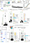
Fig. 1.. 4′-FlU is a potent broad-spectrum antiviral.
(A) Chemical structure of 4′-FlU. (B) Virus yield reduction of RSV clinical isolates 6A8, 16F10, 2-20, and recombinant recRSV-A2line19F-[mKate] [(A) or (B) antigenic subgroup]. (C) HEp-2, MDCK, BHK-T7, and BEAS-2B cell lines were assayed for reduction in cell metabolism by 4′-FlU. (D and E) recRSV-A2line19F-[FireSMASh] dose response inhibition and cytotoxicity assay with human airway epithelial (HAE) cells (D) from two donors in the presence of indicated 4′-FlU concentrations (E). (F) Dose-response inhibition of a panel of recombinant mononegaviruses by 4′-FlU. (G) Dose-response inhibition of recSARS-CoV-2-[Nluc] and virus yield reduction of alpha, gamma, and delta VoC isolates by 4′-FlU. (H) Dose-response inhibition of transiently expressed polymerase complexes from mononegaviruses MeV, RSV, NiV, or HPIV-3 by 4′-FlU (I) recRSV-A2line19F-[FireSMASh]-infected cells were treated with 10 μM of 4′-FlU and serial dilutions of exogenous nucleotides in extracellular media. Viral activity was determined by reporter activity. Symbols represent independent repeats (B), (E), (G), (H), and (I) or mean with standard deviation (C) and (F), and lines represent means. n ≥ 3, EC50s and CC50s are reported in tables S1 and S2, and all source data are provided in data S2.
Fig. 2.. 4′-FlU induces a delayed stalling of RSV and SARS-CoV-2 RdRP.
(A) SDS-PAGE with Coomassie blue staining of recombinant RSV RdRP complexes (L and P proteins). (B) Schematics of the primer extension assay. (C) Urea-PAGE fractionation of RNA transcripts produced through primer extension by the RSV RdRP in the presence of the indicated nucleotides. (n = 3). (D) Kinetic analysis of autoradiographs from (C). Nonlinear regression with the Michaelis-Menten model. Km and Vmax with 95% confidence intervals (CIs) and goodness of fit (r2) are indicated. (E to G) Urea-PAGE fractionation of RNA transcripts produced by RSV RdRP in the presence of the indicated templates and nucleotides. “Remdesivir” denotes the addition of the remdesivir active metabolite GS-443902, a well-characterized “delayed chain terminator”. 4′-FlU-TP bands in (F) to (G) were normalized to the corresponding band after UTP incorporation. Bars represent mean and error bars represent standard deviation (n = 3). (H) Purified recombinant SARS-CoV-2 RdRP complexes (nsp7, 8, and 12 proteins) “nsp12 SNN” denotes a catalytically inactive mutant. (I to K) Urea-PAGE fractionation of RNA transcripts produced by SARS-CoV-2 RdRP in the presence of the indicated templates and nucleotides. Stars denote cellular contaminants. Uncropped autoradiograph replicates are provided in data S1.
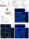
Fig. 3.. 4′-FlU is efficiently anabolized in HAE cells and is efficacious in human airway epithelium organoids.
(A to C) 4′-FlU cellular uptake and metabolism in “F1” HAE cells quantified by mass spectrometry (A). Intracellular concentration of 4′-FlU(-TP) after exposure to 20 μM 4′-FlU for 0, 1, 2, 3, 4, 6, 16, and 24 hours (B), or 24-hour incubation followed by removal of the compound for 0, 0.5, 1, 2, 3, and 6 hours before quantification (C) (n = 3). The low limit of quantitation (LLOQ) for 4′FlU (19.83 pmol/106 cells) is indicated by the dashed line. (D) HAE cells were matured at air-liquid interface (ALI). (E) Virus yield reduction of recRSV-A2line19F-[FireSMASh] was shed from the apical side in ALI HAE after incubation with serial dilutions of 4′-FlU on the basal side (n = 3). (F to H) Confocal microscopy of ALI HAE cells infected with recRSV-A2line19F-[FireSMASh], at 5 days after infection. RSV infected cells, tight junctions, and nuclei were stained with anti-RSV, anti-ZO-1, and Hoechst 34580. z-stacks of 30 × 1 μm slices with 63 × oil objective. Dotted lines, x-z and y-z stacks; scale bar, 20 μm. In all panels, symbols represent independent biological repeats and lines represent means.
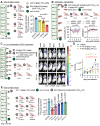
Fig. 4.. Therapeutic oral efficacy of 4′-FlU in the RSV mouse model.
(A) Balb/cJ mice were inoculated with recRSV-A2line19F-[mKate] and treated as indicated. At 4.5 days after infection, viral lung titers were determined with TCID50 titration (n = 5). (B) Balb/cJ mice were inoculated with recRSV-A2line19F-[mKate] or mock-infected, and treated as indicated. Blood samples were collected before infection and at 1.5, 2.5, 3.5, and 4.5 days after infection, and lymphocyte proportions with platelets/ml are represented over time (n = 4). (C) Balb/cJ mice were inoculated with recRSV-A2line19F-[redFirefly] and treated as indicated. In vivo luciferase activity was measured daily. (D) Total photon flux from mice lungs from (C) over time (n = 3). (E) Balb/cJ mice were inoculated with recRSV-A2line19F-[mKate] and treated as indicated. At 4.5 days after infection, viral lung titers were determined with TCID50 titration (n = 5). In all panels, symbols represent individual values, and bars or lines represent means. One-way ordinary analysis of variance (ANOVA) with Tukey’s post hoc multiple comparisons (B) and (I) or two-way ANOVA with Dunnett’s post hoc multiple comparison (C) and (G). h.p.i., hours post-infection.
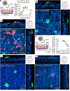
Fig. 5.. Efficacy of 4′-FlU against SARS-CoV-2 replication in HAE organoids.
(A) Multicycle growth curve of SARS-CoV-2 WA1 isolate on ALI HAE from two donors. Shed virus was harvested daily and titered by plaque assay (n = 3). (B and C) Confocal microscopy of ALI HAE cells from “F1” donor mock-infected (B) or infected (C) with SARS-CoV-2 WA1 isolate, at 3 days post-infection. SARS-CoV-2 infected cells, goblet cells, and nuclei were stained with anti-SARS-CoV-2 N immunostaining, anti-MUC5AC immunostaining, and Hoechst 34580, pseudo-colored in red, green, and blue, respectively. z-stacks of 35 μm slices (1 μm thick) with 63× objective with oil immersion. Dotted lines represent the location of x-z and y-z stacks; scale bar, 20 μm. In all panels, symbols represent independent biological repeats and lines represent means. (D) Virus yield reduction of SARS-CoV-2 WA1 clinical isolate shed from the apical side in ALI HAE after incubation with serial dilutions of 4′-FlU on the basal side (n = 3). (E and F) Confocal microscopy of ALI HAE cells infected with SARS-CoV-2 WA1 isolate, and treated with 50 μM 4′-FlU 3 days after infection. Rare ciliated cells positive for N are represented in (F).
Fig. 6.. Therapeutic oral efficacy of 4′-FlU against different SARS-CoV-2 isolates in ferrets.
(A) Single oral dose (15 or 50 mg/kg bodyweight) pharmacokinetics properties of 4′-FlU in ferret plasma (n = 3). (B) Ferrets were inoculated with SARS-CoV-2 WA1 or VoC alpha, gamma, or delta, and treated as indicated. (C) Nasal lavages were performed twice daily and viral titers were determined by plaque assay [n = 4 (WA1) or n = 3 (alpha, gamma, delta)]. (D) Viral titers in nasal turbinates at day 4 post-infection. In all panels, symbols represent individual independent biological repeats and lines show mean values. Two-way ANOVA with Sidak’s post hoc multiple comparison (C) and unpaired _t_-test (D).
Update of
- 4'-Fluorouridine is a broad-spectrum orally efficacious antiviral blocking respiratory syncytial virus and SARS-CoV-2 replication.
Sourimant J, Lieber CM, Aggarwal M, Cox RM, Wolf JD, Yoon JJ, Toots M, Ye C, Sticher Z, Kolykhalov AA, Martinez-Sobrido L, Bluemling GR, Natchus MG, Painter GR, Plemper RK. Sourimant J, et al. bioRxiv [Preprint]. 2021 May 20:2021.05.19.444875. doi: 10.1101/2021.05.19.444875. bioRxiv. 2021. PMID: 34031658 Free PMC article. Updated. Preprint.
Similar articles
- 4'-Fluorouridine is a broad-spectrum orally efficacious antiviral blocking respiratory syncytial virus and SARS-CoV-2 replication.
Sourimant J, Lieber CM, Aggarwal M, Cox RM, Wolf JD, Yoon JJ, Toots M, Ye C, Sticher Z, Kolykhalov AA, Martinez-Sobrido L, Bluemling GR, Natchus MG, Painter GR, Plemper RK. Sourimant J, et al. bioRxiv [Preprint]. 2021 May 20:2021.05.19.444875. doi: 10.1101/2021.05.19.444875. bioRxiv. 2021. PMID: 34031658 Free PMC article. Updated. Preprint. - Development of an allosteric inhibitor class blocking RNA elongation by the respiratory syncytial virus polymerase complex.
Cox RM, Toots M, Yoon JJ, Sourimant J, Ludeke B, Fearns R, Bourque E, Patti J, Lee E, Vernachio J, Plemper RK. Cox RM, et al. J Biol Chem. 2018 Oct 26;293(43):16761-16777. doi: 10.1074/jbc.RA118.004862. Epub 2018 Sep 11. J Biol Chem. 2018. PMID: 30206124 Free PMC article. - Thapsigargin Is a Broad-Spectrum Inhibitor of Major Human Respiratory Viruses: Coronavirus, Respiratory Syncytial Virus and Influenza A Virus.
Al-Beltagi S, Preda CA, Goulding LV, James J, Pu J, Skinner P, Jiang Z, Wang BL, Yang J, Banyard AC, Mellits KH, Gershkovich P, Hayes CJ, Nguyen-Van-Tam J, Brown IH, Liu J, Chang KC. Al-Beltagi S, et al. Viruses. 2021 Feb 3;13(2):234. doi: 10.3390/v13020234. Viruses. 2021. PMID: 33546185 Free PMC article. - New antiviral approaches for respiratory syncytial virus and other mononegaviruses: Inhibiting the RNA polymerase.
Fearns R, Deval J. Fearns R, et al. Antiviral Res. 2016 Oct;134:63-76. doi: 10.1016/j.antiviral.2016.08.006. Epub 2016 Aug 27. Antiviral Res. 2016. PMID: 27575793 Review. - Repurposing Probenecid to Inhibit SARS-CoV-2, Influenza Virus, and Respiratory Syncytial Virus (RSV) Replication.
Tripp RA, Martin DE. Tripp RA, et al. Viruses. 2022 Mar 15;14(3):612. doi: 10.3390/v14030612. Viruses. 2022. PMID: 35337018 Free PMC article. Review.
Cited by
- The nucleoside analog 4'-fluorouridine suppresses the replication of multiple enteroviruses by targeting 3D polymerase.
Chen Y, Li X, Han F, Ji B, Li Y, Yan J, Wang M, Fan J, Zhang S, Lu L, Zou P. Chen Y, et al. Antimicrob Agents Chemother. 2024 Jun 5;68(6):e0005424. doi: 10.1128/aac.00054-24. Epub 2024 Apr 30. Antimicrob Agents Chemother. 2024. PMID: 38687016 Free PMC article. - Henipaviruses: epidemiology, ecology, disease, and the development of vaccines and therapeutics.
Spengler JR, Lo MK, Welch SR, Spiropoulou CF. Spengler JR, et al. Clin Microbiol Rev. 2025 Mar 13;38(1):e0012823. doi: 10.1128/cmr.00128-23. Epub 2024 Dec 23. Clin Microbiol Rev. 2025. PMID: 39714175 Free PMC article. Review. - Delayed low-dose oral administration of 4'-fluorouridine inhibits pathogenic arenaviruses in animal models of lethal disease.
Welch SR, Spengler JR, Westover JB, Bailey KW, Davies KA, Aida-Ficken V, Bluemling GR, Boardman KM, Wasson SR, Mao S, Kuiper DL, Hager MW, Saindane MT, Andrews MK, Krueger RE, Sticher ZM, Jung KH, Chatterjee P, Shrivastava-Ranjan P, Lo MK, Coleman-McCray JD, Sorvillo TE, Genzer SC, Scholte FEM, Kelly JA, Jenks MH, McMullan LK, Albariño CG, Montgomery JM, Painter GR, Natchus MG, Kolykhalov AA, Gowen BB, Spiropoulou CF, Flint M. Welch SR, et al. Sci Transl Med. 2024 Nov 20;16(774):eado7034. doi: 10.1126/scitranslmed.ado7034. Epub 2024 Nov 20. Sci Transl Med. 2024. PMID: 39565871 - Developing nucleoside tailoring strategies against SARS-CoV-2 via ribonuclease targeting chimera.
Min Y, Xiong W, Shen W, Liu X, Qi Q, Zhang Y, Fan R, Fu F, Xue H, Yang H, Sun X, Ning Y, Tian T, Zhou X. Min Y, et al. Sci Adv. 2024 Apr 12;10(15):eadl4393. doi: 10.1126/sciadv.adl4393. Epub 2024 Apr 10. Sci Adv. 2024. PMID: 38598625 Free PMC article. - Synthesis of 4'-Substituted Carbocyclic Uracil Derivatives and Their Monophosphate Prodrugs as Potential Antiviral Agents.
Biteau NG, Amichai SA, Azadi N, De R, Downs-Bowen J, Lecher JC, MacBrayer T, Schinazi RF, Amblard F. Biteau NG, et al. Viruses. 2023 Feb 16;15(2):544. doi: 10.3390/v15020544. Viruses. 2023. PMID: 36851758 Free PMC article.
References
- Beigel J. H., Tomashek K. M., Dodd L. E., Mehta A. K., Zingman B. S., Kalil A. C., Hohmann E., Chu H. Y., Luetkemeyer A., Kline S., Lopez de Castilla D., Finberg R. W., Dierberg K., Tapson V., Hsieh L., Patterson T. F., Paredes R., Sweeney D. A., Short W. R., Touloumi G., Lye D. C., Ohmagari N., Oh M. D., Ruiz-Palacios G. M., Benfield T., Fätkenheuer G., Kortepeter M. G., Atmar R. L., Creech C. B., Lundgren J., Babiker A. G., Pett S., Neaton J. D., Burgess T. H., Bonnett T., Green M., Makowski M., Osinusi A., Nayak S., Lane H. C.; ACTT-1 Study Group Members , Remdesivir for the Treatment of Covid-19 - Final Report. N. Engl. J. Med. 383, 1813–1826 (2020). 10.1056/NEJMoa2007764 - DOI - PMC - PubMed
- Toots M., Yoon J.-J., Cox R. M., Hart M., Sticher Z. M., Makhsous N., Plesker R., Barrena A. H., Reddy P. G., Mitchell D. G., Shean R. C., Bluemling G. R., Kolykhalov A. A., Greninger A. L., Natchus M. G., Painter G. R., Plemper R. K., Characterization of orally efficacious influenza drug with high resistance barrier in ferrets and human airway epithelia. Sci. Transl. Med. 11, eaax5866 (2019). 10.1126/scitranslmed.aax5866 - DOI - PMC - PubMed
- Painter G. R., Bowen R. A., Bluemling G. R., DeBergh J., Edpuganti V., Gruddanti P. R., Guthrie D. B., Hager M., Kuiper D. L., Lockwood M. A., Mitchell D. G., Natchus M. G., Sticher Z. M., Kolykhalov A. A., The prophylactic and therapeutic activity of a broadly active ribonucleoside analog in a murine model of intranasal venezuelan equine encephalitis virus infection. Antiviral Res. 171, 104597 (2019). 10.1016/j.antiviral.2019.104597 - DOI - PubMed
- Sheahan T. P., Sims A. C., Zhou S., Graham R. L., Pruijssers A. J., Agostini M. L., Leist S. R., Schäfer A., Dinnon K. H. 3rd, Stevens L. J., Chappell J. D., Lu X., Hughes T. M., George A. S., Hill C. S., Montgomery S. A., Brown A. J., Bluemling G. R., Natchus M. G., Saindane M., Kolykhalov A. A., Painter G., Harcourt J., Tamin A., Thornburg N. J., Swanstrom R., Denison M. R., Baric R. S., An orally bioavailable broad-spectrum antiviral inhibits SARS-CoV-2 in human airway epithelial cell cultures and multiple coronaviruses in mice. Sci. Transl. Med. 12, eabb5883 (2020). 10.1126/scitranslmed.abb5883 - DOI - PMC - PubMed
Publication types
MeSH terms
Substances
Grants and funding
- R01 AI071002/AI/NIAID NIH HHS/United States
- R01 AI141222/AI/NIAID NIH HHS/United States
- R01 AI153400/AI/NIAID NIH HHS/United States
- R01 AI161175/AI/NIAID NIH HHS/United States
LinkOut - more resources
Full Text Sources
Other Literature Sources
Medical
Miscellaneous


