A fresh look at Cladarosymblema narrienense, a tetrapodomorph fish (Sarcopterygii: Megalichthyidae) from the Carboniferous of Australia, illuminated via X-ray tomography - PubMed (original) (raw)
A fresh look at Cladarosymblema narrienense, a tetrapodomorph fish (Sarcopterygii: Megalichthyidae) from the Carboniferous of Australia, illuminated via X-ray tomography
Alice M Clement et al. PeerJ. 2021.
Abstract
Background: The megalichthyids are one of several clades of extinct tetrapodomorph fish that lived throughout the Devonian-Permian periods. They are advanced "osteolepidid-grade" fishes that lived in freshwater swamp and lake environments, with some taxa growing to very large sizes. They bear cosmine-covered bones and a large premaxillary tusk that lies lingually to a row of small teeth. Diagnosis of the family remains controversial with various authors revising it several times in recent works. There are fewer than 10 genera known globally, and only one member definitively identified from Gondwana. Cladarosymblema narrienense Fox et al. 1995 was described from the Lower Carboniferous Raymond Formation in Queensland, Australia, on the basis of several well-preserved specimens. Despite this detailed work, several aspects of its anatomy remain undescribed.
Methods: Two especially well-preserved 3D fossils of Cladarosymblema narrienense, including the holotype specimen, are scanned using synchrotron or micro-computed tomography (µCT), and 3D modelled using specialist segmentation and visualisation software. New anatomical detail, in particular internal anatomy, is revealed for the first time in this taxon. A novel phylogenetic matrix, adapted from other recent work on tetrapodomorphs, is used to clarify the interrelationships of the megalichthyids and confirm the phylogenetic position of C. narrienense.
Results: Never before seen morphological details of the palate, hyoid arch, basibranchial skeleton, pectoral girdle and axial skeleton are revealed and described. Several additional features are confirmed or updated from the original description. Moreover, the first full, virtual cranial endocast of any tetrapodomorph fish is presented and described, giving insight into the early neural adaptations in this group. Phylogenetic analysis confirms the monophyly of the Megalichthyidae with seven genera included (Askerichthys, Cladarosymblema, Ectosteorhachis, Mahalalepis, Megalichthys, Palatinichthys, and Sengoerichthys). The position of the megalichthyids as sister group to canowindrids, crownward of "osteolepidids" (e.g.,Osteolepis and Gogonasus), but below "tristichopterids" such as Eusthenopteron is confirmed, but our findings suggest further work is required to resolve megalichthyid interrelationships.
Keywords: 3D modelling; Carboniferous; Endocast; Evolution; Megalichthyidae; Phylogenetic analysis; Sarcopterygii; Tetrapodomorph; Tomography; Vertebrate.
©2021 Clement et al.
Conflict of interest statement
The authors declare there are no competing interests.
Figures
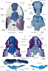
Figure 1. Micro-CT 3D rendering (116 µm pixel size) of dermal skull and braincase of Cladarosymblema narrienense (QMF 21082).
(A) Skull in dorsal view showing placement of bones on holotype; (B) braincase in ventral view; dermal skull bones, braincase and palatal bones in (C) dorsal and (D) ventral view; (E) skull and cheek in right lateral view; (F) cheek bones in mesial view.
Figure 2. Micro-CT 3D rendering (116 µm pixel size) of mandible and submandibular bones of Cladarosymblema narrienense (QMF 21082).
(A) mandibular bones in ventral view showing placement of bones on holotype; mandible in (B) dorsal; (C) lingual; and (D) labial view. (E-F), gulars and submandibular bones shown in isolation, in ventral and dorsal view.
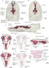
Figure 3. Micro-CT 3D (116 µm pixel size) and synchrotron rendering (12 µm pixel size) of cranial endocast and sensory lines of Cladarosymblema narrienense (QMF 21082/3).
(A) dorsal; (B) ventral; and (C) left lateral view; QMF 21083 in (D,E) dorsal view; (F,G) ventral view; (H) left lateral view showing zoomed in hypophysial fossa region.
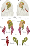
Figure 4. Micro-CT 3D rendering (116 µm pixel size) of hyoid and branchial skeleton of Cladarosymblema narrienense (QMF 21082).
(A) skull in dorsal view showing placement of bones on holotype; (B) in ventral view; (C,D) full hyoid and basibranchial skeleton including ceratobranchials as preserved in situ; (E) closeup of left hyomandibular; and right hypobranchials 1-4 in (F) ventral and (G) dorsal view.
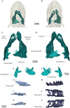
Figure 5. Micro-CT 3D rendering (116 µm pixel size) of pectoral and axial elements of Cladarosymblema narrienense (QMF 21082).
(A) ventral view, and (B) in dorsal view, showing placement of bones on holotype; pectoral girdle in (C) dorsal view; and (D) ventral view; (E, F) anocleithra in alternate views; and neural arches in (G) lateral; (I) dorsal; (K) ventral view; ring centra in H, lateral; J, dorsal; L, ventral view.
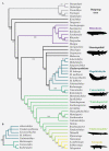
Figure 6. Parsimony analyses.
(A) 50% majority-rule consensus tree from parsimony analysis with inclusion of all taxa showing monophyly of the Megalichthyidae; (B) Canowindrid + Megalichthyid sub-set with most incomplete taxa excluded (Mahalalepis, M. mullisoni, M. laticeps, Sengoerichthys) provides greater resolution of megalichthyid phylogeny. Image silhouettes are authors own (elpistostegalid, rhizodont, megalichthyid, canowindrid) or from PhyloPic,
(Ichthyostega Image credit: Scott Hartman; Eusthenopteron Image credit: Steven Coombs (vectorized by T. Michael Keesey), Gogonasus Photo credit: Nobu Tamura (vectorized by T. Michael Keesey), CC-SA 3.0,
http://creativecommons.org/licenses/by-sa/3.0/
.
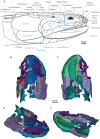
Figure 7. C. ladarosymblema narrienense.
(A) Lateral head reconstruction of Cladarosymblema narrienense, compiled from Fox et al. (1995) and new data. Colour-coded as follows: dermal skull roof (dark blue), cheek (light blue), lower jaw (pale green), opercular series (purple), and pectoral (dark green). Bones marked with “?” remain unknown in this taxon. (B–E) micro-CT 3D rendering of all segmented bones in the holotype (QMF 21082): B, dorsal view; (C) ventral view; (D) anterolaterodorsal view; and E, posteroventrolateral view.
Similar articles
- Pectoral girdle and fin anatomy of Gogonasus andrewsae Long, 1985: implications for tetrapodomorph limb evolution.
Holland T. Holland T. J Morphol. 2013 Feb;274(2):147-64. doi: 10.1002/jmor.20078. Epub 2012 Sep 29. J Morphol. 2013. PMID: 23023825 - A gigantic sarcopterygian (tetrapodomorph lobe-finned fish) from the upper Devonian of Gondwana (Eden, New South Wales, Australia).
Young B, Dunstone RL, Senden TJ, Young GC. Young B, et al. PLoS One. 2013;8(3):e53871. doi: 10.1371/journal.pone.0053871. Epub 2013 Mar 6. PLoS One. 2013. PMID: 23483884 Free PMC article. - An exceptional Devonian fish from Australia sheds light on tetrapod origins.
Long JA, Young GC, Holland T, Senden TJ, Fitzgerald EM. Long JA, et al. Nature. 2006 Nov 9;444(7116):199-202. doi: 10.1038/nature05243. Epub 2006 Oct 18. Nature. 2006. PMID: 17051154 - Reconstructing pectoral appendicular muscle anatomy in fossil fish and tetrapods over the fins-to-limbs transition.
Molnar JL, Diogo R, Hutchinson JR, Pierce SE. Molnar JL, et al. Biol Rev Camb Philos Soc. 2018 May;93(2):1077-1107. doi: 10.1111/brv.12386. Epub 2017 Nov 10. Biol Rev Camb Philos Soc. 2018. PMID: 29125205 Review. - The origin of novel features by changes in developmental mechanisms: ontogeny and three-dimensional microanatomy of polyodontode scales of two early osteichthyans.
Qu Q, Sanchez S, Zhu M, Blom H, Ahlberg PE. Qu Q, et al. Biol Rev Camb Philos Soc. 2017 May;92(2):1189-1212. doi: 10.1111/brv.12277. Epub 2016 May 19. Biol Rev Camb Philos Soc. 2017. PMID: 27194072 Review.
References
- Agassiz JLR. On the fossil fishes of Scotland. Edinburgh in 1834: John Murray, LondonReport of the fourth meeting of the british association for the advancement of science. 1835:646–648.
- Ahlberg PE, Johanson Z. Osteolepiforms and the ancestory of tetrapods. Nature. 1998;395:792–794. doi: 10.1038/27421. - DOI
- Andrews SM, Westoll TS. The postcranial skeleton of Eusthenopteron foordi Whiteaves. Transactions of the Royal Society of Edinburgh. 1970a;68:207–329. doi: 10.1017/S008045680001471X. - DOI
- Bjerring HC. Morphological observations on the exoskeletal skull roof of an osteolepiform from the Carboniferous of Scotland. Acta Zoologica. 1972;53:73–92. doi: 10.1111/j.1463-6395.1972.tb00575.x. - DOI
- Borgen UJ, Nakrem HA. Morphology, phylogeny and taxonomy of osteolepiform fish. Lethaia. 2016;61:1–514.
Grants and funding
This work was supported by the Australian Research Council (DP160102460, LE180100136, DP200103398), Flinders University Impact Seed Funding (to Alice M Clement), an NSERC Discovery Grant (to Richard Cloutier), and the following all awarded to Jing Lu: the Strategic Priority Research Program of Chinese Academy of Sciences (XDB26000000), National Science Fund for Excellent Young Scholars (42022011), and National Natural Science Foundation of China (41872023). The funders had no role in study design, data collection and analysis, decision to publish, or preparation of the manuscript.
LinkOut - more resources
Full Text Sources
