Dermal Duct Tumor: A Diagnostic Dilemma - PubMed (original) (raw)
Review
Dermal Duct Tumor: A Diagnostic Dilemma
Austinn C Miller et al. Dermatopathology (Basel). 2022.
Abstract
Poromas or poroid tumors are a group of rare, benign cutaneous neoplasms derived from the terminal eccrine or apocrine sweat gland duct. There are four poroma variants with overlapping features: dermal duct tumor (DDT), eccrine poroma, hidroacanthoma simplex, and poroid hidradenoma, of which DDT is the least common. Clinically, the variants have a nonspecific appearance and present as solitary dome-shaped papules, plaques, or nodules. They can be indistinguishable from each other and a multitude of differential diagnoses, necessitating a better understanding of the characteristics that make the diagnosis of poroid neoplasms. However, there remains a paucity of information on these lesions, especially DDTs, given their infrequent occurrence. Herein, we review the literature on DDTs with an emphasis on epidemiology, pathogenesis, clinical features, diagnosis, and management.
Keywords: DDT; dermal duct tumor; hidradenoma; poroid; poroma.
Conflict of interest statement
The authors declare no conflict of interest.
Figures
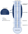
Figure 1
A simplified schematic of keratin expression in an eccrine sweat gland is shown [6,10]. The basal keratinocytes of the sweat duct ridge and lower intraepidermal acrosyringium are thought to give rise to poroid neoplasms based on the similarities in keratin expression [6,10]. K5 and K14 expression is ubiquitous throughout poromas, while K1 and K10 are found in focal aggregates. K77 is restricted to normal luminal cells embedded within the tumors.
Figure 2
Dermal duct tumor: a small, purple-brown papule at the right superior nasolabial fold.
Figure 3
Well circumscribed, dermal-based, solid and cystic tumor with no connection to the overlying epidermis (H & E, 2×).
Figure 4
Higher magnification reveals small poriod cells with round to oval nuclei and scant cytoplasm. Ductal lumen formation is present (H & E, 10×).
Figure 5
Residual DDT on punch excision (from the transected spiecimen in Figure 3) (H & E, 10×).
Similar articles
- A Large Mass over the Foot due to the Coexistence of an Eccrine Poroma and a Poroid Hidradenoma: A Case Report.
Nishikawa DRC, Silva ACLD, Yano MY, Miranda BR, Chung WT. Nishikawa DRC, et al. Rev Bras Ortop (Sao Paulo). 2021 Oct 1;59(Suppl 1):e5-e8. doi: 10.1055/s-0041-1732331. eCollection 2024 Jul. Rev Bras Ortop (Sao Paulo). 2021. PMID: 39027188 Free PMC article. - Poroma: a review of eccrine, apocrine, and malignant forms.
Sawaya JL, Khachemoune A. Sawaya JL, et al. Int J Dermatol. 2014 Sep;53(9):1053-61. doi: 10.1111/ijd.12448. Epub 2014 Apr 2. Int J Dermatol. 2014. PMID: 24697501 Review. - A Large Lumbar Adnexal Neoplasm Presenting Characteristics of Eccrine Poroma and Poroid Hidradenoma.
Michalinos A, Schizas D, Sarakinos A, Athanasiadis G, Spartalis E, Vlachodimitropoulos D, Troupis T. Michalinos A, et al. Case Rep Surg. 2017;2017:9865672. doi: 10.1155/2017/9865672. Epub 2017 Sep 19. Case Rep Surg. 2017. PMID: 29057137 Free PMC article. - From hidroacanthoma simplex to poroid hidradenoma: clinicopathologic and immunohistochemic study of poroid neoplasms and reappraisal of their histogenesis.
Battistella M, Langbein L, Peltre B, Cribier B. Battistella M, et al. Am J Dermatopathol. 2010 Jul;32(5):459-68. doi: 10.1097/DAD.0b013e3181bc91ff. Am J Dermatopathol. 2010. PMID: 20571345 - [Apocrine poroma. A relatively little known skin tumor with multilineage differentiation].
Flux K, Eckert F. Flux K, et al. Pathologe. 2014 Sep;35(5):456-61. doi: 10.1007/s00292-014-1931-1. Pathologe. 2014. PMID: 25142043 Review. German.
Cited by
- Poroid Neoplasms: A Clinicopathological Study of 13 Cases.
Efared B, Boubacar I, Ousmane Kadre KA, Abani Bako AB, Boureima HS, Amadou S, Nouhou H. Efared B, et al. Clin Pathol. 2024 Sep 12;17:2632010X241281460. doi: 10.1177/2632010X241281460. eCollection 2024 Jan-Dec. Clin Pathol. 2024. PMID: 39282157 Free PMC article. - Eccrine Poroma with Concurrent Basal Cell Carcinoma: A Rare Combination.
Shao X, Dong Y, Liu H, Wei J, Xiong X. Shao X, et al. Clin Cosmet Investig Dermatol. 2023 Oct 20;16:2965-2970. doi: 10.2147/CCID.S428611. eCollection 2023. Clin Cosmet Investig Dermatol. 2023. PMID: 37881203 Free PMC article.
References
- Ahmed Jan N., Masood S. StatPearls [Internet] Statpearls Publishing; Treasure Island, FL, USA: 2021. [(accessed on 30 October 2021)]. Poroma. Available online: http://www.ncbi.nlm.nih.gov/books/NBK560909/
Publication types
LinkOut - more resources
Full Text Sources
Miscellaneous


