Three families of Asgard archaeal viruses identified in metagenome-assembled genomes - PubMed (original) (raw)
Three families of Asgard archaeal viruses identified in metagenome-assembled genomes
Sofia Medvedeva et al. Nat Microbiol. 2022 Jul.
Abstract
Asgardarchaeota harbour many eukaryotic signature proteins and are widely considered to represent the closest archaeal relatives of eukaryotes. Whether similarities between Asgard archaea and eukaryotes extend to their viromes remains unknown. Here we present 20 metagenome-assembled genomes of Asgardarchaeota from deep-sea sediments of the basin off the Shimokita Peninsula, Japan. By combining a CRISPR spacer search of metagenomic sequences with phylogenomic analysis, we identify three family-level groups of viruses associated with Asgard archaea. The first group, verdandiviruses, includes tailed viruses of the class Caudoviricetes (realm Duplodnaviria); the second, skuldviruses, consists of viruses with predicted icosahedral capsids of the realm Varidnaviria; and the third group, wyrdviruses, is related to spindle-shaped viruses previously identified in other archaea. More than 90% of the proteins encoded by these viruses of Asgard archaea show no sequence similarity to proteins encoded by other known viruses. Nevertheless, all three proposed families consist of viruses typical of prokaryotes, providing no indication of specific evolutionary relationships between viruses infecting Asgard archaea and eukaryotes. Verdandiviruses and skuldviruses are likely to be lytic, whereas wyrdviruses potentially establish chronic infection and are released without host cell lysis. All three groups of viruses are predicted to play important roles in controlling Asgard archaea populations in deep-sea ecosystems.
© 2022. The Author(s), under exclusive licence to Springer Nature Limited.
Conflict of interest statement
Competing interests
The authors declare no competing interests.
Figures
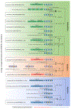
Extended Data Fig. 1 |. Matches between asgardarchaeal CRISPR spacers and viruses.
The figure is divided into three blocks (green, red and blue) corresponding to the three groups of asgardarchaeal viruses, namely, verdandiviruses, skuldviruses and wyrdviruses. CRISPR repeats and spacers are indicated as diamonds and boxes, respectively. Spacers matching asgardarchaeal viruses are shown in yellow and are connected to the names of targeted viruses with arrows. Thick vertical lines connect related repeats and the exact pairwise sequence identities are indicated.
Extended Data Fig. 2 |. Partial provirus integrated within a genomic contig of Lokiarchaeia.
The verdandivirus-derived region is boxed. Homologous genes are shown using the same colors and the key is provided on the left of the figure. Housekeeping cellular genes are shown in black and include those encoding transcription factor S (TFS) and ribosomal proteins S7e, S12e, S27e, L30e and L44e. Grey shading connects genes displaying sequence similarity at the protein level, with the percent of sequence identity depicted with different shades of grey.
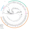
Extended Data Fig. 3 |. Maximum likelihood phylogenetic tree of Topo mini-A proteins.
Proteins of skuldviruses and wyrdviruses are shown in cyan and dark blue, respectively, whereas those encoded by other asgardarchaeal MGE are shown in red. Other Topo mini-A homologs encoded by archaea and bacteria (or their corresponding viruses) are shown in green and orange, respectively. Topo VI proteins were used as an outgroup and are colored grey. The tree was constructed using the automatic optimal model selection (LG+R5). The scale bar represents the number of substitutions per site. Circles at the nodes denote aLRT SH-like branch support values larger than 90%.
Extended Data Fig. 4 |. Sequence alignment of the major capsid proteins of selected wyrdviruses, fusellovirus SSV1 and halspivirus His1.
When available, proteins are identified with their accession numbers followed by a virus name. TMD, transmembrane domain.
Fig. 1 |. Phylogenomic tree of Asgardarchaeota.
The alignment is based on a concatenated set of 53 protein markers (subsampled to 42 sites each, resulting in a total alignment length of 2,058 sites) from 339 taxa. These taxa encompass 276 Asgardarchaeota MAGs, including 16 recovered in this study (highlighted in pink; MAGs 3H_mb2_bin20, 4H_max40_bin02,1 4H_mb2_bin40 and 7H_mb2_bin7 were removed after the alignment trimming step due to low completeness), and 63 non-Asgardarchaeota species representatives from GTDB release RS95 as an outgroup. Maximum-likelihood analysis was performed using IQ-TREE under the LG + C10 + F + G + PMSF model. The tree is rooted on the non-Asgardarchaeota group. Nodes with bootstrap statistical support >90% are indicated by grey dots. MAGs containing viral contigs and CRISPR arrays are indicated by different symbols, with the key provided in the bottom left corner.
GCA_019058445.1
contains a defective verdandivirus-derived provirus (Extended Data Fig. 2).
Fig. 2 |. Description of the Asgardarchaeota CRISPR spacer dataset.
a, Similarity of CRISPR repeats from metagenomic data (teal) and from Asgardarchaeota MAGs (red). Unique CRISPR repeat sequences are shown as nodes in the network. Node diameter is proportional to the number of spacers associated with the repeat sequence in the dataset. Identical CRISPR repeats from both metagenomic data and Asgard MAGs are connected by solid lines; dashed and dotted lines connect CRISPR repeat sequences with 95–99% and 90–95% identities, respectively. Consensus sequences for the nine major clusters are shown on the right. b, Sources of Asgardarchaeota CRISPR spacers. The number of spacers in the dataset originating from different geographical sites is indicated on the pie chart. c, Taxonomic distribution of Asgardarchaeota MAGs with CRISPR arrays for different isolation sites. Circle size is proportional to the number of spacers in the dataset.
Fig. 3 |. Diversity of verdandiviruses.
a, Genome maps of verdandiviruses. Homologous genes are shown using the same colours, with the key provided at the bottom. Also shown is the deduced schematic organization of the verdandivirus virion, with colours matching those of genes encoding the corresponding proteins. Genes encoding putative DNA-binding proteins with Zn-binding and HTH domains coloured in black and grey, respectively. Coloured circles indicate the positions of protospacers. Grey shading connects genes displaying sequence similarity at the protein level, with the percentage of sequence identity indicated by different shades of grey (see scale at the bottom). Asterisks denote complete genomes assembled as circular contigs. PAPSR, phosphoadenosine phosphosulfate reductase; TMP, tail tape measure protein; MTP, major tail protein. b, Comparison of the structural model of the MCP of verdnadivirus AsTV-10H2 with the corresponding structures of siphoviruses TW1 and HK97. The models are coloured according to their secondary structure: α-helices, dark blue; β-strands, light blue. c, Maximum-likelihood phylogeny of MCPs encoded by verdandiviruses. Taxa are coloured based on the source of origin (key at the bottom). The tree was constructed using the automatic optimal model selection (LG + G4) and is midpoint rooted. The scale bar represents the number of substitutions per site. Circles at nodes denote aLRT SH-like branch support values >90%.
Fig. 4 |. Diversity of skuldviruses.
a, Genome maps of skuldviruses and PM2 virus. Homologous genes are shown using the same colours, with the key provided at the bottom. Also shown is the deduced schematic organization of the skuldvirus virion, with colours matching those of genes encoding the corresponding proteins. Coloured circles indicate the positions of protospacers. Grey shading connects genes displaying sequence similarity at the protein level, with the percentage of sequence identity indicated by different shades of grey (scale on the right). Asterisks denote complete genomes assembled as circular contigs. RCRE, rolling circle replication endonuclease. Genome map of corticovirus PM2 is shown for comparison. b, Sequence similarity network of prokaryotic virus DJR MCPs. Protein sequences were clustered by pairwise sequence similarity using CLANS. Lines connect sequences with CLANS _P_≤ 1 × 10⁻⁴. CLANS uses P values of BLASTP comparisons calculated from Poisson distribution of high-scoring segment pairs. Different previously defined groups of DJR MCP are shown as clouds of differentially coloured circles, with the key provided on the right. Note that the Odin group has been named as such previously because some of the MCPs in this cluster originated from Odinarchaeia bin. The position of Huginnvirus described by Tamarit et al. is indicated. Subgroups PRD1, Toil and Bam35 are named after corresponding members of the family Tectiviridae. Skuldviruses are highlighted within a yellow circle. When available, MCP structures of viruses representing each group are shown next to the corresponding cluster (PDB accession numbers given in parentheses). The Skuldvirus cluster is represented by a structural model of the SkuldV1 MCP. Models are coloured according to their secondary structure: α-helices, dark blue; β-strands, light blue.
Fig. 5 |. Diversity of wyrdviruses.
Genome maps of wyrdviruses, fusellovirus SSV1 and halspivirus His1. Homologous genes are shown using the same colours, with the key provided at the bottom. Also shown is the deduced schematic organization of the skuldvirus virion with colours marching those of the genes encoding the corresponding proteins. Coloured circles indicate the positions of protospacers. Genes encoding putative DNA-binding proteins with Zn-binding and HTH domains are coloured black and grey, respectively. Grey shading connects genes displaying sequence similarity at the protein level, with the percentage of sequence identity indicated by different shades of grey (see scale on the right). Asterisks denote complete genomes assembled as circular contigs or linear contigs with terminal inverted repeats. The topology of each genome is shown next to the corresponding name: circles for circular genomes, lines for linear genomes. ThyX, thymidylate synthase X; NMAD, nucleotide modification-associated domain 1 protein; O-MTase, carbohydrate-specific methyltransferase; TFB, transcription initiation factor B; ASCH, ASC-1 homology domain; pPolB, protein-primed family B DNA polymerase. Genome maps of SSV1 and His1 viruses are shown for comparison.
Fig. 6 |. Asgard archaeal viruses and MGEs.
a, Geographical distribution of Asgard archaeal viruses and MGEs. Filled circles next to geographical locations signify the presence of corresponding virus groups in sediment samples from that location. b, Network-based analysis of shared protein clusters among Asgard archaeal viruses and prokaryotic double-stranded DNA viruses. Nodes represent viral genomes and edges represent the strength of connectivity between each genome based on shared protein clusters. Nodes representing genomes of the three groups of Asgard archaeal viruses are in shown in red, with the three groups circled within a yellow background. Nodes corresponding to other bacterial and archaeal viruses are shown in blue and orange, respectively.
Comment in
- A trove of Asgard archaeal viruses.
Alarcón-Schumacher T, Erdmann S. Alarcón-Schumacher T, et al. Nat Microbiol. 2022 Jul;7(7):931-932. doi: 10.1038/s41564-022-01148-2. Nat Microbiol. 2022. PMID: 35760838 No abstract available.
Similar articles
- Anomalous Phylogenetic Behavior of Ribosomal Proteins in Metagenome-Assembled Asgard Archaea.
Garg SG, Kapust N, Lin W, Knopp M, Tria FDK, Nelson-Sathi S, Gould SB, Fan L, Zhu R, Zhang C, Martin WF. Garg SG, et al. Genome Biol Evol. 2021 Jan 7;13(1):evaa238. doi: 10.1093/gbe/evaa238. Genome Biol Evol. 2021. PMID: 33462601 Free PMC article. - Genomes of six viruses that infect Asgard archaea from deep-sea sediments.
Rambo IM, Langwig MV, Leão P, De Anda V, Baker BJ. Rambo IM, et al. Nat Microbiol. 2022 Jul;7(7):953-961. doi: 10.1038/s41564-022-01150-8. Epub 2022 Jun 27. Nat Microbiol. 2022. PMID: 35760837 - A closed Candidatus Odinarchaeum chromosome exposes Asgard archaeal viruses.
Tamarit D, Caceres EF, Krupovic M, Nijland R, Eme L, Robinson NP, Ettema TJG. Tamarit D, et al. Nat Microbiol. 2022 Jul;7(7):948-952. doi: 10.1038/s41564-022-01122-y. Epub 2022 Jun 27. Nat Microbiol. 2022. PMID: 35760836 Free PMC article. - Asgard archaea in saline environments.
Banciu HL, Gridan IM, Zety AV, Baricz A. Banciu HL, et al. Extremophiles. 2022 Jun 28;26(2):21. doi: 10.1007/s00792-022-01266-z. Extremophiles. 2022. PMID: 35761090 Review. - Asgard archaea: Diversity, function, and evolutionary implications in a range of microbiomes.
MacLeod F, Kindler GS, Wong HL, Chen R, Burns BP. MacLeod F, et al. AIMS Microbiol. 2019 Jan 30;5(1):48-61. doi: 10.3934/microbiol.2019.1.48. eCollection 2019. AIMS Microbiol. 2019. PMID: 31384702 Free PMC article. Review.
Cited by
- A metagenomic catalogue of the ruminant gut archaeome.
Mi J, Jing X, Ma C, Shi F, Cao Z, Yang X, Yang Y, Kakade A, Wang W, Long R. Mi J, et al. Nat Commun. 2024 Nov 7;15(1):9609. doi: 10.1038/s41467-024-54025-3. Nat Commun. 2024. PMID: 39505912 Free PMC article. - Metagenomic characterization of viruses and mobile genetic elements associated with the DPANN archaeal superphylum.
Wu Z, Liu S, Ni J. Wu Z, et al. Nat Microbiol. 2024 Dec;9(12):3362-3375. doi: 10.1038/s41564-024-01839-y. Epub 2024 Oct 24. Nat Microbiol. 2024. PMID: 39448846 - Complete genomes of Asgard archaea reveal diverse integrated and mobile genetic elements.
Valentin-Alvarado LE, Shi LD, Appler KE, Crits-Christoph A, De Anda V, Adler BA, Cui ML, Ly L, Leão P, Roberts RJ, Sachdeva R, Baker BJ, Savage DF, Banfield JF. Valentin-Alvarado LE, et al. Genome Res. 2024 Oct 29;34(10):1595-1609. doi: 10.1101/gr.279480.124. Genome Res. 2024. PMID: 39406503 Free PMC article. - New Viruses Infecting Hyperthermophilic Bacterium Thermus thermophilus.
Kolesnik M, Pavlov C, Demkina A, Samolygo A, Karneyeva K, Trofimova A, Sokolova OS, Moiseenko AV, Kirsanova M, Severinov K. Kolesnik M, et al. Viruses. 2024 Sep 3;16(9):1410. doi: 10.3390/v16091410. Viruses. 2024. PMID: 39339886 Free PMC article. - Fairy: fast approximate coverage for multi-sample metagenomic binning.
Shaw J, Yu YW. Shaw J, et al. Microbiome. 2024 Aug 14;12(1):151. doi: 10.1186/s40168-024-01861-6. Microbiome. 2024. PMID: 39143609 Free PMC article.
References
- Zaremba-Niedzwiedzka K et al. Asgard archaea illuminate the origin of eukaryotic cellular complexity. Nature 541, 353–358 (2017). - PubMed
Publication types
MeSH terms
LinkOut - more resources
Full Text Sources


