Unusual causes for meralgia paresthetica: systematic review of the literature and single center experience - PubMed (original) (raw)
Review
Unusual causes for meralgia paresthetica: systematic review of the literature and single center experience
G C W de Ruiter et al. Neurosurg Rev. 2023.
Abstract
Meralgia paresthetica is often idiopathic, but sometimes symptoms may be caused by traumatic injury to the lateral femoral cutaneous nerve (LFCN) or compression of this nerve by a mass lesion. In this article the literature is reviewed on unusual causes for meralgia paresthetica, including different types of traumatic injury and compression of the LFCN by mass lesions. In addition, the experience from our center with the surgical treatment of unusual causes of meralgia paresthetica is presented. A PubMed search was performed on unusual causes for meralgia paresthetica. Specific attention was paid to factors that may have predisposed to LFCN injury and clues that may have pointed at a mass lesion. Moreover, our own database on all surgically treated cases of meralgia paresthetica between April 2014 and September 2022 was reviewed to identify unusual causes for meralgia paresthetica. A total of 66 articles was identified that reported results on unusual causes for meralgia paresthetica: 37 on traumatic injuries of the LFCN and 29 on compression of the LFCN by mass lesions. Most frequent cause of traumatic injury in the literature was iatrogenic, including different procedures around the anterior superior iliac spine, intra-abdominal procedures and positioning for surgery. In our own surgical database of 187 cases, there were 14 cases of traumatic LFCN injury and 4 cases in which symptoms were related to a mass lesion. It is important to consider traumatic causes or compression by a mass lesion in patients that present with meralgia paresthetica.
Keywords: Endometriosis; Lipoma; Mass lesion; Schwannoma; Trauma; Traumatic.
© 2023. The Author(s), under exclusive licence to Springer-Verlag GmbH Germany, part of Springer Nature.
Conflict of interest statement
The authors declare no competing interests.
Figures
Fig. 1
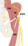
Anatomical drawing of the course of the LFCN: The nerve originates from the nerve roots L2 and L3 runs posterior to the psoas muscle (PM) in the retroperitoneal space on top of the iliac muscle (IM) and exits the pelvis through the inguinal ligament (IL), just medial to the anterior superior iliac spine (ASIS), and in the upper leg runs on top of the sartorius muscles (SM). This anatomical drawing shows the type B variant, which is most frequently encountered in patients with idiopathic meralgia paresthetica [78]
Fig. 2
Case of schwannoma inside the LFCN. A: Coronal T1-weighted MR image showing a well-circumscribed lesion inside the LFCN with homogenous gadolinium enhancement. The lesion was positioned on top of the sartorius muscle (SM). B: Intraoperative picture showing the schwannoma with bands placed around the LFCN proximal and distal to the lesion
Fig. 3
Case of lipoma inside the tensor fascia latae muscle. A: Transverse T1-weighted MR image showing a lesion of 1cm in diameter in the tensor fascia latae muscle (TFL) with high signal intensity comparable to that of subcutaneous fat (white arrow). The black arrow points at the branches of the LFCN. B: Intra-operative image with white bands placed around the separate branches of the LFCN after opening of the fascia lata
Fig. 4.
L2 Case of L2 nerve root schwannoma: A and B: sagittal and transverse T1-weighted MR images after gadolinium showing the schwannoma inside the L2 nerve root on the right side. C and D: intra-operative pictures showing the schwannoma before and after opening of the dura
Fig. 5
Case of endometriosis in the groin area: A: Transverse T1-weighted image with gadolinium showing the endometriosis lesion (white arrow) inside the inguinal ligament. B: Intra-operative image showing the LFCN (arrow) after mobilization of the endometriosis lesion. C: Picture of endometriosis lesion
Similar articles
- Meralgia paresthetica caused by hip-huggers in a patient with aberrant course of the lateral femoral cutaneous nerve.
Park JW, Kim DH, Hwang M, Bun HR. Park JW, et al. Muscle Nerve. 2007 May;35(5):678-80. doi: 10.1002/mus.20721. Muscle Nerve. 2007. PMID: 17212348 - Meralgia paresthetica: diagnosis and treatment.
Grossman MG, Ducey SA, Nadler SS, Levy AS. Grossman MG, et al. J Am Acad Orthop Surg. 2001 Sep-Oct;9(5):336-44. doi: 10.5435/00124635-200109000-00007. J Am Acad Orthop Surg. 2001. PMID: 11575913 Review. - Anatomy of the lateral femoral cutaneous nerve relevant to clinical findings in meralgia paresthetica.
Lee SH, Shin KJ, Gil YC, Ha TJ, Koh KS, Song WC. Lee SH, et al. Muscle Nerve. 2017 May;55(5):646-650. doi: 10.1002/mus.25382. Epub 2017 Jan 3. Muscle Nerve. 2017. PMID: 27543938 - Lateral Femoral Cutaneous Nerve Radiofrequency Ablation for Long-term Control of Refractory Meralgia Paresthetica.
Abd-Elsayed A, Gyorfi MJ, Ha SP. Abd-Elsayed A, et al. Pain Med. 2020 Nov 7;21(7):1433-1436. doi: 10.1093/pm/pnz372. Pain Med. 2020. PMID: 32022852 - Meralgia Paresthetica.
Solomons JNT, Sagir A, Yazdi C. Solomons JNT, et al. Curr Pain Headache Rep. 2022 Jul;26(7):525-531. doi: 10.1007/s11916-022-01053-7. Epub 2022 May 27. Curr Pain Headache Rep. 2022. PMID: 35622311 Review.
Cited by
- Meralgia Paresthetica-An Approach Specific Neurological Complication in Patients Undergoing DAA Total Hip Replacement: Anatomical and Clinical Considerations.
Almasi J, Ambrus R, Steno B. Almasi J, et al. Life (Basel). 2024 Jan 20;14(1):151. doi: 10.3390/life14010151. Life (Basel). 2024. PMID: 38276280 Free PMC article.
References
Publication types
MeSH terms
LinkOut - more resources
Full Text Sources
Miscellaneous


