Insertion signal sequence fused to minimal peptides elicits specific CD8+ T-cell responses and prolongs survival of thymoma-bearing mice - PubMed (original) (raw)
. 1994 Aug 1;54(15):4155-61.
Affiliations
- PMID: 7518351
- PMCID: PMC2254935
Insertion signal sequence fused to minimal peptides elicits specific CD8+ T-cell responses and prolongs survival of thymoma-bearing mice
B R Minev et al. Cancer Res. 1994.
Abstract
CD8+ T-lymphocytes (TCD8+) recognize minimal peptides of 8-10 residues which are the products of intracellularly processed proteins and are presented at the cell surface by major histocompatibility complex class I molecules. An important step in this process is the translocation of processed proteins from the cytosol across the endoplasmic reticulum membrane, mediated by transporter associated with antigen-processing proteins or alternatively by endoplasmic reticulum-insertion signal sequences located at the NH2-terminus of the precursor molecules. We report here that the addition of an endoplasmic reticulum-insertion signal sequence at the NH2-terminus of TCD8+ epitopes from chicken ovalbumin (amino acids 257-264) or a naturally occurring tumor antigen expressed by the murine mastocytoma P815 (P1A amino acids 35-43) significantly enhanced the priming of specific TCD8+ in vivo. The signal sequence did not enhance peptide immunogenicity by merely increasing the hydrophobicity of the peptide, since ovalbumin amino acids 257-264 peptide with the signal sequence at its COOH-terminus did not demonstrate enhanced efficacy. The signal sequence did not act as a helper epitope, since TCD8+ responses were not diminished in class II-deficient transgenic mice or in mice depleted of CD4+ T-cells in vivo. Importantly, a single immunization with the fusion peptide significantly prolonged survival of mice challenged with E.G7OVA, a thymoma transfected with the complementary DNA of chicken ovalbumin.
Figures
Fig. 1
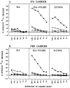
ESOVA but not OVAES or OVA enhances priming in vivo. Specific anti-OVA immune response was elicited by immunization with synthetic peptides in IFA (A) or PBS (B). C57BL/6N mice were immunized with 200 _μ_g/mouse ESOVA (●), OVAES (▽), OVA (▼), or PBS (□). After 10 days, recipient spleen cells were stimulated against OVA minimal peptide in vitro for 6 days and their cytotoxic activity was determined with EL-4 cells (left), EL-4 cells pulsed with OVA257–264 for 90 min (middle), or E.G7OVA cells (right).
Fig. 2
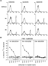
ESOVA enhances priming in vivo in mice depleted of TCD4+. A, FACS analysis of splenocytes isolated from normal nondepleted (left column), TCD4+-depleted (middle column), or TCD8+-depleted (right column) mice. Mice were given i.v. injections, three times at intervals of 6 days, of antibody GK1.5 (anti-CD4) or 2.43 (anti-CD8). FACS analysis was performed on the day of secondary stimulation of splenocytes, using fluorescein isothiocyanate-labeled anti-CD4 and anti-CD8 antibodies. B, cytotoxic activity in nondepleted (left), TCD4+-depleted (middle), or TCD8+-depleted (right) mice. C57BL/6N mice were depleted of TCD4+ or TCD8+ as described in the text. The mice were immunized with ESOVA (●), OVAES (▽), OVA (▼), or PBS (□). After 10 days, recipient spleen cells were stimulated against OVA minimal peptide in vitro for 6 days and their cytotoxic activity was determined with EL-4 cells pulsed with OVA257–264 for 90 min.
Fig. 3
ESOVA enhances priming in vivo in the absence of MHC class II. MHC class II-deficient transgenic mice were immunized with ESOVA (●), OVA (▼), or PBS (□). After 10 days, recipient spleen cells were stimulated in vitro for 6 days against OVA minimal peptide and their cytotoxic activity was determined with EL-4 cells (left) or EL-4 cells pulsed with OVA257–264 for 90 min (right).
Fig. 4
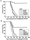
Immunoprotection of C57BL/6N mice challenged with E.G7OVA cells by a single immunization with ESOVA in IFA (A) or PBS (B). Mice were immunized with ESOVA, OVAES, OVA, or PBS 10 days before the challenge with 104 E.G7OVA cells.
Fig. 5
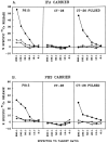
ESP1A but not P1A enhances priming in vivo. Specific anti-P1A immune response was elicited by immunization with synthetic peptides in IFA (A) or PBS (B). DBA/2 mice were immunized with 200 _μ_g/mouse ESP1A (●), P1A (▼), or PBS (▽). After 10 days, recipient spleen cells were stimulated against P1A minimal peptide in vitro for 6 days and their cytotoxic activity was determined with P815 cells (left), CT-26 cells (middle), or CT-26 cells pulsed with P1A35–43 for 90 min (right).
Similar articles
- Minimal determinant expressed by a recombinant vaccinia virus elicits therapeutic antitumor cytolytic T lymphocyte responses.
McCabe BJ, Irvine KR, Nishimura MI, Yang JC, Spiess PJ, Shulman EP, Rosenberg SA, Restifo NP. McCabe BJ, et al. Cancer Res. 1995 Apr 15;55(8):1741-7. Cancer Res. 1995. PMID: 7536130 Free PMC article. - Effective induction of anti-tumor immune responses with oligomannose-coated liposome targeting to intraperitoneal phagocytic cells.
Ikehara Y, Shiuchi N, Kabata-Ikehara S, Nakanishi H, Yokoyama N, Takagi H, Nagata T, Koide Y, Kuzushima K, Takahashi T, Tsujimura K, Kojima N. Ikehara Y, et al. Cancer Lett. 2008 Feb 18;260(1-2):137-45. doi: 10.1016/j.canlet.2007.10.038. Cancer Lett. 2008. PMID: 18077084 - Augmented induction of CD8+ cytotoxic T-cell response and antitumour resistance by T helper type 1-inducing peptide.
Kikuchi T, Uehara S, Ariga H, Tokunaga T, Kariyone A, Tamura T, Takatsu K. Kikuchi T, et al. Immunology. 2006 Jan;117(1):47-58. doi: 10.1111/j.1365-2567.2005.02262.x. Immunology. 2006. PMID: 16423040 Free PMC article. - An essential role of antigen-presenting cell/T-helper type 1 cell-cell interactions in draining lymph node during complete eradication of class II-negative tumor tissue by T-helper type 1 cell therapy.
Chamoto K, Wakita D, Narita Y, Zhang Y, Noguchi D, Ohnishi H, Iguchi T, Sakai T, Ikeda H, Nishimura T. Chamoto K, et al. Cancer Res. 2006 Feb 1;66(3):1809-17. doi: 10.1158/0008-5472.CAN-05-2246. Cancer Res. 2006. PMID: 16452242 - Primary T-cell and activated macrophage response associated with tumor protection using peptide/poly-N-acetyl glucosamine vaccination.
Maitre N, Brown JM, Demcheva M, Kelley JR, Lockett MA, Vournakis J, Cole DJ. Maitre N, et al. Clin Cancer Res. 1999 May;5(5):1173-82. Clin Cancer Res. 1999. PMID: 10353754
Cited by
- Early detection of tumour immune-rejection using magnetic resonance imaging.
Hu DE, Beauregard DA, Bearchell MC, Thomsen LL, Brindle KM. Hu DE, et al. Br J Cancer. 2003 Apr 7;88(7):1135-42. doi: 10.1038/sj.bjc.6600814. Br J Cancer. 2003. PMID: 12671716 Free PMC article. - Minimal determinant expressed by a recombinant vaccinia virus elicits therapeutic antitumor cytolytic T lymphocyte responses.
McCabe BJ, Irvine KR, Nishimura MI, Yang JC, Spiess PJ, Shulman EP, Rosenberg SA, Restifo NP. McCabe BJ, et al. Cancer Res. 1995 Apr 15;55(8):1741-7. Cancer Res. 1995. PMID: 7536130 Free PMC article. - Developing recombinant and synthetic vaccines for the treatment of melanoma.
Restifo NP, Rosenberg SA. Restifo NP, et al. Curr Opin Oncol. 1999 Jan;11(1):50-7. doi: 10.1097/00001622-199901000-00012. Curr Opin Oncol. 1999. PMID: 9914879 Free PMC article. Review. - Cutting edge: CD4+ T cell control of CD8+ T cell reactivity to a model tumor antigen.
Surman DR, Dudley ME, Overwijk WW, Restifo NP. Surman DR, et al. J Immunol. 2000 Jan 15;164(2):562-5. doi: 10.4049/jimmunol.164.2.562. J Immunol. 2000. PMID: 10623795 Free PMC article. - The promise of nucleic acid vaccines.
Restifo NP, Ying H, Hwang L, Leitner WW. Restifo NP, et al. Gene Ther. 2000 Jan;7(2):89-92. doi: 10.1038/sj.gt.3301117. Gene Ther. 2000. PMID: 10673713 Free PMC article. Review.
References
- Greenberg PD. Adoptive T cell therapy of tumors: mechanisms operative in the recognition and elimination of tumor cells. Adv. Immunol. 1991;49:281–355. - PubMed
- Rosenberg SA, Spiess P, Lafreniere R. A new approach to the adoptive immunotherapy of cancer with tumor-infiltrating lymphocytes. Science (Washington DC) 1986;233:1318–1321. - PubMed
- Townsend A, Bodmer H. Antigen recognition of class I restricted T lymphocytes. Annu. Rev. Immunol. 1989;7:601–624. - PubMed
- Yewdell JW, Bennink JR. Cell biology of antigen processing and presentation to MHC class I molecule-restricted T lymphocytes. Annu. Rev. Immunol. 1992;52:1–123. - PubMed
- Rammensee HG. MHC class I structure and function. Semin. Immunol. 1993;5:73–145.
MeSH terms
Substances
LinkOut - more resources
Full Text Sources
Other Literature Sources
Medical
Research Materials


