Transgenic expression of embryonic MAP2 in adult mouse brain: implications for neuronal polarization - PubMed (original) (raw)
Transgenic expression of embryonic MAP2 in adult mouse brain: implications for neuronal polarization
K M Marsden et al. J Neurosci. 1996.
Abstract
The major neuronal microtubule-associated protein MAP2 is selectively localized in dendrites, where its expression is under strong developmental regulation. To learn more about its potential effects on neuronal morphogenesis and its sorting within the neuronal cytoplasm, we have raised transgenic mice that express high levels of the embryonic form, MAP2c, in the adult brain. One transgenic line expressed higher levels of MAP2c than endogenous adult MAP2. This had no detectable effect on either the arrangement or morphology of neurons, suggesting that although MAP2c is necessary for neuronal morphogenesis it is not involved in its regulation. Like endogenous adult MAP2, transgenic MAP2c was present in dendrites but not axons, indicating that the signal responsible for its cytoplasmic sorting is contained within the 1.5 kb of its coding sequence. In situ hybridization with specific probes showed that transgenic MAP2c mRNA was limited to cell bodies. Thus, the dendritic localization of MAP2c protein cannot be the result of previous transport of its mRNA but must depend on a signal associated with the protein itself. Furthermore, because the amino acid sequence of MAP2c is present in all forms of MAP2, this signal is also contained within adult high-M(r) MAP2 protein. This raises the possibility that, rather than the conventional scheme of mRNA sorting preceding protein localization, the transport of adult MAP2 mRNA into dendrites could depend on it being part of a translation complex in which the targeting signal is on the nascent protein.
Figures
Fig. 1.
Expression of embryonic MAP2c in the brains of adult transgenic mice. A, B, Western blot analysis of MAP2 expression in transgenic and nontransgenic mice. Each lane contains 40 μg of microtubule protein stained with monoclonal antibodies against MAP2 (A) or the myc epitope (B). Lane 1, Control nontransgenic mouse;lane 2, a nontransgenic littermate to transgenic mice;lanes 3, 4, two independent transgenic mouse lines. Positions are indicated for endogenous adult high_M_r MAP2 (2b), embryonic MAP2 (2c), and tubulin (T).
Fig. 2.
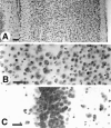
Cresyl violet-stained sections from the brain of a transgenic mouse expressing high levels of myc MAP2c. No abnormalities in either cell number or arrangement were detectable in either cerebral cortex (A, B) or hippocampus (C). Positions are indicated for endogenous adult high_M_r MAP2 (2b), embryonic MAP2 (2c), and tubulin (T). Scale bars, 50 μm.
Fig. 3.
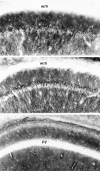
Immunohistochemical staining of sections from the CA1 region of the hippocampus from the brain of a transgenic mouse expressing high levels of MAP2c. A, Stained with monoclonal antibodies against the myc epitope to visualize transgenically expressed MAP2c. B, Neighboring section stained with monoclonal antibody AP14 that selectively labels the adult high_M_r forms of MAP2. C, Neighboring section stained with rabbit polyclonal antiserum against microtubule-associated protein tau (Goedert et al., 1989) that selectively labels tau in axons. The layer of pyramidal neuron cell bodies is labeled py, and the overlying white matter axon tracts (alveus and corpus callosum) wm. Scale bar, 100 μm.
Fig. 4.
Dendritic localization of MAP2c in cultured hippocampal neurons from transgenic mice. Cultures established at embryonic day 16 and grown for 15 d were fixed and double-immunofluorescence-stained with anti-c-myc antibody for transgenic MAP2c (rhodamine; A) and rabbit antibody against tau (fluorescein; B). In A some dendrites are identified by arrowheads. Preparations were also made using fluorescein to label anti-c-myc and rhodamine to label tau with identical results. Scale bar, 50 μm.
Fig. 5.
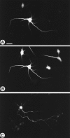
Location of myc-MAP2c in primary hippocampal neurons transfected in vitro. Cultures established at embryonic day 16 grown for 11 d were fixed and double-immunofluorescence-stained for the myc epitope of MAP2c (rhodamine; A) and for MAP2 (fluorescein; B). In the strongly expressing cell in A, transfected MAP2c shows the same dendritic localization as the endogenous adult MAP2 shown in_B_. A soluble test protein (CAT) expressed using the same promoter and transfection procedure is distributed throughout dendrites and axons (C). Scale bar, 50 μm.
Fig. 6.
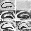
Localization of MAP2 and tubulin mRNAs in transgenic mouse hippocampus by in situ hybridization. Sections from brains of either transgenic (left) or control (right) mice were incubated with specific35S-labeled oligonucleotide probes, and the distribution of their target mRNAs was determined by autoradiography. Probes specific for the MAP2c-specific splice junction (A,B) gave specific labeling only in transgenic brain sections, and this labeling was limited to cell body layer in both the CA region (labeled CA1) and dentate gyrus (dg). By contrast, probes specific for endogenous high_M_r MAP2 mRNA labeled both cell bodies and the adjacent dendrite-rich neuropil in both transgenic (C) and control (D) brains. A tubulin control probe labeled only cell body layers of CA1 and dentate gyrus neurons (E,F). Scale bar, 0.5 mm.
Fig. 7.
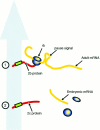
Diagrammatic representation of possible mechanisms for MAP2 transport signals. The large gray _arrow_represents the putative dendrite-directed transport mechanism.1, A transport signal (green box) near the N terminus of the protein emerges during translation so that the translation complex, including the adult high-_M_r protein (2b protein) and mRNA (Adult mRNA) and ribosomes (rb) are cotransported. A pause signal unique to the high-M_r mRNA (shown as a_loop) may act as an additional feature mediating mRNA transport. 2, MAP2c mRNA (Embryonic mRNA), either because it is shorter or because it lacks a pause signal, completes its translation in the cell body so that only the protein is carried into the dendrite.
Similar articles
- Embryonic MAP2 lacks the cross-linking sidearm sequences and dendritic targeting signal of adult MAP2.
Papandrikopoulou A, Doll T, Tucker RP, Garner CC, Matus A. Papandrikopoulou A, et al. Nature. 1989 Aug 24;340(6235):650-2. doi: 10.1038/340650a0. Nature. 1989. PMID: 2770869 - The low molecular weight form of microtubule-associated protein 2 is transported into both axons and dendrites.
Meichsner M, Doll T, Reddy D, Weisshaar B, Matus A. Meichsner M, et al. Neuroscience. 1993 Jun;54(4):873-80. doi: 10.1016/0306-4522(93)90581-y. Neuroscience. 1993. PMID: 8341422 - Phosphorylation of microtubule-associated protein 2 (MAP2) and its relevance for the regulation of the neuronal cytoskeleton function.
Sánchez C, Díaz-Nido J, Avila J. Sánchez C, et al. Prog Neurobiol. 2000 Jun;61(2):133-68. doi: 10.1016/s0301-0082(99)00046-5. Prog Neurobiol. 2000. PMID: 10704996 Review. - Molecular characterization of microtubule-associated proteins tau and MAP2.
Goedert M, Crowther RA, Garner CC. Goedert M, et al. Trends Neurosci. 1991 May;14(5):193-9. doi: 10.1016/0166-2236(91)90105-4. Trends Neurosci. 1991. PMID: 1713721 Review.
Cited by
- The role of microtubule-associated protein 2c in the reorganization of microtubules and lamellipodia during neurite initiation.
Dehmelt L, Smart FM, Ozer RS, Halpain S. Dehmelt L, et al. J Neurosci. 2003 Oct 22;23(29):9479-90. doi: 10.1523/JNEUROSCI.23-29-09479.2003. J Neurosci. 2003. PMID: 14573527 Free PMC article. - Isoform specificity in the relationship of actin to dendritic spines.
Kaech S, Fischer M, Doll T, Matus A. Kaech S, et al. J Neurosci. 1997 Dec 15;17(24):9565-72. doi: 10.1523/JNEUROSCI.17-24-09565.1997. J Neurosci. 1997. PMID: 9391011 Free PMC article. - Identification of a cis-acting dendritic targeting element in MAP2 mRNAs.
Blichenberg A, Schwanke B, Rehbein M, Garner CC, Richter D, Kindler S. Blichenberg A, et al. J Neurosci. 1999 Oct 15;19(20):8818-29. doi: 10.1523/JNEUROSCI.19-20-08818.1999. J Neurosci. 1999. PMID: 10516301 Free PMC article. - Differential intracellular sorting of immediate early gene mRNAs depends on signals in the mRNA sequence.
Wallace CS, Lyford GL, Worley PF, Steward O. Wallace CS, et al. J Neurosci. 1998 Jan 1;18(1):26-35. doi: 10.1523/JNEUROSCI.18-01-00026.1998. J Neurosci. 1998. PMID: 9412483 Free PMC article. - Mitotic impairment by doublecortin is diminished by doublecortin mutations found in patients.
Couillard-Despres S, Uyanik G, Ploetz S, Karl C, Koch H, Winkler J, Aigner L. Couillard-Despres S, et al. Neurogenetics. 2004 Jun;5(2):83-93. doi: 10.1007/s10048-004-0176-1. Epub 2004 Mar 25. Neurogenetics. 2004. PMID: 15045646
References
- Albala JS, Kress Y, Liu W-K, Weidenheim K, Yen S-HC, Shafit Zagardo B. Human microtubule-associated protein-2c localizes to dendrites and axons in fetal spinal motor neurons. J Neurochem. 1995;95:2480–2490. - PubMed
- Bernhardt R, Matus A. Light and electron microscopic studies of the distribution of microtubule-associated protein 2 in rat brain: a difference between dendritic and axonal cytoskeletons. J Comp Neurol. 1984;226:203–221. - PubMed
Publication types
MeSH terms
Substances
LinkOut - more resources
Full Text Sources
Other Literature Sources

