Collateral sprouting of uninjured primary afferent A-fibers into the superficial dorsal horn of the adult rat spinal cord after topical capsaicin treatment to the sciatic nerve - PubMed (original) (raw)
Collateral sprouting of uninjured primary afferent A-fibers into the superficial dorsal horn of the adult rat spinal cord after topical capsaicin treatment to the sciatic nerve
R J Mannion et al. J Neurosci. 1996.
Abstract
That terminals of uninjured primary sensory neurons terminating in the dorsal horn of the spinal cord can collaterally sprout was first suggested by Liu and Chambers (1958), but this has since been disputed. Recently, horseradish peroxidase conjugated to the B subunit of cholera toxin (B-HRP) and intracellular HRP injections have shown that sciatic nerve section or crush produces a long-lasting rearrangement in the organization of primary afferent central terminals, with A-fibers sprouting into lamina II, a region that normally receives only C-fiber input (Woolf et al., 1992). The mechanism of this A-fiber sprouting has been thought to involve injury-induced C-fiber transganglionic degeneration combined with myelinated A-fibers being conditioned into a regenerative growth state. In this study, we ask whether C-fiber degeneration and A-fiber conditioning are both necessary for the sprouting of A-fibers into lamina II. Local application of the C-fiber-specific neurotoxin capsaicin to the sciatic nerve has previously been shown to result in C-fiber damage and degenerative atrophy in lamina II. We have used B-HRP to transganglionically label A-fiber central terminals and have shown that 2 weeks after topical capsaicin treatment to the sciatic nerve, the pattern of B-HRP staining in the dorsal horn is indistinguishable from that seen after axotomy, with lamina II displaying novel staining in the identical region containing capsaicin-treated C-fiber central terminals. These results suggest that after C-fiber injury, uninjured A-fiber central terminals can collaterally sprout into lamina II of the dorsal horn. This phenomenon may help to explain the pain associated with C-fiber neuropathy.
Figures
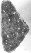
Fig. 1.
Electron microscopic photomontage of a transverse section through distal sciatic nerve 2 weeks after topical capsaicin treatment. Scale bar, 200 μm.
Fig. 2.
Bright-field photomicrograph of a 50 μm transverse section through the spinal cord showing TMP labeling in lamina II of the L5 dorsal horn 1 week after unilateral topical capsaicin treatment to the sciatic nerve. Note the absence of staining of the medial three-quarters of lamina II (arrows) ipsilateral to the treatment. Scale bar, 200 μm.
Fig. 3.
Bright-field photomicrographs of 50 μm transverse sections through the dorsal horn showing B-HRP labeling of central terminals of the sciatic nerve in a sham-operated animal at the L5 (a) and L3 (b) level. Note that lamina II is devoid of any B-HRP reaction product (arrows). Territory occupied by saphenous afferent central terminals can be seen to interrupt B-HRP labeling in the center of the dorsal horn in_b_. In a capsaicin-treated animal at L5 (c) and L3 (d), lamina II now displays dense B-HRP staining (arrows). Scale bar, 200 μm.
Fig. 4.
Schematic horizontal map of the somatotopic distribution at the lamina II level in the lumbar enlargement (L3–L6 segments) of B-HRP staining 2 weeks after peripheral axotomy, TMP depletion 1 week after topical capsaicin treatment to the sciatic nerve, and B-HRP staining 2 weeks after topical capsaicin treatment to the sciatic nerve. The pictures are essentially indistinguishable.
Similar articles
- Intact sciatic myelinated primary afferent terminals collaterally sprout in the adult rat dorsal horn following section of a neighbouring peripheral nerve.
Doubell TP, Mannion RJ, Woolf CJ. Doubell TP, et al. J Comp Neurol. 1997 Mar 31;380(1):95-104. doi: 10.1002/(sici)1096-9861(19970331)380:1<95::aid-cne7>3.0.co;2-o. J Comp Neurol. 1997. PMID: 9073085 - Perineural capsaicin induces the uptake and transganglionic transport of choleratoxin B subunit by nociceptive C-fiber primary afferent neurons.
Oszlács O, Jancsó G, Kis G, Dux M, Sántha P. Oszlács O, et al. Neuroscience. 2015 Dec 17;311:243-52. doi: 10.1016/j.neuroscience.2015.10.042. Epub 2015 Oct 28. Neuroscience. 2015. PMID: 26520849 - A-fiber sprouting in spinal cord dorsal horn is attenuated by proximal nerve stump encapsulation.
White FA, Kocsis JD. White FA, et al. Exp Neurol. 2002 Oct;177(2):385-95. doi: 10.1006/exnr.2002.7996. Exp Neurol. 2002. PMID: 12429185 - Effect of neonatal capsaicin and infraorbital nerve section on whisker-related patterns in the rat trigeminal nucleus.
Waite PM, de Permentier PJ. Waite PM, et al. J Comp Neurol. 1997 Sep 8;385(4):599-615. doi: 10.1002/(sici)1096-9861(19970908)385:4<599::aid-cne6>3.0.co;2-z. J Comp Neurol. 1997. PMID: 9302107 Review.
Cited by
- Stereological and somatotopic analysis of the spinal microglial response to peripheral nerve injury.
Beggs S, Salter MW. Beggs S, et al. Brain Behav Immun. 2007 Jul;21(5):624-33. doi: 10.1016/j.bbi.2006.10.017. Epub 2006 Dec 16. Brain Behav Immun. 2007. PMID: 17267172 Free PMC article. - Resiniferatoxin induces paradoxical changes in thermal and mechanical sensitivities in rats: mechanism of action.
Pan HL, Khan GM, Alloway KD, Chen SR. Pan HL, et al. J Neurosci. 2003 Apr 1;23(7):2911-9. doi: 10.1523/JNEUROSCI.23-07-02911.2003. J Neurosci. 2003. PMID: 12684478 Free PMC article. - Chronic hypersensitivity for inflammatory nociceptor sensitization mediated by the epsilon isozyme of protein kinase C.
Aley KO, Messing RO, Mochly-Rosen D, Levine JD. Aley KO, et al. J Neurosci. 2000 Jun 15;20(12):4680-5. doi: 10.1523/JNEUROSCI.20-12-04680.2000. J Neurosci. 2000. PMID: 10844037 Free PMC article. - The Role of Capsaicin-induced Acute Inactivation of C-fibers on Tactile Learning in Rat.
Rahmani M, Rajabi S, Allahtavakoli M, Roohbakhsh A, Sheibani V, Shamsizadeh A. Rahmani M, et al. Iran J Basic Med Sci. 2013 Feb;16(2):129-33. Iran J Basic Med Sci. 2013. PMID: 24298379 Free PMC article. - Systems and Circuits Linking Chronic Pain and Circadian Rhythms.
Warfield AE, Prather JF, Todd WD. Warfield AE, et al. Front Neurosci. 2021 Jul 2;15:705173. doi: 10.3389/fnins.2021.705173. eCollection 2021. Front Neurosci. 2021. PMID: 34276301 Free PMC article. Review.
References
- Ainsworth A, Hall P, Wall PD, Allt G, Lynn Mackenzie M, Gibson S, Polak JM. Effects of capsaicin applied locally to adult peripheral nerve II: anatomy and enzyme and peptide chemistry of peripheral nerve and spinal cord. Pain. 1981;11:379–388. - PubMed
- Alvarez FJ, Morris HR, Priestley JV. Sub-populations of smaller diameter trigeminal primary afferent neurons defined by expression of calcitonin gene-related peptide and the cell surface oligosaccharide recognized by monoclonal antibody LA4. J Neurocytol. 1991;20:716–731. - PubMed
- Apfel SC, Wright D, Dormia C, Snider WD, Kessler JA. Nerve growth factor stimulates BDNF mRNA expression in the peripheral nervous system. Soc Neurosci Abstr. 1995;419:14. - PubMed
- Arvidsson J, Ygge J, Grant G. Cell loss in lumbar dorsal root ganglia and transganglionic degeneration after sciatic nerve resection in the rat. Brain Res. 1986;373:15–21. - PubMed
Publication types
MeSH terms
Substances
Grants and funding
- F32 NS010161/NS/NINDS NIH HHS/United States
- NS11255/NS/NINDS NIH HHS/United States
- R01 NS010161/NS/NINDS NIH HHS/United States
- P01 NS011255/NS/NINDS NIH HHS/United States
- NS10161/NS/NINDS NIH HHS/United States
LinkOut - more resources
Full Text Sources


