Transgenic mice carrying the dominant rhodopsin mutation P347S: evidence for defective vectorial transport of rhodopsin to the outer segments - PubMed (original) (raw)
Transgenic mice carrying the dominant rhodopsin mutation P347S: evidence for defective vectorial transport of rhodopsin to the outer segments
T Li et al. Proc Natl Acad Sci U S A. 1996.
Abstract
To explore the pathogenic mechanism of dominant mutations affecting the carboxyl terminus of rhodopsin that cause retinitis pigmentosa, we generated five lines of transgenic mice carrying the proline-347 to serine (P347S) mutation. The severity of photoreceptor degeneration correlated with the levels of transgene expression in these lines. Visual function as measured by the electroretinogram was approximately normal at an early age when there was little histologic evidence of photoreceptor degeneration, but it deteriorated as photoreceptors degenerated. Immunocytochemical staining showed the mutant rhodopsin predominantly in the outer segments prior to histologically evident degeneration, a finding supported by quantitation of signal intensities in different regions of the photoreceptor cells by confocal microscopy. A distinct histopathologic abnormality was the accumulation of submicrometer-sized vesicles extracellularly near the junction between inner and outer segments. The extracellular vesicles were bound by a single membrane that apparently contained rhodopsin as revealed by ultrastructural immunocytochemical staining with anti-rhodopsin antibodies. The outer segments, although shortened, contained well-packed discs. Proliferation of the endoplasmic reticulum as reported in Drosophila expressing dominant rhodopsin mutations was not observed. The accumulation of rhodopsinladen vesicles likely represents aberrant transport of rhodopsin from the inner segments to the nascent disc membranes of the outer segments. It is possible that photoreceptor degeneration occurs because of a failure to renew outer segments at a normal rate, thereby leading to a progressive shortening of outer segments, or because of the loss of cellular contents to the extracellular space, or because of both.
Figures
Figure 3
Labeling intensity histograms along the expanse of the retina plotted from digitized images under a confocal microscope. The retinas were from the P347S C1 line and the NHR-E line at 20 days of age. The vertical axis represents signal intensity, saturating at 250 arbitrary units. The horizontal axis extends left to right from the basal RPE toward the inner retina. The approximate locations of the retinal layers are marked at the top. Abbreviations are the same as in Fig. 2.
Figure 1
ERG recordings and matching histology of transgenic and control mice. The ages are indicated in postnatal days (e.g., p30). Arrows indicate the measurement of a-wave implicit times. A range of 18–28 msec is typically observed for wt mice. (Bar = 25 μm.)
Figure 2
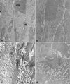
Localization of mutant rhodopsin by immunofluorescence. For each genotype labeled at the bottom of the figure, the left panel is an immunofluorescence image, and the right is the same view photographed under Hoffman interference contrast optics. The P347S retina was from the C1 line, the NHR-E retina was from a heterozygote, and the wt retina was from a C57BL/6 mouse. All mice were 20 days of age. OS, outer segments; IS, inner segments; ONL, outer nuclear layer; OPL, outer plexiform layer; INL, inner nuclear layer. (Bar = 25 μm.)
Figure 4
Transmission electron micrographs of the apical inner segments and basal outer segments of photoreceptor cells from 20-day-old retinas. (a) normal morphology in NHR-E retina shown as a control. (b) P347S C1 heterozygote. (c) P347S C1 homozygote. (d) P347S A1. OS, outer segments; M, mitochondria. (Bar = 1 μm.)
Figure 5
Scanning electron micrographs showing the region of outer retina. (a) Normal morphology in a wt retina shown as a control. Photoreceptor nuclei appear at the very bottom of the photograph. Extending upwards are well-defined inner segments, the thinner connecting cilia, and the outer segments. (b) P347S A1 retina. Note that the inner and outer segments were masked by surrounding vesicles. (Bar = 1 μm.)
Figure 6
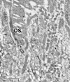
Ultrastructural immunocytochemical examination of a 20-day-old P347S C1 retina. Staining was with rho 4D2 antibody and gold-labeled secondary antibody. A packet of vesicles in the center of view is stained (arrow), but at lower intensity than the outer segments (OS). (Bar = 1 μm.)
Similar articles
- Effect of vitamin A supplementation on rhodopsin mutants threonine-17 --> methionine and proline-347 --> serine in transgenic mice and in cell cultures.
Li T, Sandberg MA, Pawlyk BS, Rosner B, Hayes KC, Dryja TP, Berson EL. Li T, et al. Proc Natl Acad Sci U S A. 1998 Sep 29;95(20):11933-8. doi: 10.1073/pnas.95.20.11933. Proc Natl Acad Sci U S A. 1998. PMID: 9751768 Free PMC article. - Glycosylation of rhodopsin is necessary for its stability and incorporation into photoreceptor outer segment discs.
Murray AR, Vuong L, Brobst D, Fliesler SJ, Peachey NS, Gorbatyuk MS, Naash MI, Al-Ubaidi MR. Murray AR, et al. Hum Mol Genet. 2015 May 15;24(10):2709-23. doi: 10.1093/hmg/ddv031. Epub 2015 Jan 30. Hum Mol Genet. 2015. PMID: 25637522 Free PMC article. - Retinal degeneration in humanized mice expressing mutant rhodopsin under the control of the endogenous murine promoter.
Liu X, Jia R, Meng X, Li Y, Yang L. Liu X, et al. Exp Eye Res. 2022 Feb;215:108893. doi: 10.1016/j.exer.2021.108893. Epub 2021 Dec 14. Exp Eye Res. 2022. PMID: 34919893 - Defective trafficking of rhodopsin and its role in retinal degenerations.
Hollingsworth TJ, Gross AK. Hollingsworth TJ, et al. Int Rev Cell Mol Biol. 2012;293:1-44. doi: 10.1016/B978-0-12-394304-0.00006-3. Int Rev Cell Mol Biol. 2012. PMID: 22251557 Review.
Cited by
- The primary cilium as a novel extracellular sensor in bone.
Hoey DA, Chen JC, Jacobs CR. Hoey DA, et al. Front Endocrinol (Lausanne). 2012 Jun 13;3:75. doi: 10.3389/fendo.2012.00075. eCollection 2012. Front Endocrinol (Lausanne). 2012. PMID: 22707948 Free PMC article. - Tmem138 is localized to the connecting cilium essential for rhodopsin localization and outer segment biogenesis.
Guo D, Ru J, Xie L, Wu M, Su Y, Zhu S, Xu S, Zou B, Wei Y, Liu X, Liu Y, Liu C. Guo D, et al. Proc Natl Acad Sci U S A. 2022 Apr 12;119(15):e2109934119. doi: 10.1073/pnas.2109934119. Epub 2022 Apr 8. Proc Natl Acad Sci U S A. 2022. PMID: 35394880 Free PMC article. - Severe retinal degeneration caused by a novel rhodopsin mutation.
Liu H, Wang M, Xia CH, Du X, Flannery JG, Ridge KD, Beutler B, Gong X. Liu H, et al. Invest Ophthalmol Vis Sci. 2010 Feb;51(2):1059-65. doi: 10.1167/iovs.09-3585. Epub 2009 Sep 9. Invest Ophthalmol Vis Sci. 2010. PMID: 19741247 Free PMC article. - Mutation analysis of codons 345 and 347 of rhodopsin gene in Indian retinitis pigmentosa patients.
Dikshit M, Agarwal R. Dikshit M, et al. J Genet. 2001 Aug;80(2):111-6. doi: 10.1007/BF02728336. J Genet. 2001. PMID: 11910130 - PRCD is essential for high-fidelity photoreceptor disc formation.
Spencer WJ, Ding JD, Lewis TR, Yu C, Phan S, Pearring JN, Kim KY, Thor A, Mathew R, Kalnitsky J, Hao Y, Travis AM, Biswas SK, Lo WK, Besharse JC, Ellisman MH, Saban DR, Burns ME, Arshavsky VY. Spencer WJ, et al. Proc Natl Acad Sci U S A. 2019 Jun 25;116(26):13087-13096. doi: 10.1073/pnas.1906421116. Epub 2019 Jun 12. Proc Natl Acad Sci U S A. 2019. PMID: 31189593 Free PMC article.
References
- Daiger S, Sullivan L, Rodriguez J. Behav Brain Sci. 1995;18:452–467.
- Rosenfeld P J, Cowley G S, McGee T L, Sandberg M A, Berson E L, Dryja T P. Nat Genet. 1992;1:209–213. - PubMed
- Sung C H, Davenport C M, Nathans J. J Biol Chem. 1993;268:26645–26649. - PubMed
- Kaushal S, Khorana H G. Biochemistry. 1994;33:6121–6128. - PubMed
Publication types
MeSH terms
Substances
LinkOut - more resources
Full Text Sources
Other Literature Sources
Molecular Biology Databases


