Network of tangential pathways for neuronal migration in adult mammalian brain - PubMed (original) (raw)
Network of tangential pathways for neuronal migration in adult mammalian brain
F Doetsch et al. Proc Natl Acad Sci U S A. 1996.
Abstract
Cells in the brains of adult mammals continue to proliferate in the subventricular zone (SVZ) throughout the lateral wall of the lateral ventricle. Here we show, using whole mount dissections of this wall from adult mice, that the SVZ is organized as an extensive network of chains of neuronal precursors. These chains are immunopositive to the polysialylated form of NCAM, a molecule present at sites of plasticity, and TuJ1, an early neuronal marker. The majority of the chains are oriented along the rostrocaudal axis and many join the rostral migratory stream that terminates in the olfactory bulb. Using focal microinjections of DII and transplantation of SVZ cells carrying a neuron-specific reporter gene, we demonstrate that cells originating at different rostrocaudal levels of the SVZ migrate rostrally and reach the olfactory bulb where they differentiate into neurons. Our results reveal an extensive network of pathways for the tangential chain migration of neuronal precursors throughout the lateral wall of the lateral ventricle in the adult mammalian brain.
Figures
Figure 1
Network of PSA-NCAM immunopositive chains in the lateral wall of the lateral ventricle of adult mice. Dorsal is up and rostral is left. (A) Camera lucida drawing of PSA-NCAM immunopositive chains in whole mounts of this wall. The dorsal group of chains on the wall of the anterior horn are connected to the RMS, but the tissue broke at this point and others (dashed line) during processing. The outline corresponds to the colored area in_C_. The rectangle indicates the area shown in_B_. (B) Photomicrograph of PSA-NCAM-immunopositive chains shown in marked rectangle in camera lucida drawing in A. Notice a group of chains (arrow) that are disorganized and end in the central region of the anterior horn. (C) Schematic sagittal view of adult mouse brain showing in different colors the different regions of the lateral wall of the lateral ventricle. The medially located anterior horn (red) is connected to the laterally located inferior horn (yellow) through an intermediate bridge (orange). Black arrow indicates direction of migration of neuronal precursors in the RMS to the olfactory bulb, where cells disperse radially (thin arrows) to reach the granular and glomerular layers (12). Numbers indicate anterior-posterior (A-P) stereotaxic coordinates (measured in mm). OB, olfactory bulb; M, foramen of Monro; NC, neocortex; CB, cerebellum; cc, corpus callosum. (A, bar = 250 μm; B, bar = 100 μm.)
Figure 2
Cells in chains are neuronal precursors. Double immunostaining for TuJ1 (A, fluorescein) and PSA-NCAM (B, rhodamine) of part of the SVZ in the lateral wall of the lateral ventricle (caudal anterior horn). (Bar = 30 microns.)
Figure 3
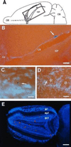
NSE-LacZ-transgenic SVZ cells grafted into caudal SVZ of nontransgenic host migrate to the olfactory bulb where they differentiate into granular and periglomerular neurons. (A) Schematic drawing of horizontal hemisection indicating the position of the graft (asterisk) in the lateral wall of the lateral ventricle (LV) and the position of photomicrograph shown in_B_ (rectangle). (B) Horizontal section of brain 17 (A-P −1.5, Table 1) showing graft site (arrow) relative to lateral ventricle (LV), corpus callosum (cc), and hippocampus (HP). (C) LacZ-positive granule neuron in the olfactory bulb 30 days after transplantation. Arrow indicates the Hoechst counterstained nucleus. (D) LacZ-positive periglomerular neuron (arrow) in the olfactory bulb 30 days after transplantation. The centers of glomeruli are indicated by stars. (E) Hoechst-stained horizontal olfactory bulb section from brain 17 with superimposed red dots indicating the distribution of LacZ-positive granular neurons and one LacZ-positive periglomerular neuron. OB, olfactory bulb; CB, cerebellum; gl, glomerular layer; epl, external plexiform layer; gcl, granule cell layer. (B, bar = 250 μm;C, bar = 50 μm; D, bar = 120 μm; E, bar = 400 μm.)
Figure 4
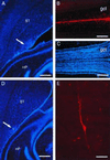
Microinjection of DiI into caudal SVZ results in labeled cells in the olfactory bulb. (A) DiI microinjection site at A-P −1 (arrow) in the SVZ at the level of the anterior hippocampus (horizontal section). (B) In this same brain, 30 days after injection, many DiI-labeled cells have reached the core of the olfactory bulb (the rostral extension of the RMS) and have migrated into the granule cell layer. (C) Hoechst 33258 counterstaining of section shown in B, to indicate olfactory bulb anatomy. (D) DiI microinjection site at A-P −1.5 (arrow) in the SVZ at the level of the hippocampus. (E) DiI-labeled granule neuron in the olfactory bulb of brain shown in D. HP, hippocampus, ST, striatum, gcl, granule cell layer. (A and D, bar = 450 microns; B and C, bar = 400 microns; bar = 20 microns.)
Similar articles
- Adult subventricular zone neuronal precursors continue to proliferate and migrate in the absence of the olfactory bulb.
Kirschenbaum B, Doetsch F, Lois C, Alvarez-Buylla A. Kirschenbaum B, et al. J Neurosci. 1999 Mar 15;19(6):2171-80. doi: 10.1523/JNEUROSCI.19-06-02171.1999. J Neurosci. 1999. PMID: 10066270 Free PMC article. - Embryonic (PSA) N-CAM reveals chains of migrating neuroblasts between the lateral ventricle and the olfactory bulb of adult mice.
Rousselot P, Lois C, Alvarez-Buylla A. Rousselot P, et al. J Comp Neurol. 1995 Jan 2;351(1):51-61. doi: 10.1002/cne.903510106. J Comp Neurol. 1995. PMID: 7896939 - Subventricular zone-derived neuronal progenitors migrate into the subcortical forebrain of postnatal mice.
De Marchis S, Fasolo A, Puche AC. De Marchis S, et al. J Comp Neurol. 2004 Aug 23;476(3):290-300. doi: 10.1002/cne.20217. J Comp Neurol. 2004. PMID: 15269971 - Architecture and cell types of the adult subventricular zone: in search of the stem cells.
García-Verdugo JM, Doetsch F, Wichterle H, Lim DA, Alvarez-Buylla A. García-Verdugo JM, et al. J Neurobiol. 1998 Aug;36(2):234-48. doi: 10.1002/(sici)1097-4695(199808)36:2<234::aid-neu10>3.0.co;2-e. J Neurobiol. 1998. PMID: 9712307 Review. - The Role of Adult-Born Neurons in the Constantly Changing Olfactory Bulb Network.
Malvaut S, Saghatelyan A. Malvaut S, et al. Neural Plast. 2016;2016:1614329. doi: 10.1155/2016/1614329. Epub 2015 Dec 29. Neural Plast. 2016. PMID: 26839709 Free PMC article. Review.
Cited by
- Morphological Signatures of Neurogenesis and Neuronal Migration in Hypothalamic Vasopressinergic Magnocellular Nuclei of the Adult Rat.
Zhang L, Zetter MA, Hernández VS, Hernández-Pérez OR, Jáuregui-Huerta F, Krabichler Q, Grinevich V. Zhang L, et al. Int J Mol Sci. 2024 Jun 26;25(13):6988. doi: 10.3390/ijms25136988. Int J Mol Sci. 2024. PMID: 39000096 Free PMC article. - A fluorescent perilipin 2 knock-in mouse model reveals a high abundance of lipid droplets in the developing and adult brain.
Madsen S, Delgado AC, Cadilhac C, Maillard V, Battiston F, Igelbüscher CM, De Neck S, Magrinelli E, Jabaudon D, Telley L, Doetsch F, Knobloch M. Madsen S, et al. Nat Commun. 2024 Jun 28;15(1):5489. doi: 10.1038/s41467-024-49449-w. Nat Commun. 2024. PMID: 38942786 Free PMC article. - Neuraminidase inhibition promotes the collective migration of neurons and recovery of brain function.
Matsumoto M, Matsushita K, Hane M, Wen C, Kurematsu C, Ota H, Bang Nguyen H, Quynh Thai T, Herranz-Pérez V, Sawada M, Fujimoto K, García-Verdugo JM, Kimura KD, Seki T, Sato C, Ohno N, Sawamoto K. Matsumoto M, et al. EMBO Mol Med. 2024 Jun;16(6):1228-1253. doi: 10.1038/s44321-024-00073-7. Epub 2024 May 24. EMBO Mol Med. 2024. PMID: 38789599 Free PMC article. - Threonine fuels glioblastoma through YRDC-mediated codon-biased translational reprogramming.
Wu X, Yuan H, Wu Q, Gao Y, Duan T, Yang K, Huang T, Wang S, Yuan F, Lee D, Taori S, Plute T, Heissel S, Alwaseem H, Isay-Del Viscio M, Molina H, Agnihotri S, Hsu DJ, Zhang N, Rich JN. Wu X, et al. Nat Cancer. 2024 Jul;5(7):1024-1044. doi: 10.1038/s43018-024-00748-7. Epub 2024 Mar 22. Nat Cancer. 2024. PMID: 38519786 - Thyroid hormone action in adult neurogliogenic niches: the known and unknown.
Valcárcel-Hernández V, Mayerl S, Guadaño-Ferraz A, Remaud S. Valcárcel-Hernández V, et al. Front Endocrinol (Lausanne). 2024 Mar 7;15:1347802. doi: 10.3389/fendo.2024.1347802. eCollection 2024. Front Endocrinol (Lausanne). 2024. PMID: 38516412 Free PMC article. Review.
References
- Allen E. J Comp Neurol. 1912;22:547–568.
- Smart I. J Comp Neurol. 1961;116:325–348.
- Privat A, Leblond C P. J Comp Neurol. 1972;146:277–302. - PubMed
- Levison S W, Goldman J E. Neuron. 1993;10:201–212. - PubMed
- Blakemore W F, Jolly D R. J Neurocytol. 1972;1:69–84. - PubMed
Publication types
MeSH terms
Grants and funding
- R01 NS028478/NS/NINDS NIH HHS/United States
- R37 NS028478/NS/NINDS NIH HHS/United States
- NICH-NS32116/NS/NINDS NIH HHS/United States
- NINDS-NS28478/NS/NINDS NIH HHS/United States
LinkOut - more resources
Full Text Sources
Other Literature Sources
Research Materials
Miscellaneous


