D-serine as a neuromodulator: regional and developmental localizations in rat brain glia resemble NMDA receptors - PubMed (original) (raw)
D-serine as a neuromodulator: regional and developmental localizations in rat brain glia resemble NMDA receptors
M J Schell et al. J Neurosci. 1997.
Abstract
D-Serine is localized in mammalian brain to a discrete population of glial cells near NMDA receptors, suggesting that D-serine is an endogenous agonist of the receptor-associated glycine site. To explore this possibility, we have compared the immunohistochemical localizations of D-serine, glycine, and NMDA receptors in rat brain. In the telencephalon, D-serine is concentrated in protoplasmic astrocytes, which are abundant in neuropil in close vicinity to NMDA receptor 2A/B subunits. Ultrastructural examination of the CA1 region of hippocampus reveals D-serine in the cytosolic matrix of astrocytes that ensheath neurons and blood vessels, whereas NR2A/B is concentrated in dendritic spines. By contrast, glycine immunoreactivity in telencephalon is the lowest in brain. During postnatal week 2, D-serine levels in cerebellum are comparable to those in adult cerebral cortex but fall to undetectable levels by day 26. During week 2, we observe parallel ontogeny of D-serine in Bergmann glia and NR2A/B in Purkinje cells, suggesting a role for astrocytic D-serine in NMDA receptor-mediated synaptogenesis. D-Serine in the radial processes of Bergmann glia is also well positioned to regulate NMDA receptor-dependent granule cell migration. In the inner granule layer, D-serine is found transiently in protoplasmic astrocytes surrounding glomeruli, where it could regulate development of the mossy fiber/granule cell synapse. D-Serine seems to be the endogenous ligand of glycine sites in the telencephalon and developing cerebellum, whereas glycine predominates in the adult cerebellum, olfactory bulb, and hindbrain.
Figures
Fig. 1.
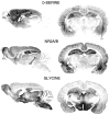
P21 serial brain sections stained for
d
-serine, NR2A/B, or glycine. Am, Amygdala;Cl, claustrum; Cx, cortex;EPL, external plexiform layer; Hb, habenula; Hp, hippocampus; Hy, hypothalamus; PM, pons/medulla; Sn, substantia nigra; Sp, spinal cord; WM, white matter; VNL, vomeronasal nerve layer.
Fig. 2.
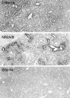
P21 amygdala stained for
d
-serine, NR2A/B, or glycine. Both
d
-serine and NR2A/B appear concentrated near blood vessels.
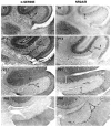
Fig. 3.
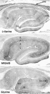
P21 hippocampus stained for
d
-serine, NR2A/B, or glycine. DG, Dentate gyrus;Hi, hilus; L, stratum lacunosum molecular; Lu, stratum lucidum of CA3 region;Mol, molecular layer of dentate gyrus; O, stratum oriens; P, stratum pyramidale; R, stratum radiatum; S, subiculum; WM, white matter.
Fig. 4.
Detailed comparison of
d
-serine and NR2A/B in P21 hippocampal CA1 region. A, B,
d
-Serine concentrates in the glia of molecular layers, especially near blood vessels (asterisks), whereas NR2A/B is found in pyramidal neurons and all layers of neuropil.C, D, Higher-power magnification of regions near blood vessels.
Fig. 5.
Ultrastructural comparison of
d
-serine and NR2A/B in hippocampal CA1 region. Brain sections were stained with the immunoperoxidase technique and then processed for electron microscopy.
d
-Serine concentrates in the cytosolic matrix of astrocytes (Ast) in neuropil and in foot processes ensheathing blood vessels (BV), whereas endothelial cells (En) are unstained. NR2A/B concentrates in dendritic spines (arrows). 10,000× magnification.
Fig. 6.
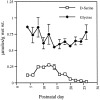
Levels of free
d
-serine and glycine in cerebellum during postnatal development. Cerebella were analyzed by HPLC for free amino acids. Values are mean ± SEM;n = 3.
Fig. 7.
Transient staining for
d
-serine and NR2A/B in developing cerebellum. The cell bodies of
d
-serine glia (Ast) are well labeled by P7, when Purkinje cells (P) begin to stain for NR2A/B. One week later, both
d
-serine and NR2A/B concentrate in the molecular layer, with
d
-serine in Bergmann glia (BG) and NR2A/B throughout the dendritic tree of Purkinje cells. In the P14 granule layer, many protoplasmic astrocytes stain intensely for
d
-serine, whereas a few Golgi neurons (Go) are lightly stained for NR2A/B. By P21, staining for both has decreased, but substantial amounts of
d
-serine persist in the radial process of BG and in the cell bodies of protoplasmic astrocytes (Ast). NR2A/B at P21 has become less prominent in Purkinje cells and has appeared in some basket cell pinceau (Pi). In mature adults,
d
-serine occurs weakly in Bergmann glia cell bodies, whereas NR2A/B is restricted to basket cell pinceau.
Fig. 8.
High magnification of adult cerebellum near Purkinje cell bodies.
d
-Serine is restricted to Bergmann glia (BG) in the molecular layer (Mol), especially in glial cell bodies that reside between Purkinje cells (P). The granule layer (Gr) is not stained for
d
-serine. NR2A/B still occurs in a minority of Purkinje cell dendrites, but the most intense staining occurs in basket cell pinceau (Pi), which also stain for glycine. Golgi neurons (Go), whose cell bodies reside in the granule layer, are the major glycinergic element of the cerebellum.
Fig. 9.
Models depicting the proposed modulatory roles for
d
-serine and glycine in the CA1 region of hippocampus (left) and the AOB (ACCES. OLF BULB, right).
d
-Serine is black; glycine is gray. Stars indicate localizations of NMDA receptors. In the hippocampus,
d
-serine-containing protoplasmic astrocytes (Ast) are localized near NMDA receptors located on pyramidal cell (Py) dendrites, whereas glycinergic cells are rare. In the AOB, both
d
-serine and glycine appear concentrated near NMDA receptors located on mitral cells (Mi), with the
d
-serine found in superficial bulbar glia (SBG) surrounding the vomeronasal nerve (VN) and in protoplasmic astrocytes (Ast) in the plexiform layer. Glycine concentrates in interneurons (I) and periglomerular cells (PG).
Fig. 10.
Models contrasting the proposed roles for
d
-serine and glycine in developing and adult cerebellum.
d
-Serine is black; glycine is_gray_. Stars indicate localizations of NMDA receptors. In developing molecular layer (left), astrocytic
d
-serine is found in Bergmann glia (BG), which ensheath Purkinje cells expressing NMDA receptors and also guide migrating granule cells (Gr) expressing NMDA receptors.
d
-Serine released from Bergmann glial processes might synergize with glutamate released by parallel fibers (PF) and climbing fibers (CF). In the developing inner granule layer, protoplasmic astrocytes (Ast) might release
d
-serine near the developing glomerular synapse to synergize with glutamate from mossy fibers (MF), whereas glycinergic basket (Ba) and Golgi neurons (Go) have not yet established connections with NMDA receptor-containing synapses. In contrast, in adult cerebellum (right), no
d
-serine is present, and NMDA receptors have disappeared from Purkinje cells. NMDA receptor-associated glycine sites located on the basket cell pinceau and granule cells might be modulated exclusively by glycinergic basket (Ba) and Golgi (Go) neurons.
Similar articles
- D-serine, an endogenous synaptic modulator: localization to astrocytes and glutamate-stimulated release.
Schell MJ, Molliver ME, Snyder SH. Schell MJ, et al. Proc Natl Acad Sci U S A. 1995 Apr 25;92(9):3948-52. doi: 10.1073/pnas.92.9.3948. Proc Natl Acad Sci U S A. 1995. PMID: 7732010 Free PMC article. - D-serine is an endogenous ligand for the glycine site of the N-methyl-D-aspartate receptor.
Mothet JP, Parent AT, Wolosker H, Brady RO Jr, Linden DJ, Ferris CD, Rogawski MA, Snyder SH. Mothet JP, et al. Proc Natl Acad Sci U S A. 2000 Apr 25;97(9):4926-31. doi: 10.1073/pnas.97.9.4926. Proc Natl Acad Sci U S A. 2000. PMID: 10781100 Free PMC article. - D-amino acids as putative neurotransmitters: focus on D-serine.
Snyder SH, Kim PM. Snyder SH, et al. Neurochem Res. 2000 May;25(5):553-60. doi: 10.1023/a:1007586314648. Neurochem Res. 2000. PMID: 10905615 Review. - D-Serine as a glial modulator of nerve cells.
Miller RF. Miller RF. Glia. 2004 Aug 15;47(3):275-283. doi: 10.1002/glia.20073. Glia. 2004. PMID: 15252817 Review. - Immunohistochemical localization of N-methyl-D-aspartate receptor NR1, NR2A, NR2B and NR2C/D subunits in the adult mammalian cerebellum.
Thompson CL, Drewery DL, Atkins HD, Stephenson FA, Chazot PL. Thompson CL, et al. Neurosci Lett. 2000 Apr 7;283(2):85-8. doi: 10.1016/s0304-3940(00)00930-7. Neurosci Lett. 2000. PMID: 10739881
Cited by
- Amino acid analyses of the exosome-eluted fractions from human serum by HPLC with fluorescence detection.
Onozato M, Tanaka Y, Arita M, Sakamoto T, Ichiba H, Sadamoto K, Kondo M, Fukushima T. Onozato M, et al. Pract Lab Med. 2018 Apr 22;12:e00099. doi: 10.1016/j.plabm.2018.e00099. eCollection 2018 Nov. Pract Lab Med. 2018. PMID: 30014016 Free PMC article. - Localization of D-serine and serine racemase in neurons and neuroglias in mouse brain.
Ding X, Ma N, Nagahama M, Yamada K, Semba R. Ding X, et al. Neurol Sci. 2011 Apr;32(2):263-7. doi: 10.1007/s10072-010-0422-2. Epub 2010 Oct 2. Neurol Sci. 2011. PMID: 20890627 - AMPA receptor mediated D-serine release from retinal glial cells.
Sullivan SJ, Miller RF. Sullivan SJ, et al. J Neurochem. 2010 Dec;115(6):1681-9. doi: 10.1111/j.1471-4159.2010.07077.x. Epub 2010 Nov 19. J Neurochem. 2010. PMID: 20969576 Free PMC article. - Rational and Translational Implications of D-Amino Acids for Treatment-Resistant Schizophrenia: From Neurobiology to the Clinics.
de Bartolomeis A, Vellucci L, Austin MC, De Simone G, Barone A. de Bartolomeis A, et al. Biomolecules. 2022 Jun 29;12(7):909. doi: 10.3390/biom12070909. Biomolecules. 2022. PMID: 35883465 Free PMC article. Review. - EphrinBs regulate D-serine synthesis and release in astrocytes.
Zhuang Z, Yang B, Theus MH, Sick JT, Bethea JR, Sick TJ, Liebl DJ. Zhuang Z, et al. J Neurosci. 2010 Nov 24;30(47):16015-24. doi: 10.1523/JNEUROSCI.0481-10.2010. J Neurosci. 2010. PMID: 21106840 Free PMC article.
References
- Altman J. Postnatal development of the cerebellar cortex in the rat. II. Phases in the maturation of Purkinje cells and of the molecular layer. J Comp Neurol. 1972;145:399–463. - PubMed
- Campistron G, Buijs RM, Geffard M. Glycine neurons in the brain and spinal cord: antibody production and immunocytochemical localization. Brain Res. 1986;376:400–405. - PubMed
- Chouinard ML, Gaitan D, Wood PL. Presence of the N-methyl-d-aspartate-associated glycine receptor agonist, d-serine, in human temporal cortex: comparison of normal, Parkinson, and Alzheimer tissues. J Neurochem. 1993;61:1561–1564. - PubMed
- Corrigan JJ. d-amino acids in animals. Science. 1969;164:142–149. - PubMed
- Cotman CW, Monaghan DT, Ottersen OP, Storm-Mathisen J. Anatomical organization of excitatory amino acid receptors and their pathways. Trends Neurosci. 1987;10:273–280.
Publication types
MeSH terms
Substances
Grants and funding
- DA 00074/DA/NIDA NIH HHS/United States
- 5RO1DA04431/DA/NIDA NIH HHS/United States
- R37 MH018501/MH/NIMH NIH HHS/United States
- R01 MH018501/MH/NIMH NIH HHS/United States
- K05 DA000074/DA/NIDA NIH HHS/United States
- MH 18501/MH/NIMH NIH HHS/United States
LinkOut - more resources
Full Text Sources
Other Literature Sources
Miscellaneous
