Prostate enlargement in mice due to fetal exposure to low doses of estradiol or diethylstilbestrol and opposite effects at high doses - PubMed (original) (raw)
Prostate enlargement in mice due to fetal exposure to low doses of estradiol or diethylstilbestrol and opposite effects at high doses
F S vom Saal et al. Proc Natl Acad Sci U S A. 1997.
Abstract
On the basis of results of studies using high doses of estrogens, exposure to estrogen during fetal life is known to inhibit prostate development. However, it is recognized in endocrinology that low concentrations of a hormone can stimulate a tissue, while high concentrations can have the opposite effect. We report here that a 50% increase in free-serum estradiol in male mouse fetuses (released by a maternal Silastic estradiol implant) induced a 40% increase in the number of developing prostatic glands during fetal life; subsequently, in adulthood, the number of prostatic androgen receptors per cell was permanently increased by 2-fold, and the prostate was enlarged by 30% (due to hyperplasia) relative to untreated males. However, as the free serum estradiol concentration in male fetuses was increased from 2- to 8-fold, adult prostate weight decreased relative to males exposed to the 50% increase in estradiol. As a model for fetal exposure to man-made estrogens, pregnant mice were fed diethylstilbestrol (DES) from gestation days 11 to 17. Relative to controls, DES doses of 0.02, 0.2, and 2.0 ng per g of body weight per day increased adult prostate weight, whereas a 200-ng-per-g dose decreased adult prostate weight in male offspring. Our findings suggest that a small increase in estrogen may modulate the action of androgen in regulating prostate differentiation, resulting in a permanent increase in prostatic androgen receptors and prostate size. For both estradiol and DES, prostate weight first increased then decreased with dose, resulting in an inverted-U dose-response relationship.
Figures
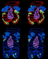
Figure 1
Two stereo pair images (convergent, or cross-eyed viewing) of computer-assisted, serial-section reconstructions showing the dorsal portion of the prostate from two mouse fetuses. The prostate from a control male with 0.21 pg/ml free serum estradiol (blue urethra) is shown below. The top prostate is reconstructed from a male fetus exposed to 0.32 pg/ml free serum estradiol (red urethra). Glandular buds that form into the dorsal (green), lateral (yellow), and dorsocranial (blue) glands in the adult prostate can be seen as outgrowths of the fetal urogenital sinus (ventral buds are not visible). The utriculus (pink) is the remnant of the regressing embryonic female reproductive tract (Müllerian ducts). Compared with controls, estradiol significantly increased the number of prostatic glandular buds and caused a reduction in the size of the lumen of the urethra, which passes through the prostate.
Figure 2
Mean (+ SEM) prostate weight (mg) in 8-month-old male mice produced by mothers implanted s.c. with Silastic capsules containing 0, 25, 100, 200, or 300 μg of estradiol from day 13 to day 19 of pregnancy. The free serum estradiol concentration (in pg/ml) in male fetuses on gestation day 18 in response to these doses of estradiol (controls = 0.21 pg/ml) is shown in relation to adult prostate weight. Group means that differed significantly are indicated by different letters, while groups with the same letter did not differ significantly.
Figure 3
For males exposed during fetal life to 0.21 pg/ml free estradiol (controls) and 0.32 pg/ml free estradiol (25 μg estradiol dose group), mean (+ SEM) total DNA (in μg), androgen receptors per mg of DNA (per cell), and androgen receptors per mg of protein are shown (these were determined after the prostate was weighed). * Statistically significant.
Figure 4
Mean (+ SEM) prostate weight (mg) in 8-month-old CF-1 male mice produced by females fed different doses of DES from day 11 to day 17 of pregnancy. Group means that differed significantly are indicated by different letters.
Similar articles
- Low-dose bioactivity of xenoestrogens in animals: fetal exposure to low doses of methoxychlor and other xenoestrogens increases adult prostate size in mice.
Welshons WV, Nagel SC, Thayer KA, Judy BM, Vom Saal FS. Welshons WV, et al. Toxicol Ind Health. 1999 Jan-Mar;15(1-2):12-25. doi: 10.1177/074823379901500103. Toxicol Ind Health. 1999. PMID: 10188188 - Uterine responsiveness to estradiol and DNA methylation are altered by fetal exposure to diethylstilbestrol and methoxychlor in CD-1 mice: effects of low versus high doses.
Alworth LC, Howdeshell KL, Ruhlen RL, Day JK, Lubahn DB, Huang TH, Besch-Williford CL, vom Saal FS. Alworth LC, et al. Toxicol Appl Pharmacol. 2002 Aug 15;183(1):10-22. doi: 10.1006/taap.2002.9459. Toxicol Appl Pharmacol. 2002. PMID: 12217638 - Alterations of maternal estrogen levels during gestation affect the skeleton of female offspring.
Migliaccio S, Newbold RR, Bullock BC, Jefferson WJ, Sutton FG Jr, McLachlan JA, Korach KS. Migliaccio S, et al. Endocrinology. 1996 May;137(5):2118-25. doi: 10.1210/endo.137.5.8612556. Endocrinology. 1996. PMID: 8612556 - Estrogenic environmental chemicals and drugs: mechanisms for effects on the developing male urogenital system.
Taylor JA, Richter CA, Ruhlen RL, vom Saal FS. Taylor JA, et al. J Steroid Biochem Mol Biol. 2011 Oct;127(1-2):83-95. doi: 10.1016/j.jsbmb.2011.07.005. Epub 2011 Jul 30. J Steroid Biochem Mol Biol. 2011. PMID: 21827855 Free PMC article. Review.
Cited by
- High butter-fat diet and bisphenol A additively impair male rat spermatogenesis.
Tarapore P, Hennessy M, Song D, Ying J, Ouyang B, Govindarajah V, Leung YK, Ho SM. Tarapore P, et al. Reprod Toxicol. 2017 Mar;68:191-199. doi: 10.1016/j.reprotox.2016.09.008. Epub 2016 Sep 19. Reprod Toxicol. 2017. PMID: 27658648 Free PMC article. - Sexually dimorphic nonreproductive behaviors as indicators of endocrine disruption.
Weiss B. Weiss B. Environ Health Perspect. 2002 Jun;110 Suppl 3(Suppl 3):387-91. doi: 10.1289/ehp.02110s3387. Environ Health Perspect. 2002. PMID: 12060833 Free PMC article. Review. - The toxic origins of disease.
Gross L. Gross L. PLoS Biol. 2007 Jul;5(7):e193. doi: 10.1371/journal.pbio.0050193. Epub 2007 Jun 26. PLoS Biol. 2007. PMID: 17594178 Free PMC article. - Bipotential effects of estrogen on growth hormone synthesis and storage in vitro.
Childs GV, Iruthayanathan M, Akhter N, Unabia G, Whitehead-Johnson B. Childs GV, et al. Endocrinology. 2005 Apr;146(4):1780-8. doi: 10.1210/en.2004-1111. Epub 2004 Dec 23. Endocrinology. 2005. PMID: 15618363 Free PMC article. - Role of estrogen receptor alpha and beta expression and signaling on cognitive function during aging.
Foster TC. Foster TC. Hippocampus. 2012 Apr;22(4):656-69. doi: 10.1002/hipo.20935. Epub 2011 Apr 27. Hippocampus. 2012. PMID: 21538657 Free PMC article. Review.
References
- Sharpe R M. J Endocrinol. 1993;136:357–360. - PubMed
- vom Saal F S, Montano M M, Wang M H. In: Chemically-Induced Alterations in Sexual and Functional Development: The Wildlife/Human Connection. Colborn T, Clement C, editors. Vol. 21. Princeton: Princeton Scientific; 1992. pp. 17–83.
- Pakarinen P, Huhtaniemi I. J Mol Endocrinol. 1989;3:139–144. - PubMed
Publication types
MeSH terms
Substances
LinkOut - more resources
Full Text Sources
Research Materials


