P-selectin glycoprotein ligand-1 (PSGL-1) on T helper 1 but not on T helper 2 cells binds to P-selectin and supports migration into inflamed skin - PubMed (original) (raw)
P-selectin glycoprotein ligand-1 (PSGL-1) on T helper 1 but not on T helper 2 cells binds to P-selectin and supports migration into inflamed skin
E Borges et al. J Exp Med. 1997.
Abstract
We have shown recently that mouse Th1 cells but not Th2 cells are selectively recruited into inflamed sites of a delayed-type hypersensitivity (DTH) reaction of the skin. This migration was blocked by monoclonal antibodies (mAb) against P- and E-selectin. Here we show that Th1 cells bind to P-selectin via the P-selectin glycoprotein ligand-1 (PSGL-1). This is the only glycoprotein ligand that was detectable by affinity isolation with a P-selectin-Ig fusion protein. Binding of Th1 cells to P-selectin, as analyzed by flow cytometry and in cell adhesion assays, was completely blocked by antibodies against PSGL-1. The same antibodies blocked partially the migration of Th1 cells into cutaneous DTH reactions. This blocking activity, in combination with that of a mAb against E-selectin, was additive. PSGL-1 on Th2 cells, although expressed at similar levels as on Th1 cells, did not support binding to P-selectin. Thus, the P-selectin-binding form of PSGL-1 distinguishes Th1 cells from Th2 cells. Furthermore, PSGL-1 is relevant for the entry of Th1 cells into inflamed areas of the skin. This is the first demonstration for the importance of PSGL-1 for mouse leukocyte recruitment in vivo.
Figures
Figure 1
PSGL-1 from Th1 cells but not from Th2 cells can be affinity isolated by P-selectin–IgG. Equal numbers of Th1 or Th2 cells were surface biotinylated, and detergent extracts were incubated with either immobilized human Ig as control (Co), P-selectin–IgG (P), or affinity-purified antibodies against PSGL-1 (α). Specifically bound proteins were either directly electrophoresed (lanes 1–5 and 8) or subjected to a reprecipitation (as indicated) with affinity-purified anti–PSGL-1 rabbit antibodies (lane 6) or immobilized nonimmune rabbit antibodies (lane 7). Isolated proteins were separated on 6% polyacrylamide gels under reducing conditions. The material in lanes 6 and 7 corresponds to twice as many cells as used for lanes 1–5 and 8. The front of the gel is marked by an arrow on the left. Molecular mass markers (in kD) are indicated on the left.
Figure 2
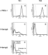
FACS® analysis of Th1 and Th2 cells with P-selectin–Ig and antibodies against PSGL-1. Th1 and Th2 cells (as indicated) were analyzed by flow cytometry either with affinitypurified rabbit antibodies against mouse PSGL-1 (A and B, solid lines) or with P-selectin–Ig (C and D, solid lines). Dotted lines show negative control staining either with nonimmune rabbit IgG (A and B) or with human IgG (C and D). E shows the staining of Th1 cells with P-selectin–IgG after preincubation of the cells either with nonimmune rabbit IgG (faint line) or with affinitypurified rabbit anti–PSGL-1 antibodies (bold line). P-selectin–Ig was detected with PE-conjugated F(ab′)2 donkey anti–human IgG, and rabbit antibodies were detected with FITC-conjugated goat anti–rabbit IgG.
Figure 3
Adhesion of Th1 and Th2 cells to immobilized P-selectin–Ig. Cell adhesion assays were performed with Th2 or Th1 cells (as indicated) in 96-well microtiter plates coated with human IgG (hIgG) or P-selectin– IgG (P-Sel). Before the assay, cells were incubated with HBSS(--), HBSS with 50 μg/ml rabbit nonimmune antibodies (Co), or HBSS with 50 μg/ ml affinity-purified antibodies against PSGL-1 (α PSGL). Bound cells were counted by computer-aided image analysis in three randomly chosen areas of defined size (per well) in three different wells for each determination. The depicted experiment represents one of three similar experiments.
Figure 4
Partial inhibition of Th1 cell immigration into inflamed skin by antibodies against PSGL-1. Radiolabeled Th1 cells were injected together with PBS (no Ab) or the same buffer containing 100 μg of nonimmune rabbit IgG Fab fragments (Co Fab), 100 μg of affinity-purified anti–PSGL-1 Fab fragments (PSGL-1), 200 μg of mAb RB40 against mouse P-selectin (P-Sel), 200 μg of mAb UZ4 against mouse E-selectin (E-Sel). Immigration of cells into the noninflamed control skin region of the same mice is depicted as solid bars. For each determination, four mice were analyzed. Experiments shown by the left graph were performed with a different preparation of Th1 cells than the experiments depicted by the right graph. Numbers on the left refer to the percentage of injected cells that were found in the analyzed skin area of 2.5 cm2.
Similar articles
- P-Selectin glycoprotein ligand 1 (PSGL-1) is a physiological ligand for E-selectin in mediating T helper 1 lymphocyte migration.
Hirata T, Merrill-Skoloff G, Aab M, Yang J, Furie BC, Furie B. Hirata T, et al. J Exp Med. 2000 Dec 4;192(11):1669-76. doi: 10.1084/jem.192.11.1669. J Exp Med. 2000. PMID: 11104809 Free PMC article. - CD43 collaborates with P-selectin glycoprotein ligand-1 to mediate E-selectin-dependent T cell migration into inflamed skin.
Matsumoto M, Shigeta A, Furukawa Y, Tanaka T, Miyasaka M, Hirata T. Matsumoto M, et al. J Immunol. 2007 Feb 15;178(4):2499-506. doi: 10.4049/jimmunol.178.4.2499. J Immunol. 2007. PMID: 17277158 - Rolling of Th1 cells via P-selectin glycoprotein ligand-1 stimulates LFA-1-mediated cell binding to ICAM-1.
Atarashi K, Hirata T, Matsumoto M, Kanemitsu N, Miyasaka M. Atarashi K, et al. J Immunol. 2005 Feb 1;174(3):1424-32. doi: 10.4049/jimmunol.174.3.1424. J Immunol. 2005. PMID: 15661900 - Structure and function of P-selectin glycoprotein ligand-1.
Moore KL. Moore KL. Leuk Lymphoma. 1998 Mar;29(1-2):1-15. doi: 10.3109/10428199809058377. Leuk Lymphoma. 1998. PMID: 9638971 Review. - Migration of helper T-lymphocyte subsets into inflamed tissues.
Lukacs NW. Lukacs NW. J Allergy Clin Immunol. 2000 Nov;106(5 Suppl):S264-9. doi: 10.1067/mai.2000.110160. J Allergy Clin Immunol. 2000. PMID: 11080742 Review.
Cited by
- Prevention of leukocyte migration to inflamed skin with a novel fluorosugar modifier of cutaneous lymphocyte-associated antigen.
Dimitroff CJ, Kupper TS, Sackstein R. Dimitroff CJ, et al. J Clin Invest. 2003 Oct;112(7):1008-18. doi: 10.1172/JCI19220. J Clin Invest. 2003. PMID: 14523038 Free PMC article. - Flexible programs of chemokine receptor expression on human polarized T helper 1 and 2 lymphocytes.
Sallusto F, Lenig D, Mackay CR, Lanzavecchia A. Sallusto F, et al. J Exp Med. 1998 Mar 16;187(6):875-83. doi: 10.1084/jem.187.6.875. J Exp Med. 1998. PMID: 9500790 Free PMC article. - Diverse inflammatory cytokines induce selectin ligand expression on murine CD4 T cells via p38α MAPK.
Ebel ME, Awe O, Kaplan MH, Kansas GS. Ebel ME, et al. J Immunol. 2015 Jun 15;194(12):5781-8. doi: 10.4049/jimmunol.1500485. Epub 2015 May 4. J Immunol. 2015. PMID: 25941329 Free PMC article. - Populations of human T lymphocytes that traverse the vascular endothelium stimulated by Borrelia burgdorferi are enriched with cells that secrete gamma interferon.
Gergel EI, Furie MB. Gergel EI, et al. Infect Immun. 2004 Mar;72(3):1530-6. doi: 10.1128/IAI.72.3.1530-1536.2004. Infect Immun. 2004. PMID: 14977959 Free PMC article. - Both Th1 and Th2 cells require P-selectin glycoprotein ligand-1 for optimal rolling on inflamed endothelium.
Mangan PR, O'Quinn D, Harrington L, Bonder CS, Kubes P, Kucik DF, Bullard DC, Weaver CT. Mangan PR, et al. Am J Pathol. 2005 Dec;167(6):1661-75. doi: 10.1016/S0002-9440(10)61249-7. Am J Pathol. 2005. PMID: 16314478 Free PMC article.
References
- Lasky LA. Selectin-carbohydrate interactions and the initiation of the inflammatory response. Annu Rev Biochem. 1995;64:113–139. - PubMed
- Springer TA. Traffic signals on endothelium for lymphocyte recirculation and leukocyte emigration. Ann Rev Physiol. 1995;57:827–872. - PubMed
- Picker LJ, Kishimoto TK, Smith CW, Warnock RA, Butcher EC. ELAM-1 is an adhesion molecule for skin-homing T cells. Nature (Lond) 1991;349:796–799. - PubMed
- Shimizu Y, Shaw S, Graber N, Gopal TV, Horgan KJ, Van Seventer GA, Newman W. Activation-independent binding of human memory T cells to adhesion molecule ELAM-1. Nature (Lond) 1991;349:799–802. - PubMed
Publication types
MeSH terms
Substances
LinkOut - more resources
Full Text Sources
Other Literature Sources


