Uniform vascular-endothelial-cell-specific gene expression in both embryonic and adult transgenic mice - PubMed (original) (raw)
Uniform vascular-endothelial-cell-specific gene expression in both embryonic and adult transgenic mice
T M Schlaeger et al. Proc Natl Acad Sci U S A. 1997.
Abstract
TIE2 is a vascular endothelial-specific receptor tyrosine kinase essential for the regulation of vascular network formation and remodeling. Previously, we have shown that the 1.2-kb 5' flanking region of the TIE2 promoter is capable of directing beta-galactosidase reporter gene expression specifically into a subset of endothelial cells (ECs) of transgenic mouse embryos. However, transgene activity was restricted to early embryonic stages and not detectable in adult mice. Herein we describe the identification and characterization of an autonomous endothelial-specific enhancer in the first intron of the mouse TIE2 gene. Furthermore, combination of the TIE2 promoter with an intron fragment containing this enhancer allows it to target reporter gene expression specifically and uniformly to virtually all vascular ECs throughout embryogenesis and adulthood. To our knowledge, this is the first time that an in vivo expression system has been assembled by which heterologous genes can be targeted exclusively to the ECs of the entire vasculature. This should be a valuable tool to address the function of genes during physiological and pathological processes of vascular ECs in vivo. Furthermore, we were able to identify a short region critical for enhancer function in vivo that contains putative binding sites for Ets-like transcription factors. This should, therefore, allow us to determine the molecular mechanisms underlying the vascular-EC-specific expression of the TIE2 gene.
Figures
Figure 3
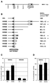
(A) Structure of the TIE2 locus, including the first exon (shaded box), flanked by upstream and intronic sequences [open box, _Xho_I–_Kpn_I fragment (XK); solid box, _Nco_I–_Xba_I fragment (NcXb)]. (B) Structure of LacZ reporter gene constructs and their respective relative activities (HH− = 100%) in transient transfections of BAECs. Values significantly (P < 0.05; Student’s t test) above 100% have been marked with an asterisk. (C) Activity (in percent of tk core promoter activity) of HH− and HHXK in BAECs and HEK293 cells. (D) Activity of tkXK (tk promoter and 1.7-kb enhancer) and tkNcXb (tk promoter and 303-bp enhancer fragment) in BAECs.
Figure 1
Whole-mount 5-bromo-4-chloro-3-indolyl β-
d
-galactopyranoside-stained embryos at E11.5. (a) An embryo transgenic for the construct HH− shows the reduced staining, as described (4). (b) An embryo transgenic for the construct HHNS shows specific staining in virtually all vessels. TG, number of transgenic embryos analyzed; ES, number showing the endothelial-specific staining shown in the respective picture; ET, number showing ectopic staining; NO, number showing no staining at all.
Figure 2
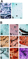
LacZ staining of an embryo at later embryonic stage (A) and in adult tissues (B). (Aa) In situ hybridization of embryos at E14.5 with LacZ probe. (b) Adjacent section of a, immunostained with anti-PECAM antibody. Identical staining pattern of a and b confirms specific and uniform endothelial expression of LacZ. ∗, Heart; arrow, dorsal aorta. (B) LacZ staining of adult brain (a, b), eye (c), heart (d), kidney medulla (e), kidney cortex (f), intestine (g), and spleen (h, i). In brain (a), virtually all vessels are stained. In higher magnification of the thinner sections of the brain (b), clearly demonstrates the EC staining (arrowheads). In eye (c), uniform and strong LacZ staining of the capillary plexus is indicated by arrowheads. In heart (d), uniform LacZ staining of the myocardial vasculature is evident. LacZ staining was also observed in endocardium (data not shown). In kidney cortex (f), all of the glomerular microvasculatures (arrows) are strongly LacZ-positive. Other microvasculatures are difficult to see with this low magnification unless they are clustered (arrowheads), but higher magnification confirmed the complete staining of all microvasculatures. In intestine (g), LacZ-positive mesenteric vasculature is indicated (arrow). In spleen, low magnification (h) and higher magnification (i) clearly demonstrates the uniform and EC-specific LacZ staining. (Bar = 100 μm.)
Figure 4
Base sequence of the core enhancer. Sequences matching know transcription factor binding site consensus sequences are underlined: CF1 (consensus, ANATGG), αINF2 (AARKGA), GATA (WGATAR), Sp1 (KRGGCKRRK), NF-S (YGTCAGC), Ets1 (SMGGAWGY), CP2-γ (AGCCACT), and PEA3 (AGGAAR); the CACA microsatellite and a putative bZIP transcription factor binding site (see text) are also underlined. Furthermore, the octameric palindrome (see text) is marked by three asterisks.
Similar articles
- Octamer-dependent in vivo expression of the endothelial cell-specific TIE2 gene.
Fadel BM, Boutet SC, Quertermous T. Fadel BM, et al. J Biol Chem. 1999 Jul 16;274(29):20376-83. doi: 10.1074/jbc.274.29.20376. J Biol Chem. 1999. PMID: 10400661 - Endothelial cell-specific expression of Cre recombinase in transgenic mice.
Li WL, Cheng X, Tan XH, Zhang JS, Sun YS, Chen L, Yang X. Li WL, et al. Yi Chuan Xue Bao. 2005 Sep;32(9):909-15. Yi Chuan Xue Bao. 2005. PMID: 16201233 - Role of the Ets transcription factors in the regulation of the vascular-specific Tie2 gene.
Dube A, Akbarali Y, Sato TN, Libermann TA, Oettgen P. Dube A, et al. Circ Res. 1999 May 28;84(10):1177-85. doi: 10.1161/01.res.84.10.1177. Circ Res. 1999. PMID: 10347092 - Role of ets factors in the activity and endothelial cell specificity of the mouse Tie gene promoter.
Iljin K, Dube A, Kontusaari S, Korhonen J, Lahtinen I, Oettgen P, Alitalo K. Iljin K, et al. FASEB J. 1999 Feb;13(2):377-86. doi: 10.1096/fasebj.13.2.377. FASEB J. 1999. PMID: 9973326 - Mef2c is activated directly by Ets transcription factors through an evolutionarily conserved endothelial cell-specific enhancer.
De Val S, Anderson JP, Heidt AB, Khiem D, Xu SM, Black BL. De Val S, et al. Dev Biol. 2004 Nov 15;275(2):424-34. doi: 10.1016/j.ydbio.2004.08.016. Dev Biol. 2004. PMID: 15501228
Cited by
- Endothelial pentraxin 3 contributes to murine ischemic acute kidney injury.
Chen J, Matzuk MM, Zhou XJ, Lu CY. Chen J, et al. Kidney Int. 2012 Dec;82(11):1195-207. doi: 10.1038/ki.2012.268. Epub 2012 Aug 15. Kidney Int. 2012. PMID: 22895517 Free PMC article. - Selective endothelial overexpression of arginase II induces endothelial dysfunction and hypertension and enhances atherosclerosis in mice.
Vaisman BL, Andrews KL, Khong SM, Wood KC, Moore XL, Fu Y, Kepka-Lenhart DM, Morris SM Jr, Remaley AT, Chin-Dusting JP. Vaisman BL, et al. PLoS One. 2012;7(7):e39487. doi: 10.1371/journal.pone.0039487. Epub 2012 Jul 19. PLoS One. 2012. PMID: 22829869 Free PMC article. - Endothelial-specific knockdown of interleukin-1 (IL-1) type 1 receptor differentially alters CNS responses to IL-1 depending on its route of administration.
Ching S, Zhang H, Belevych N, He L, Lai W, Pu XA, Jaeger LB, Chen Q, Quan N. Ching S, et al. J Neurosci. 2007 Sep 26;27(39):10476-86. doi: 10.1523/JNEUROSCI.3357-07.2007. J Neurosci. 2007. PMID: 17898219 Free PMC article. - Mechanistic insights into autocrine and paracrine roles of endothelial GABA signaling in the embryonic forebrain.
Choi YK, Vasudevan A. Choi YK, et al. Sci Rep. 2019 Nov 7;9(1):16256. doi: 10.1038/s41598-019-52729-x. Sci Rep. 2019. PMID: 31700116 Free PMC article. - Identification of an octamer element required for in vivo expression of the TIE1 gene in endothelial cells.
Boutet SC, Quertermous T, Fadel BM. Boutet SC, et al. Biochem J. 2001 Nov 15;360(Pt 1):23-9. doi: 10.1042/0264-6021:3600023. Biochem J. 2001. PMID: 11695988 Free PMC article.
References
- Risau W, Flamme I. Annu Rev Cell Dev Biol. 1995;11:73–91. - PubMed
- Risau W. FASEB J. 1995;9:926–933. - PubMed
- Folkman J. Nat Med. 1995;1:27–31. - PubMed
- Schlaeger T M, Qin Y, Fujiwara Y, Magram J, Sato T N. Development (Cambridge, UK) 1995;121:1089–1098. - PubMed
Publication types
MeSH terms
Substances
LinkOut - more resources
Full Text Sources
Other Literature Sources
Molecular Biology Databases
Research Materials
Miscellaneous


