Induction of maturation of human blood dendritic cell precursors by measles virus is associated with immunosuppression - PubMed (original) (raw)
Induction of maturation of human blood dendritic cell precursors by measles virus is associated with immunosuppression
J J Schnorr et al. Proc Natl Acad Sci U S A. 1997.
Abstract
As well as inducing a protective immune response against reinfection, acute measles is associated with a marked suppression of immune functions against superinfecting agents and recall antigens, and this association is the major cause of the current high morbidity and mortality rate associated with measles virus (MV) infections. Dendritic cells (DCs) are antigen-presenting cells crucially involved in the initiation of primary and secondary immune responses, so we set out to define the interaction of MV with these cells. We found that both mature and precursor human DCs generated from peripheral blood monocytic cells express the major MV protein receptor CD46 and are highly susceptible to infection with both MV vaccine (ED) and wild-type (WTF) strains, albeit with different kinetics. Except for the down-regulation of CD46, the expression pattern of functionally important surface antigens on mature DCs was not markedly altered after MV infection. However, precursor DCs up-regulated HLA-DR, CD83, and CD86 within 24 h of WTF infection and 72 h after ED infection, indicating their functional maturation. In addition, interleukin 12 synthesis was markedly enhanced after both ED and WTF infection in DCs. On the other hand, MV-infected DCs strongly interfered with mitogen-dependent proliferation of freshly isolated peripheral blood lymphocytes in vitro. These data indicate that the differentiation of effector functions of DCs is not impaired but rather is stimulated by MV infection. Yet, mature, activated DCs expressing MV surface antigens do give a negative signal to inhibit lymphocyte proliferation and thus contribute to MV-induced immunosuppression.
Figures
Figure 1
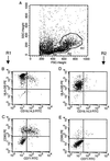
Dot blot analysis of IMS-DCs isolated from peripheral blood. After a 24-h culture in the presence of granulocyte–macrophage colony-stimulating factor and IL-4, cell morphology (A) and protein expression (B_–_E) for mature DCs [region 1 (R1); B and _C_] and precursor DCs [region 2 (R2); D and _E_] were analyzed. For double immunofluorescence staining, mAb against HLA-DR and a mixture of CD3-, CD14-, and CD19-specific antibodies (B and D) or antibodies against CD83 and CD71 (C and E) were used.
Figure 2
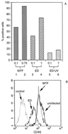
Expression of MV surface proteins F and H and CD46 on infected MCM-DCs. (A) Cells were infected after 8 days in culture with MV-WTF (lanes 1 and 2, moi 0.1 and 0.75) or MV-ED (lanes 3 and 4, moi 0.1 and 1) or were incubated with UV-irradiated MV (ED-UV, lanes 5 and 6, corresponding to mois of 0.1 and 1) for 24 h, stained using a mixture of mAb against MV surface proteins F and H and subsequently analyzed by FACScanning. (B) CD46 expression on uninfected and MV-infected cells was measured by indirect immunofluorescence with a monoclonal anti-CD46 antibody (13/42). Cells were infected with MV-WTF or MV-ED at an moi of 0.1 as indicated for 24 h. An anti-CD19 antibody was used as negative control.
Figure 4
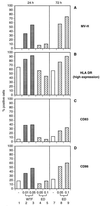
Surface protein expression of MV-infected pre-DCs. IMS-DCs were left uninfected (A_–_D, each lanes 1 and 7) or were infected for 24 h (A_–_D; WTF: lanes 2 and 3, moi 0.01 and 0.05; ED: lanes 4 and 5, moi 0.05 and 0.1) or 72 h (A_–_D; ED: each lanes 8 and 9, moi 0.05 and 0.1). Surface expression of MV-H, HLA-DR, CD86, and CD83 on pre-DCs gated in R2 (Fig. 1, R2) was determined using specific antibodies. Because of the rapid infection kinetics, WTF-infected cells could not be analyzed after 72 h.
Figure 3
Expression of HLA-DR on the surface of IMS-DCs after MV infection. IMS-DCs were double-stained for HLA-DR and CD19/CD14/CD3 expression 24 h postinfection with MV WTF moi 0.01 (B and G) and moi 0.05 (C and H) or MV ED moi 0.05 (D and I) and moi 0.1 (E and J) or were left uninfected (A and F). Mature DCs (R1; F_–_J) and pre-DC (R2; A_–_E) were gated and analyzed in analogy to Fig. 1.
Figure 5
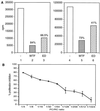
Inhibition of proliferation induced by MV-infected IMS-DCs. (A) IMS-DCs were infected with MV-WTF (lanes 2 and 5) or ED (lanes 3 and 6) at an moi of 0.1 for 48 h or were left uninfected (lanes 1 and 4). After UV irradiation, IMS-DCs (both infected and uninfected) were washed and cocultivated with a 4-fold excess of PHA-stimulated autologous (lanes 1 to 3) or allogeneic (lanes 4 to 6) PBLs for an additional 3 days. Proliferation rates were defined after a 16-h labeling period and are indicated as counts per minute (cpm). Inhibition of the proliferative response of PBLs by cocultivation with WTF- (lanes 2 and 5) or ED-infected (lanes 3 and 6) IMS-DCs compared with cocultivation with uninfected IMS-DCs (lanes 1 and 4) is indicated by %. (B) IMS-DCs were infected with MV-ED (moi 1) for 48 h (or left uninfected), UV-irradiated, and cocultivated with PBLs in the presence of PHA in the PC/RC ratios indicated for further 48 h, followed by a 16-h labeling.
Similar articles
- Measles virus induces abnormal differentiation of CD40 ligand-activated human dendritic cells.
Servet-Delprat C, Vidalain PO, Bausinger H, Manié S, Le Deist F, Azocar O, Hanau D, Fischer A, Rabourdin-Combe C. Servet-Delprat C, et al. J Immunol. 2000 Feb 15;164(4):1753-60. doi: 10.4049/jimmunol.164.4.1753. J Immunol. 2000. PMID: 10657621 - Susceptibility of human dendritic cells (DCs) to measles virus (MV) depends on their activation stages in conjunction with the level of CDw150: role of Toll stimulators in DC maturation and MV amplification.
Murabayashi N, Kurita-Taniguchi M, Ayata M, Matsumoto M, Ogura H, Seya T. Murabayashi N, et al. Microbes Infect. 2002 Jul;4(8):785-94. doi: 10.1016/s1286-4579(02)01598-8. Microbes Infect. 2002. PMID: 12270725 - Measles virus interacts with human SLAM receptor on dendritic cells to cause immunosuppression.
Hahm B, Arbour N, Oldstone MB. Hahm B, et al. Virology. 2004 Jun 1;323(2):292-302. doi: 10.1016/j.virol.2004.03.011. Virology. 2004. PMID: 15193925 Free PMC article. - Dendritic cells and measles virus infection.
Schneider-Schaulies S, Klagge IM, ter Meulen V. Schneider-Schaulies S, et al. Curr Top Microbiol Immunol. 2003;276:77-101. doi: 10.1007/978-3-662-06508-2_4. Curr Top Microbiol Immunol. 2003. PMID: 12797444 Review. - Membrane dynamics and interactions in measles virus dendritic cell infections.
Avota E, Koethe S, Schneider-Schaulies S. Avota E, et al. Cell Microbiol. 2013 Feb;15(2):161-9. doi: 10.1111/cmi.12025. Epub 2012 Sep 28. Cell Microbiol. 2013. PMID: 22963539 Review.
Cited by
- HIV-1-infected monocyte-derived dendritic cells do not undergo maturation but can elicit IL-10 production and T cell regulation.
Granelli-Piperno A, Golebiowska A, Trumpfheller C, Siegal FP, Steinman RM. Granelli-Piperno A, et al. Proc Natl Acad Sci U S A. 2004 May 18;101(20):7669-74. doi: 10.1073/pnas.0402431101. Epub 2004 May 5. Proc Natl Acad Sci U S A. 2004. PMID: 15128934 Free PMC article. - Roles of macrophages in measles virus infection of genetically modified mice.
Roscic-Mrkic B, Schwendener RA, Odermatt B, Zuniga A, Pavlovic J, Billeter MA, Cattaneo R. Roscic-Mrkic B, et al. J Virol. 2001 Apr;75(7):3343-51. doi: 10.1128/JVI.75.7.3343-3351.2001. J Virol. 2001. PMID: 11238860 Free PMC article. - Consequences of Fas-mediated human dendritic cell apoptosis induced by measles virus.
Servet-Delprat C, Vidalain PO, Azocar O, Le Deist F, Fischer A, Rabourdin-Combe C. Servet-Delprat C, et al. J Virol. 2000 May;74(9):4387-93. doi: 10.1128/jvi.74.9.4387-4393.2000. J Virol. 2000. PMID: 10756053 Free PMC article. - A Morbillivirus Infection Shifts DC Maturation Toward a Tolerogenic Phenotype to Suppress T Cell Activation.
Rodríguez-Martín D, García-García I, Martín V, Rojas JM, Sevilla N. Rodríguez-Martín D, et al. J Virol. 2022 Sep 28;96(18):e0124022. doi: 10.1128/jvi.01240-22. Epub 2022 Sep 12. J Virol. 2022. PMID: 36094317 Free PMC article. - Viruses, dendritic cells and the lung.
Peebles RS Jr, Graham BS. Peebles RS Jr, et al. Respir Res. 2001;2(4):245-9. doi: 10.1186/rr63. Epub 2001 Jun 27. Respir Res. 2001. PMID: 11686890 Free PMC article. Review.
References
- Besser G M, Davis J, Duncan C, Kirk B, Kuper S W. Br J Haematol. 1967;13:189–193. - PubMed
- Griffin D E, Ward B J, Jauregui E, Johnston R T, Vaisberg A. N Engl J Med. 1989;320:1667–1672. - PubMed
- Griffin D E, Moench T R, Johnson R T, Lindo de Soriano I, Vaisberg A. Clin Immunol Immunopathol. 1986;40:305–312. - PubMed
- Griffin D E. In: Current Topics of Microbiology and Immunolgy: Measles Virus. ter Meulen V, Billeter M A, editors. Berlin: Springer; 1995. pp. 117–134.
Publication types
MeSH terms
Substances
LinkOut - more resources
Full Text Sources
Research Materials


