Abnormal dendritic spines in fragile X knockout mice: maturation and pruning deficits - PubMed (original) (raw)
Abnormal dendritic spines in fragile X knockout mice: maturation and pruning deficits
T A Comery et al. Proc Natl Acad Sci U S A. 1997.
Abstract
Fragile X syndrome arises from blocked expression of the fragile X mental retardation protein (FMRP). Golgi-impregnated mature cerebral cortex from fragile X patients exhibits long, thin, tortuous postsynaptic spines resembling spines observed during normal early neocortical development. Here we describe dendritic spines in Golgi-impregnated cerebral cortex of transgenic fragile X gene (Fmr1) knockout mice that lack expression of the protein. Dendritic spines on apical dendrites of layer V pyramidal cells in occipital cortex of fragile X knockout mice were longer than those in wild-type mice and were often thin and tortuous, paralleling the human syndrome and suggesting that FMRP expression is required for normal spine morphological development. Moreover, spine density along the apical dendrite was greater in the knockout mice, which may reflect impaired developmental organizational processes of synapse stabilization and elimination or pruning.
Figures
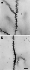
Figure 1
Golgi-Cox impregnated apical dendrites in transgenic and wild-type mice. (A) Segment of apical dendrite from layer V pyramidal neuron in Fmr1 knockout mouse demonstrating both the increased incidence of long, thin dendritic spines and the increased spine density. (B) Apical dendrite of layer V pyramidal neuron from wild-type mouse. (Bar = 10 μm.)
Figure 2
Mean dendritic length (and SEM) of spines on apical dendrites of layer V pyramidal neurons in visual cortex from knockout (KO) and wild-type mice. Dendritic spines in Fmr1 transgenic mice were significantly longer than those in the wild-type animals (one-tailed t test; t = 2.25, df = 6, P = 0.033).
Figure 3
Number of spines in each reticule-based length category on apical dendrites of layer V pyramidal neurons in visual cortex from knockout (KO) and wild-type mice. Knockout mice have fewer short spines and more long spines (χ2 = 46.29, 3 df, P < .0005).
Figure 4
Mean spine density (and SEM) distributions in fragile X knockout (KO) and wild-type mice. Overall spine density along apical dendrites of layer V pyramidal cells is significantly greater in knockout mice than in wild-type controls (F1,33 = 63.3, P < 0.0001). Dotted lines in graphs indicate reduced numbers of dendrites in the analysis due to some apical dendrites having been truncated by the section plane.
Similar articles
- Dendritic spine and dendritic field characteristics of layer V pyramidal neurons in the visual cortex of fragile-X knockout mice.
Irwin SA, Idupulapati M, Gilbert ME, Harris JB, Chakravarti AB, Rogers EJ, Crisostomo RA, Larsen BP, Mehta A, Alcantara CJ, Patel B, Swain RA, Weiler IJ, Oostra BA, Greenough WT. Irwin SA, et al. Am J Med Genet. 2002 Aug 1;111(2):140-6. doi: 10.1002/ajmg.10500. Am J Med Genet. 2002. PMID: 12210340 - Abnormal dendrite and spine morphology in primary visual cortex in the CGG knock-in mouse model of the fragile X premutation.
Berman RF, Murray KD, Arque G, Hunsaker MR, Wenzel HJ. Berman RF, et al. Epilepsia. 2012 Jun;53 Suppl 1(0 1):150-60. doi: 10.1111/j.1528-1167.2012.03486.x. Epilepsia. 2012. PMID: 22612820 Free PMC article. - Abnormal dendritic spine characteristics in the temporal and visual cortices of patients with fragile-X syndrome: a quantitative examination.
Irwin SA, Patel B, Idupulapati M, Harris JB, Crisostomo RA, Larsen BP, Kooy F, Willems PJ, Cras P, Kozlowski PB, Swain RA, Weiler IJ, Greenough WT. Irwin SA, et al. Am J Med Genet. 2001 Jan 15;98(2):161-7. doi: 10.1002/1096-8628(20010115)98:2<161::aid-ajmg1025>3.0.co;2-b. Am J Med Genet. 2001. PMID: 11223852 - Dendritic spine structural anomalies in fragile-X mental retardation syndrome.
Irwin SA, Galvez R, Greenough WT. Irwin SA, et al. Cereb Cortex. 2000 Oct;10(10):1038-44. doi: 10.1093/cercor/10.10.1038. Cereb Cortex. 2000. PMID: 11007554 Review. - Synaptic regulation of protein synthesis and the fragile X protein.
Greenough WT, Klintsova AY, Irwin SA, Galvez R, Bates KE, Weiler IJ. Greenough WT, et al. Proc Natl Acad Sci U S A. 2001 Jun 19;98(13):7101-6. doi: 10.1073/pnas.141145998. Proc Natl Acad Sci U S A. 2001. PMID: 11416194 Free PMC article. Review.
Cited by
- Increasing our understanding of human cognition through the study of Fragile X Syndrome.
Cook D, Nuro E, Murai KK. Cook D, et al. Dev Neurobiol. 2014 Feb;74(2):147-77. doi: 10.1002/dneu.22096. Epub 2013 Jul 30. Dev Neurobiol. 2014. PMID: 23723176 Free PMC article. Review. - WNT signaling in neuronal maturation and synaptogenesis.
Rosso SB, Inestrosa NC. Rosso SB, et al. Front Cell Neurosci. 2013 Jul 4;7:103. doi: 10.3389/fncel.2013.00103. eCollection 2013. Front Cell Neurosci. 2013. PMID: 23847469 Free PMC article. - Long-term behavioral effects of prenatal stress in the Fmr1-knock-out mouse model for fragile X syndrome.
Petroni V, Subashi E, Premoli M, Memo M, Lemaire V, Pietropaolo S. Petroni V, et al. Front Cell Neurosci. 2022 Oct 27;16:917183. doi: 10.3389/fncel.2022.917183. eCollection 2022. Front Cell Neurosci. 2022. PMID: 36385949 Free PMC article. - Deficit in motor training-induced clustering, but not stabilization, of new dendritic spines in FMR1 knock-out mice.
Reiner BC, Dunaevsky A. Reiner BC, et al. PLoS One. 2015 May 7;10(5):e0126572. doi: 10.1371/journal.pone.0126572. eCollection 2015. PLoS One. 2015. PMID: 25950728 Free PMC article. - Genetic background mutations drive neural circuit hyperconnectivity in a fragile X syndrome model.
Kennedy T, Rinker D, Broadie K. Kennedy T, et al. BMC Biol. 2020 Jul 30;18(1):94. doi: 10.1186/s12915-020-00817-0. BMC Biol. 2020. PMID: 32731855 Free PMC article.
References
- Hagerman R J, Cronister A, editors. Fragile X Syndrome: Diagnosis, Treatment, and Research. 2nd Ed. Baltimore: Johns Hopkins Univ. Press; 1996.
- Warren S T, Nelson D L. J Am Med Assoc. 1994;271:536–542. - PubMed
- Verheij C, Bakker C E, de Graaff E, Keulemans J, Willemsen R, Verkerk A J, Galjaard H, Reuser A J, Hoogeveen A T, Oostra B A. Nature (London) 1993;363:722–724. - PubMed
- Verkerk A J M H, Pieretti M, Sutcliffe J S, Fu Y H, Kuhl D P A, et al. Cell. 1991;65:905–914. - PubMed
Publication types
MeSH terms
LinkOut - more resources
Full Text Sources
Other Literature Sources
Medical
Molecular Biology Databases


