Genetic influence on neurogenesis in the dentate gyrus of adult mice - PubMed (original) (raw)
Comparative Study
Genetic influence on neurogenesis in the dentate gyrus of adult mice
G Kempermann et al. Proc Natl Acad Sci U S A. 1997.
Abstract
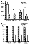
To address genetic influences on hippocampal neurogenesis in adult mice, we compared C57BL/6, BALB/c, CD1(ICR), and 129Sv/J mice to examine proliferation, survival, and differentiation of newborn cells in the dentate gyrus. Proliferation was highest in C57BL/6; the survival rate of newborn cells was highest in CD1. In all strains approximately 60% of surviving newborn cells had a neuronal phenotype, but 129/SvJ produced more astrocytes. Over 6 days C57BL/6 produced 0.36% of their total granule cell number of 239,000 as new neurons, BALB/c 0.30% of 242,000, CD1 (ICR) 0.32% of 351,000, and 129/SvJ 0.16% of 280,000. These results show that different aspects of adult hippocampal neurogenesis are differentially influenced by the genetic background.
Figures
Figure 1
Proliferation (A) and survival (B) of cells in the subgranular zone of C57BL/6 mice. BrdU-labeled cells 1 day after the last injection of BrdU (A) have dark irregular shaped nuclei (see Inset). Four weeks later (B) the number of BrdU-positive cells has decreased and the remaining cells have more rounded nuclei, sometimes with the typical chromatin structure of granule cells (see also insert and compare with C_–_F). Absolute granule cell numbers were determined stereologically using a 15 × 15 μm counting frame superimposed on a video image of Hoechst-33342-stained sections (C_–_F). No differences in the appearance of the granule cells can be noted, and the neuronal density in the granule cell layer is similar (see Table 2). Phenotypes of surviving newborn cells 4 weeks after the last injection were examined by means of immunofluorescence and confocal microscopy. Cells were categorized as to whether they showed double-labeling for BrdU (green) and granule cell marker calbindin (red) (G), BrdU and astrocytic marker GFAP (blue) (H), or for BrdU and neither of the two other markers (I). Quantification of phenotype distribution is found in Fig. 2_B_. (Bar in A = 75 μm for A and B and 25 μm for the Insets in A and B.) Counting frames in C_–_F are 15 × 15 μm. (Bars in G_–_I = 15 μm.)
Figure 2
Quantification of BrdU-positive cells (A) and phenotype distribution (B). Numbers of BrdU-positive cells were determined 1 day after the last injection to assess proliferative activity (open bars in A) and 4 weeks later to address survival of newborn cells (hatched bars in A). Numbers are totals per granule cell layer (means ± SE). At 4 weeks cells were also examined for the expression of phenotypic marker proteins (B): calbindin for granule cells and GFAP for astrocytes (see Fig. 1 G_–_I). A total of 50 BrdU-positive cells per animal were analyzed. Statistical analysis is given in Table 1.
Similar articles
- Environmental stimulation of 129/SvJ mice causes increased cell proliferation and neurogenesis in the adult dentate gyrus.
Kempermann G, Brandon EP, Gage FH. Kempermann G, et al. Curr Biol. 1998 Jul 30-Aug 13;8(16):939-42. doi: 10.1016/s0960-9822(07)00377-6. Curr Biol. 1998. PMID: 9707406 - Differences in immunoreactivities of Ki-67 and doublecortin in the adult hippocampus in three strains of mice.
Kim JS, Jung J, Lee HJ, Kim JC, Wang H, Kim SH, Shin T, Moon C. Kim JS, et al. Acta Histochem. 2009;111(2):150-6. doi: 10.1016/j.acthis.2008.05.002. Epub 2008 Jul 22. Acta Histochem. 2009. PMID: 18649926 - Genetic influence on neurogenesis in the dentate gyrus of two strains of adult mice.
Schauwecker PE. Schauwecker PE. Brain Res. 2006 Nov 20;1120(1):83-92. doi: 10.1016/j.brainres.2006.08.086. Epub 2006 Sep 26. Brain Res. 2006. PMID: 16999941 - Adult neurogenesis in the mammalian dentate gyrus.
Abbott LC, Nigussie F. Abbott LC, et al. Anat Histol Embryol. 2020 Jan;49(1):3-16. doi: 10.1111/ahe.12496. Epub 2019 Sep 30. Anat Histol Embryol. 2020. PMID: 31568602 Review. - [Activated microglial cells trigger neurogenesis following neuronal loss in the dentate gyrus of adult mice].
Ogita K, Yoneyama M, Hasebe S, Shiba T. Ogita K, et al. Nihon Shinkei Seishin Yakurigaku Zasshi. 2012 Nov;32(5-6):281-5. Nihon Shinkei Seishin Yakurigaku Zasshi. 2012. PMID: 23373316 Review. Japanese.
Cited by
- Therapeutic Options of Crystallin Mu and Protein Disulfide Isomerase A3 for Cuprizone-Induced Demyelination in Mouse Hippocampus.
Hahn KR, Kwon HJ, Kim DW, Hwang IK, Yoon YS. Hahn KR, et al. Neurochem Res. 2024 Nov;49(11):3078-3093. doi: 10.1007/s11064-024-04227-4. Epub 2024 Aug 20. Neurochem Res. 2024. PMID: 39164609 Free PMC article. - Modelling adult neurogenesis in the aging rodent hippocampus: a midlife crisis.
Arellano JI, Rakic P. Arellano JI, et al. Front Neurosci. 2024 Jun 3;18:1416460. doi: 10.3389/fnins.2024.1416460. eCollection 2024. Front Neurosci. 2024. PMID: 38887368 Free PMC article. - Chronic Variable Stress and Cafeteria Diet Combination Exacerbate Microglia and c-fos Activation but Not Experimental Anxiety or Depression in a Menopause Model.
Vega-Rivera NM, Estrada-Camarena E, Azpilcueta-Morales G, Cervantes-Anaya N, Treviño S, Becerril-Villanueva E, López-Rubalcava C. Vega-Rivera NM, et al. Int J Mol Sci. 2024 Jan 25;25(3):1455. doi: 10.3390/ijms25031455. Int J Mol Sci. 2024. PMID: 38338735 Free PMC article. - Erythropoietin re-wires cognition-associated transcriptional networks.
Singh M, Zhao Y, Gastaldi VD, Wojcik SM, Curto Y, Kawaguchi R, Merino RM, Garcia-Agudo LF, Taschenberger H, Brose N, Geschwind D, Nave KA, Ehrenreich H. Singh M, et al. Nat Commun. 2023 Aug 21;14(1):4777. doi: 10.1038/s41467-023-40332-8. Nat Commun. 2023. PMID: 37604818 Free PMC article. - Engineered Mesenchymal Stem Cells Over-Expressing BDNF Protect the Brain from Traumatic Brain Injury-Induced Neuronal Death, Neurological Deficits, and Cognitive Impairments.
Choi BY, Hong DK, Kang BS, Lee SH, Choi S, Kim HJ, Lee SM, Suh SW. Choi BY, et al. Pharmaceuticals (Basel). 2023 Mar 13;16(3):436. doi: 10.3390/ph16030436. Pharmaceuticals (Basel). 2023. PMID: 36986535 Free PMC article.
References
- Altman J, Das G D. J Comp Neurol. 1965;124:319–335. - PubMed
- Kaplan M S, Hinds J W. Science. 1977;197:1092–1094. - PubMed
- Stanfield B B, Trice J E. Exp Brain Res. 1988;72:399–406. - PubMed
- Cameron H A, Woolley C S, McEwen B S, Gould E. Neuroscience. 1993;56:337–344. - PubMed
Publication types
MeSH terms
LinkOut - more resources
Full Text Sources
Other Literature Sources
Molecular Biology Databases

