Ultrastructural localization of zinc transporter-3 (ZnT-3) to synaptic vesicle membranes within mossy fiber boutons in the hippocampus of mouse and monkey - PubMed (original) (raw)
Ultrastructural localization of zinc transporter-3 (ZnT-3) to synaptic vesicle membranes within mossy fiber boutons in the hippocampus of mouse and monkey
H J Wenzel et al. Proc Natl Acad Sci U S A. 1997.
Abstract
Zinc transporter-3 (ZnT-3), a member of a growing family of mammalian zinc transporters, is expressed in regions of the brain that are rich in histochemically reactive zinc (as revealed by the Timm's stain), including entorhinal cortex, amygdala, and hippocampus. ZnT-3 protein is most abundant in the zinc-enriched mossy fibers that project from the dentate granule cells to hilar and CA3 pyramidal neurons. We show here by electron microscopy that ZnT-3 decorates the membranes of all clear, small, round synaptic vesicles (SVs) in the mossy fiber boutons of both mouse and monkey. Furthermore, up to 60-80% of these SVs contain Timm's-stainable zinc. The coincidence of ZnT-3 on the membranes of SVs that accumulate zinc, and its homology with known zinc transporters, suggest that ZnT-3 is responsible for the transport of zinc into SVs, and hence for the ability of these neurons to release zinc upon excitation.
Figures
Figure 1
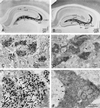
Localization of histochemically reactive zinc (A, C, E) and IR for ZnT-3 protein (B, D, F) in the mouse hippocampus. (A) Transverse section of the hippocampus demonstrating intense reaction product in the mossy fiber (MF) system (dark band labeled “mf”) after Timm’s staining procedure; counterstained with cresyl violet. Note the lighter laminar staining in s. radiatum (rad) and oriens (or) of CA1. (B) Transverse section of the hippocampus reacted with an affinity-purified antiserum against ZnT-3 protein. IR is intense in the zinc-containing MF projections; arrows indicate ZnT-3 staining in s. lucidum (luc), pyramidale (pyr) and oriens (or) of CA3. The dentate outer (oml) and inner (iml) molecular layers, and s. radiatum (rad) and oriens (or) of CA1 also appear lightly immunoreactive for ZnT-3. (C) Electron micrograph from the dentate hilus (indicated area in A) showing MF boutons (mfb) with high density of silver granules. (D) Electron micrograph from the dentate hilus (indicated area in B) showing intense ZnT-3 IR in MFBs (mfb). Note the probable interneuron terminal (A) without ZnT-3 IR, making a symmetric synaptic contact (arrowhead) onto a dendrite (d). (E, F) Higher magnification of MFBs from CA3 s. lucidum (indicated areas in A and B) to demonstrate the vesicular localization of zinc (E) and ZnT-3 IR on synaptic vesicle membranes (F). Other abbreviations: CA1, CA3 hippocampal regions; DG, dentate gyrus; hi, hilus; M, mitochondrion; S, spine; arrowhead, synaptic contact.
Figure 2
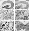
Localization of histochemically reactive zinc (A, C, E) and IR for ZnT-3 protein (B, D, F) in the monkey hippocampus. (A) Frontal section of the hippocampus demonstrating intense Timm’s stain for vesicular zinc in the MF projections (mf) within hilus and CA3. Lighter staining is seen in s. radiatum and oriens and subiculum (Sub). (B) Frontal section of the monkey hippocampus. ZnT-3 IR is present in the zinc-rich MF projections (mf) in hilus and CA3 region; lighter IR is seen in the dentate iml, in CA1 s. radiatum (rad) and oriens (or) and in subiculum. (C) Electron micrograph from the dentate hilus (indicated area in A) showing MFBs labeled with silver granules. (D) Electron micrograph from the s. lucidum (indicated area in B) demonstrating ZnT-3 IR in MFBs that surround a dendrite (d). (E) Higher magnification of part of a giant MFB in synaptic contact with spines in CA3 s. lucidum, showing dark silver granules located in the clear round SVs. (F) Higher magnification of a MFB in synaptic contact with a giant spine (arrowheads), also making nonsynaptic contacts onto a dendritic shaft (d, arrows). ZnT-3 IR is localized to the membranes of clear, round SVs within the bouton. Abbreviations as in Fig. 1.
Figure 3
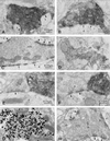
Electron micrographs from the mouse and monkey (E) hippocampus showing ZnT-3 IR in terminals of glutamatergic projections. (A) ZnT-3 localization on clear, round SVs in a MFB in synaptic contact with spines (arrowheads). (B) ZnT-3 positive MFB making nonsynaptic contacts (arrows) onto a dendrite (d) in CA3 s. lucidum. (C) Control section of the CA3 s. lucidum in which the antibody against ZnT-3 protein was omitted; SVs in MFB (mfb) are devoid of ZnT-3 IR. The MFB forms synaptic contacts (arrowheads) with spines (s) and nonsynaptic contacts with a dendrite (d; arrows). (D) Synaptic bouton (SB) in the outer molecular layer of the dentate gyrus showing ZnT-3 IR on SV membranes. (E) and (F) Hilar dendrites (d) from the monkey (E) and mouse (F) hippocampus, contacted by MFBs with intense ZnT-3 IR. Adjacent presumably GABA-ergic terminals make symmetric synaptic contacts (arrowheads) and lack ZnT-3 IR on their SVs. (G) Part of a large MFB showing vesicular zinc (large silver granules) and ImmunoGold (Janssen) particles (10 nm, ○) corresponding to ZnT-3 protein labeled by a postembedding ImmunoGold procedure. (H) MFB with ImmunoGold-labeling for glutamate (10 nm particles, ○) demonstrating glutamate as the neurotransmitter of MF projections. Abbreviations as in Fig. 1.
Similar articles
- ZnT-3, a putative transporter of zinc into synaptic vesicles.
Palmiter RD, Cole TB, Quaife CJ, Findley SD. Palmiter RD, et al. Proc Natl Acad Sci U S A. 1996 Dec 10;93(25):14934-9. doi: 10.1073/pnas.93.25.14934. Proc Natl Acad Sci U S A. 1996. PMID: 8962159 Free PMC article. - Elimination of zinc from synaptic vesicles in the intact mouse brain by disruption of the ZnT3 gene.
Cole TB, Wenzel HJ, Kafer KE, Schwartzkroin PA, Palmiter RD. Cole TB, et al. Proc Natl Acad Sci U S A. 1999 Feb 16;96(4):1716-21. doi: 10.1073/pnas.96.4.1716. Proc Natl Acad Sci U S A. 1999. PMID: 9990090 Free PMC article. - Fate of the hippocampal mossy fiber projection after destruction of its postsynaptic targets with intraventricular kainic acid.
Nadler JV, Perry BW, Gentry C, Cotman CW. Nadler JV, et al. J Comp Neurol. 1981 Mar 10;196(4):549-69. doi: 10.1002/cne.901960404. J Comp Neurol. 1981. PMID: 7204671 - Mammalian zinc transporters.
McMahon RJ, Cousins RJ. McMahon RJ, et al. J Nutr. 1998 Apr;128(4):667-70. doi: 10.1093/jn/128.4.667. J Nutr. 1998. PMID: 9521625 Review. - Integrative aspects of zinc transporters.
Cousins RJ, McMahon RJ. Cousins RJ, et al. J Nutr. 2000 May;130(5S Suppl):1384S-7S. doi: 10.1093/jn/130.5.1384S. J Nutr. 2000. PMID: 10801948 Review.
Cited by
- RIM-BP2 regulates Ca2+ channel abundance and neurotransmitter release at hippocampal mossy fiber terminals.
Miyano R, Sakamoto H, Hirose K, Sakaba T. Miyano R, et al. Elife. 2024 Feb 8;12:RP90799. doi: 10.7554/eLife.90799. Elife. 2024. PMID: 38329474 Free PMC article. - The ZIP3 Zinc Transporter Is Localized to Mossy Fiber Terminals and Is Required for Kainate-Induced Degeneration of CA3 Neurons.
Bogdanovic M, Asraf H, Gottesman N, Sekler I, Aizenman E, Hershfinkel M. Bogdanovic M, et al. J Neurosci. 2022 Mar 30;42(13):2824-2834. doi: 10.1523/JNEUROSCI.0908-21.2022. Epub 2022 Feb 15. J Neurosci. 2022. PMID: 35169020 Free PMC article. - Modulation of neuronal signal transduction and memory formation by synaptic zinc.
Sindreu C, Storm DR. Sindreu C, et al. Front Behav Neurosci. 2011 Nov 9;5:68. doi: 10.3389/fnbeh.2011.00068. eCollection 2011. Front Behav Neurosci. 2011. PMID: 22084630 Free PMC article. - Axonal transport of zinc transporter 3 and zinc containing organelles in the rodent adrenergic system.
Wang ZY, Dahlström A. Wang ZY, et al. Neurochem Res. 2008 Dec;33(12):2472-9. doi: 10.1007/s11064-008-9798-2. Epub 2008 Aug 20. Neurochem Res. 2008. PMID: 18712599 Review. - Metal ions and synaptic transmission: think zinc.
Huang EP. Huang EP. Proc Natl Acad Sci U S A. 1997 Dec 9;94(25):13386-7. doi: 10.1073/pnas.94.25.13386. Proc Natl Acad Sci U S A. 1997. PMID: 9391032 Free PMC article. Review. No abstract available.
References
- Haug F-M. Histochemie. 1967;8:355–368. - PubMed
- Pérez-Clausell J, Danscher G. Brain Res. 1985;337:91–98. - PubMed
- Frederickson C J. Int Rev Neurobiol. 1989;31:145–238. - PubMed
- Slomianka L. Neuroscience. 1992;48:325–352. - PubMed
- Danscher G. Histochemistry. 1981;71:1–16. - PubMed
Publication types
MeSH terms
Substances
LinkOut - more resources
Full Text Sources
Molecular Biology Databases
Miscellaneous


