Cytosolic Gag p24 as an index of productive entry of human immunodeficiency virus type 1 - PubMed (original) (raw)
Cytosolic Gag p24 as an index of productive entry of human immunodeficiency virus type 1
V Maréchal et al. J Virol. 1998 Mar.
Abstract
We have investigated the cellular uptake of Gag p24 shortly after exposure of cells to human immunodeficiency virus (HIV) particles. In the absence of envelope glycoprotein on virions or of viral receptors or coreceptors at the cell surface, p24 was incorporated in intracellular vesicles but not detected in the cytosolic subcellular fraction. When appropriate envelope-receptor interactions could occur, the nonspecific vesicular uptake was still intense and cytosolic p24 represented 10 to 40% of total intracellular p24. The measurement of cytosolic p24 early after exposure to HIV type 1 is a reliable assay for investigating virus entry and early events leading to authentic cell infection.
Figures
FIG. 1
Confocal microscopy analysis of intracellular p24 after cell exposure to HIV-1. HeLa cells (middle panel) or P4 cells (right panel) were exposed to HIV-1NL43 for 2 h at 4°C, washed, warmed to 37°C for 30 min, fixed, and labeled with anti-Gag MAbs. Left panel, control HeLa cells not exposed to HIV-1.
FIG. 2
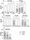
p24 levels in subcellular extracts of cells exposed to HIV-1. HeLa, P4, or P4C5 cells were exposed to strain NL43 (lymphotropic) or JRCSF (macrophage tropic) or to HIV particles devoid of envelope protein (NL43Δenv), respectively, for 30 min at 4°C. Cells were extensively washed and were incubated at 37°C for 1 h. Cells were then treated with pronase, and p24 amounts in subcellular fractions were measured. Total intracellular p24 levels (in picograms) are indicated over the bars. The percentages of cytosolic and vesicular p24 are shown inside the bars. (a) Dose-response analysis for NL43; (b) analysis of the entry of HIV-1 strains with different tropisms (input, 300 ng of p24); (c) analysis of the entry in CEM cells (input, 400 ng of p24). Data are representative of at least three experiments.
FIG. 3
Effect of bafilomycin A1 on HIV-1 infection efficiency and entry into P4 cells. Cells were either left untreated or treated with 0.5 μM bafilomycin A1 during virus exposure. (a) Infection was assessed by measuring β-galactosidase activity in cell extracts. Viral input corresponded to 1 ng of p24. (b) Intracellular p24 levels were measured in the cytosolic and vesicular fractions. Total intracellular p24 levels (in picograms) are indicated over the bars. The percentages of cytosolic and vesicular p24 are shown inside the bars. Viral input corresponded to 300 ng of p24 during 1 h at 37°C. HIV/VSV is a VSV-G pseudotype of NL43. Data are from one of three representative experiments.
Similar articles
- Human immunodeficiency virus type 1 entry into macrophages mediated by macropinocytosis.
Maréchal V, Prevost MC, Petit C, Perret E, Heard JM, Schwartz O. Maréchal V, et al. J Virol. 2001 Nov;75(22):11166-77. doi: 10.1128/JVI.75.22.11166-11177.2001. J Virol. 2001. PMID: 11602756 Free PMC article. - Human immunodeficiency virus type 1 Vpu has a CD4- and an envelope glycoprotein-independent function.
Geraghty RJ, Panganiban AT. Geraghty RJ, et al. J Virol. 1993 Jul;67(7):4190-4. doi: 10.1128/JVI.67.7.4190-4194.1993. J Virol. 1993. PMID: 8510220 Free PMC article. - Gag-Gag interactions in the C-terminal domain of human immunodeficiency virus type 1 p24 capsid antigen are essential for Gag particle assembly.
Zhang WH, Hockley DJ, Nermut MV, Morikawa Y, Jones IM. Zhang WH, et al. J Gen Virol. 1996 Apr;77 ( Pt 4):743-51. doi: 10.1099/0022-1317-77-4-743. J Gen Virol. 1996. PMID: 8627263 - Human immunodeficiency virus type-1 and chemokines: beyond competition for common cellular receptors.
Stantchev TS, Broder CC. Stantchev TS, et al. Cytokine Growth Factor Rev. 2001 Jun-Sep;12(2-3):219-43. doi: 10.1016/s1359-6101(00)00033-2. Cytokine Growth Factor Rev. 2001. PMID: 11325604 Review.
Cited by
- HIV-1 Nef inhibits ruffles, induces filopodia, and modulates migration of infected lymphocytes.
Nobile C, Rudnicka D, Hasan M, Aulner N, Porrot F, Machu C, Renaud O, Prévost MC, Hivroz C, Schwartz O, Sol-Foulon N. Nobile C, et al. J Virol. 2010 Mar;84(5):2282-93. doi: 10.1128/JVI.02230-09. Epub 2009 Dec 16. J Virol. 2010. PMID: 20015995 Free PMC article. - Segregation of CD4 and CXCR4 into distinct lipid microdomains in T lymphocytes suggests a mechanism for membrane destabilization by human immunodeficiency virus.
Kozak SL, Heard JM, Kabat D. Kozak SL, et al. J Virol. 2002 Feb;76(4):1802-15. doi: 10.1128/jvi.76.4.1802-1815.2002. J Virol. 2002. PMID: 11799176 Free PMC article. - Sodium lauryl sulfate abrogates human immunodeficiency virus infectivity by affecting viral attachment.
Bestman-Smith J, Piret J, Désormeaux A, Tremblay MJ, Omar RF, Bergeron MG. Bestman-Smith J, et al. Antimicrob Agents Chemother. 2001 Aug;45(8):2229-37. doi: 10.1128/AAC.45.8.2229-2237.2001. Antimicrob Agents Chemother. 2001. PMID: 11451679 Free PMC article. - Involvement of clathrin-mediated endocytosis in human immunodeficiency virus type 1 entry.
Daecke J, Fackler OT, Dittmar MT, Kräusslich HG. Daecke J, et al. J Virol. 2005 Feb;79(3):1581-94. doi: 10.1128/JVI.79.3.1581-1594.2005. J Virol. 2005. PMID: 15650184 Free PMC article.
References
- Amara A, Le Gall S, Schwartz O, Salamero J, Montes M, Loetscher P, Baggioloni M, Virelizier J L, Arenzana-Seisdedos F. HIV co-receptor down-regulation as anti-viral principle. SDF-1a dependent internalization of the chemokine receptor CXCR4 contributes to inhibition of HIV replication. J Exp Med. 1997;186:139–146. - PMC - PubMed
- Charneau P, Mirabeau G, Roux P, Paulous S, Buc H, Clavel F. HIV-1 reverse transcription: a termination step at the center of the genome. J Mol Biol. 1994;241:651–662. - PubMed
Publication types
MeSH terms
Substances
LinkOut - more resources
Full Text Sources


