Neural precursor differentiation into astrocytes requires signaling through the leukemia inhibitory factor receptor - PubMed (original) (raw)
Neural precursor differentiation into astrocytes requires signaling through the leukemia inhibitory factor receptor
S A Koblar et al. Proc Natl Acad Sci U S A. 1998.
Abstract
The differentiation of precursor cells into neurons or astrocytes in the developing brain has been thought to be regulated in part by growth factors. We show here that neural precursors isolated from the developing forebrain of mice that are deficient in the gene for the low-affinity leukemia inhibitory factor receptor (LIFR-/-) fail to generate astrocytes expressing glial fibrillary acidic protein (GFAP) when cultured in vitro. Precursors from mice heterozygous for the null allele show normal levels of GFAP expression. These findings support the in vivo findings that show extremely low levels of GFAP mRNA in brains of embryonic day 19 LIFR-/- mice. In addition, monolayers of neural cells from LIFR-/- mice are far less able to support the neuronal differentiation of normal neural precursors than are monolayers from heterozygous or wild-type animals, indicating that endogenous signaling through the LIFR is required for the expression of both functional and phenotypic markers of astrocyte differentiation. LIFR-/- precursors are not irreversibly blocked from differentiating into astrocytes: they express GFAP after long-term passaging or stimulation with bone morphogenetic protein-2. These findings strongly implicate the LIF family of cytokines in the regulation of astrocyte differentiation and indeed the LIF-deficient animals show a significant reduction in the number of GFAP cells in the hippocampus. However, because this reduction is only partial it suggests that LIF may not be the predominant endogenous ligand signaling through the LIFR.
Figures
Figure 1
Low levels of GFAP mRNA in LIFR−/− brain. mRNA was prepared from the brain, including forebrain, midbrain, and hindbrain of E19 littermates, and the presence of GFAP mRNA was determined by PCR.
Figure 2
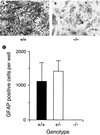
Failure of LIFR−/− neural cells to express GFAP. Neuroepithelial cells from E12 forebrain were plated in vitro at a density of 2.5 × 10⁴ per 200 mm2 into multiwell plates (Falcon 3047) and cultured for 20 days in the presence of serum. Cultures then were stained by immunoperoxidase for the presence of GFAP. The LIFR+/+ cultures contained large numbers of GFAP-positive cells (A and C) whereas the LIFR−/− cultures (B and C) contained few if any (<2 cells per well in every case). The data shown in C are the mean and SEM obtained from three separate experiments, each with three replicates.
Similar articles
- Regulation of neural stem cell differentiation in the forebrain.
Bartlett PF, Brooker GJ, Faux CH, Dutton R, Murphy M, Turnley A, Kilpatrick TJ. Bartlett PF, et al. Immunol Cell Biol. 1998 Oct;76(5):414-8. doi: 10.1046/j.1440-1711.1998.00762.x. Immunol Cell Biol. 1998. PMID: 9797460 - LIF receptor signaling modulates neural stem cell renewal.
Pitman M, Emery B, Binder M, Wang S, Butzkueven H, Kilpatrick TJ. Pitman M, et al. Mol Cell Neurosci. 2004 Nov;27(3):255-66. doi: 10.1016/j.mcn.2004.07.004. Mol Cell Neurosci. 2004. PMID: 15519241 - Leukaemia inhibitory factor or related factors promote the differentiation of neuronal and astrocytic precursors within the developing murine spinal cord.
Richards LJ, Kilpatrick TJ, Dutton R, Tan SS, Gearing DP, Bartlett PF, Murphy M. Richards LJ, et al. Eur J Neurosci. 1996 Feb;8(2):291-9. doi: 10.1111/j.1460-9568.1996.tb01213.x. Eur J Neurosci. 1996. PMID: 8714700 - [Function, molecular structure and gene expression regulation of receptor for D-factor/LIF].
Tomida M. Tomida M. Nihon Rinsho. 1992 Aug;50(8):1956-61. Nihon Rinsho. 1992. PMID: 1433987 Review. Japanese. - Role of leukemia inhibitory factor during mammalian development.
Shellard J, Perreau J, Brûlet P. Shellard J, et al. Eur Cytokine Netw. 1996 Dec;7(4):699-712. Eur Cytokine Netw. 1996. PMID: 9010672 Review.
Cited by
- A positive autoregulatory loop of Jak-STAT signaling controls the onset of astrogliogenesis.
He F, Ge W, Martinowich K, Becker-Catania S, Coskun V, Zhu W, Wu H, Castro D, Guillemot F, Fan G, de Vellis J, Sun YE. He F, et al. Nat Neurosci. 2005 May;8(5):616-25. doi: 10.1038/nn1440. Epub 2005 Apr 24. Nat Neurosci. 2005. PMID: 15852015 Free PMC article. - Interleukin-11 potentiates oligodendrocyte survival and maturation, and myelin formation.
Zhang Y, Taveggia C, Melendez-Vasquez C, Einheber S, Raine CS, Salzer JL, Brosnan CF, John GR. Zhang Y, et al. J Neurosci. 2006 Nov 22;26(47):12174-85. doi: 10.1523/JNEUROSCI.2289-06.2006. J Neurosci. 2006. PMID: 17122042 Free PMC article. - Activation of STAT3 signaling in axotomized neurons and reactive astrocytes after fimbria-fornix transection.
Schubert KO, Naumann T, Schnell O, Zhi Q, Steup A, Hofmann HD, Kirsch M. Schubert KO, et al. Exp Brain Res. 2005 Sep;165(4):520-31. doi: 10.1007/s00221-005-2330-x. Epub 2005 Jul 1. Exp Brain Res. 2005. PMID: 15991029 - DREAM mediates cAMP-dependent, Ca2+-induced stimulation of GFAP gene expression and regulates cortical astrogliogenesis.
Cebolla B, Fernández-Pérez A, Perea G, Araque A, Vallejo M. Cebolla B, et al. J Neurosci. 2008 Jun 25;28(26):6703-13. doi: 10.1523/JNEUROSCI.0215-08.2008. J Neurosci. 2008. PMID: 18579744 Free PMC article. - Region-specific differentiation potential of adult rat spinal cord neural stem/precursors and their plasticity in response to in vitro manipulation.
Kulbatski I, Tator CH. Kulbatski I, et al. J Histochem Cytochem. 2009 May;57(5):405-23. doi: 10.1369/jhc.2008.951814. Epub 2009 Jan 5. J Histochem Cytochem. 2009. PMID: 19124840 Free PMC article.
References
- Kilpatrick T J, Bartlett P F. Neuron. 1993;10:255–265. - PubMed
- Kilpatrick T J, Richards L R, Bartlett P F. Mol Cell Neurosci. 1995;6:2–15. - PubMed
- Turner D L, Cepko C L. Nature (London) 1987;328:131–136. - PubMed
- Temple S. Nature (London) 1989;340:471–473. - PubMed
Publication types
MeSH terms
Substances
LinkOut - more resources
Full Text Sources
Other Literature Sources
Medical
Molecular Biology Databases
Miscellaneous

