Analysis of Gag-specific cytotoxic T lymphocytes in simian immunodeficiency virus-infected rhesus monkeys by cell staining with a tetrameric major histocompatibility complex class I-peptide complex - PubMed (original) (raw)
Analysis of Gag-specific cytotoxic T lymphocytes in simian immunodeficiency virus-infected rhesus monkeys by cell staining with a tetrameric major histocompatibility complex class I-peptide complex
M J Kuroda et al. J Exp Med. 1998.
Abstract
A tetrameric recombinant major histocompatibility complex (MHC) class I-peptide complex was used as a staining reagent in flow cytometric analyses to quantitate and define the phenotype of Gag-specific cytotoxic T lymphocytes (CTLs) in the peripheral blood of simian immunodeficiency virus macaque (SIVmac)-infected rhesus monkeys. The heavy chain of the rhesus monkey MHC class I molecule Mamu-A*01 and beta2-microglobulin were refolded in the presence of an SIVmac Gag synthetic peptide (p11C, C-M) representing the optimal nine-amino acid peptide of Mamu-A*01-restricted predominant CTL epitope to create a tetrameric Mamu-A*01/p11C, C-M complex. Tetrameric Mamu-A*01/p11C, C-M complex bound to T cells of SIVmac-infected, Mamu-A*01(+), but not uninfected, Mamu-A*01(+), or infected, Mamu-A*01(-) rhesus monkeys. Specific staining of peripheral blood mononuclear cells (PBMC) from SIVmac-infected, Mamu-A*01(+) rhesus monkeys was only found in the cluster of differentiation (CD)8alpha/beta+ T lymphocyte subset and the percentage of CD8alpha/beta+ T cells in the peripheral blood of four SIVmac-infected, Mamu-A*01+ rhesus monkeys staining with this complex ranged from 0.7 to 10.3%. Importantly, functional SIVmac Gag p11C-specific CTL activity was seen in sorted and expanded tetrameric Mamu-A*01/p11C, C-M complex-binding, but not nonbinding, CD8alpha/beta+ T cells. Furthermore, the percentage of CD8alpha/beta+ T cells binding this tetrameric Mamu-A*01/p11C, C-M complex correlated well with p11C-specific cytotoxic activity as measured in both bulk and limiting dilution effector frequency assays. Finally, phenotypic characterization of the cells binding this tetrameric complex indicated that this lymphocyte population is heterogeneous. These studies indicate the power of this approach for examining virus-specific CTLs in in vivo settings.
Figures
Figure 1
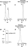
Tetrameric Mamu-A*01/p11C, C–M complex formation. (A) Gel filtration profile of Mamu-A*01 monomer refolded with β2m and p11C, C–M. (B) SDS/PAGE of the 43-kD peak shown in A immunoprecipitated with anti–human heavy chain mAb BB7.7. The positions of β2m and Mamu-A*01 are shown with arrows. (C) Gel filtration profile of biotinylated Mamu-A*01/p11C, C–M monomer. (D) Gel filtration profile of tetrameric Mamu-A*01/p11C, C–M avidin complex.
Figure 2
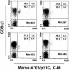
Tetrameric Mamu-A*01/p11C, C–M complex binds only to the CD8α/β+ subset of T cells from PBMCs of four SIVmac-infected, Mamu-A*01+ rhesus monkeys. CD3+ T cells were analyzed for binding of CD8α/β and tetrameric Mamu-A*01/p11C, C–M complex.
Figure 3
Tetrameric Mamu-A*01/p11C, C–M complex binds specifically to CD8α/β+ T cells from PBMCs of SIVmac-infected, Mamu-A*01+ rhesus monkeys. PBMCs of three groups of monkeys, three monkeys per group, were assessed: SIVmac− Mamu-A*01+ monkeys, SIVmac+ Mamu-A*01− monkeys, and SIVmac+ Mamu-A*01+ monkeys. Flow cytometric analysis was performed on gated CD8α/β+CD3+ T cells stained with FITC-coupled tetrameric Mamu-A*01/p11C, C–M complex.
Figure 4
Tetrameric Mamu-A*01/p11C, C–M complex binds p11C-specific CD8α/β+ CTLs. Freshly isolated PBMCs from SIVmac-infected, Mamu-A*01+ rhesus monkey 403 were stained with FITC-coupled tetrameric Mamu-A*01/p11C, C–M complex. CD8α/β+, Mamu-A*01/ p11C, C–M complex positive and CD8α/β+, Mamu-A*01/p11C, C–M complex negative cells were sorted by flow cytometry and expanded after Con A stimulation for 10 d in IL-2–containing medium. The cells were again stained and analyzed by flow cytometry, and the p11C-specific CTL activity of each cell population was assessed.
Figure 5
Tetrameric Mamu-A*01/p11C, C–M complex staining of a lymphocyte population correlates with its p11C-specific CTL activity. Whole blood specimen from a Mamu-A*01+, SIVmac-infected rhesus monkey 403 were stained with FITC-coupled tetrameric Mamu-A*01/p11C, C–M complex. Cells were expanded for 12 d in IL-2–containing medium, either after stimulation with Con A or with p11C-pulsed irradiated autologous PBMCs. The cells were again stained and analyzed by flow cytometry, and the p11C-specific CTL activity of each cell population was assessed.
Figure 6
Phenotypic characterization of tetrameric Mamu-A*01/ p11C, C–M complex–binding CD8α/β+ T cells. A whole blood specimen from a Mamu-A*01+, SIVmac-infected rhesus monkey 403 was stained with FITC-coupled tetrameric Mamu-A*01/p11C, C–M complex and four different PE-coupled mAbs (CD11a, CD28, CD45RA, and HLA-DR). (A) Mean fluorescence (MF) values are indicated for the CD11a staining. Upper left (UL) is the mean fluorescence of the CD11a positive and Mamu-A*01/p11C, C–M negative CD8α/β+ T cell population. Upper right (UR) is the mean fluorescence of the CD11a positive and Mamu-A*01/p11C, C–M positive CD8α/β+ T cell population. (B– D) Percentages of CD28, CD45RA, MHC class II DR, and tetrameric Mamu-A*01/p11C, C–M complex positive or negative CD8α/β+ T cells are indicated.
Similar articles
- Cytotoxic T lymphocytes do not appear to select for mutations in an immunodominant epitope of simian immunodeficiency virus gag.
Chen ZW, Shen L, Miller MD, Ghim SH, Hughes AL, Letvin NL. Chen ZW, et al. J Immunol. 1992 Dec 15;149(12):4060-6. J Immunol. 1992. PMID: 1460291 - Simian immunodeficiency virus-specific cytotoxic T lymphocytes in rhesus monkeys: characterization and vaccine induction.
Letvin NL, Miller MD, Shen L, Chen ZW, Yasutomi Y. Letvin NL, et al. Semin Immunol. 1993 Jun;5(3):215-23. doi: 10.1006/smim.1993.1025. Semin Immunol. 1993. PMID: 8394161 Review. - Cytotoxic T lymphocytes specific for the simian immunodeficiency virus.
Letvin NL, Schmitz JE, Jordan HL, Seth A, Hirsch VM, Reimann KA, Kuroda MJ. Letvin NL, et al. Immunol Rev. 1999 Aug;170:127-34. doi: 10.1111/j.1600-065x.1999.tb01334.x. Immunol Rev. 1999. PMID: 10566147 Review.
Cited by
- Relatively low level of antigen-specific monocytes detected in blood from untreated tuberculosis patients using CD4+ T-cell receptor tetramers.
Huang Y, Huang Y, Fang Y, Wang J, Li Y, Wang N, Zhang J, Gao M, Huang L, Yang F, Wang C, Lin S, Yao Y, Ren L, Chen Y, Du X, Xie D, Wu R, Zhang K, Jiang L, Yu X, Lai X. Huang Y, et al. PLoS Pathog. 2012;8(11):e1003036. doi: 10.1371/journal.ppat.1003036. Epub 2012 Nov 29. PLoS Pathog. 2012. PMID: 23209409 Free PMC article. Clinical Trial. - Differences in time of virus appearance in the blood and virus-specific immune responses in intravenous and intrarectal primary SIVmac251 infection of rhesus macaques; a pilot study.
Stevceva L, Tryniszewska E, Hel Z, Nacsa J, Kelsall B, Washington Parks R, Franchini G. Stevceva L, et al. BMC Infect Dis. 2001;1:9. doi: 10.1186/1471-2334-1-9. Epub 2001 Jul 27. BMC Infect Dis. 2001. PMID: 11504564 Free PMC article. - Elicitation of high-frequency cytotoxic T-lymphocyte responses against both dominant and subdominant simian-human immunodeficiency virus epitopes by DNA vaccination of rhesus monkeys.
Barouch DH, Craiu A, Santra S, Egan MA, Schmitz JE, Kuroda MJ, Fu TM, Nam JH, Wyatt LS, Lifton MA, Krivulka GR, Nickerson CE, Lord CI, Moss B, Lewis MG, Hirsch VM, Shiver JW, Letvin NL. Barouch DH, et al. J Virol. 2001 Mar;75(5):2462-7. doi: 10.1128/JVI.75.5.2462-2467.2001. J Virol. 2001. PMID: 11160750 Free PMC article. - Impact of vaccine-induced mucosal high-avidity CD8+ CTLs in delay of AIDS viral dissemination from mucosa.
Belyakov IM, Kuznetsov VA, Kelsall B, Klinman D, Moniuszko M, Lemon M, Markham PD, Pal R, Clements JD, Lewis MG, Strober W, Franchini G, Berzofsky JA. Belyakov IM, et al. Blood. 2006 Apr 15;107(8):3258-64. doi: 10.1182/blood-2005-11-4374. Epub 2005 Dec 22. Blood. 2006. PMID: 16373659 Free PMC article. - Recombinant Mycobacterium bovis BCG prime-recombinant adenovirus boost vaccination in rhesus monkeys elicits robust polyfunctional simian immunodeficiency virus-specific T-cell responses.
Cayabyab MJ, Korioth-Schmitz B, Sun Y, Carville A, Balachandran H, Miura A, Carlson KR, Buzby AP, Haynes BF, Jacobs WR, Letvin NL. Cayabyab MJ, et al. J Virol. 2009 Jun;83(11):5505-13. doi: 10.1128/JVI.02544-08. Epub 2009 Mar 18. J Virol. 2009. PMID: 19297477 Free PMC article.
References
- Altman JD, Moss PAH, Goulder PJR, Barouch DH, McHeyzer-Williams MG, Bell JI, McMichael AJ, Davis MM. Phenotypic analysis of antigen-specific T lymphocytes. Science. 1996;274:94–96. - PubMed
- Rinaldo C, Huang XL, Fan ZF, Ding M, Beltz L, Logar A, Panicali D, Mazzara G, Liebmann J, Cottrill M, Gupta P. High levels of anti-human immunodeficiency virus type 1 (HIV-1) memory cytotoxic T-lymphocyte activity and low viral load are associated with lack of disease in HIV-1–infected long-term nonprogressors. J Virol. 1995;69:5838–5842. - PMC - PubMed
Publication types
MeSH terms
Substances
Grants and funding
- P51 RR000167/RR/NCRR NIH HHS/United States
- R01 AI020729/AI/NIAID NIH HHS/United States
- AI-20729/AI/NIAID NIH HHS/United States
- R37 AI020729/AI/NIAID NIH HHS/United States
- AI-35166/AI/NIAID NIH HHS/United States
- AI-43068/AI/NIAID NIH HHS/United States
- P51 RR000168/RR/NCRR NIH HHS/United States
- K26 RR000168/RR/NCRR NIH HHS/United States
LinkOut - more resources
Full Text Sources
Other Literature Sources
Research Materials


