Glutamate receptor targeting to synaptic populations on Purkinje cells is developmentally regulated - PubMed (original) (raw)
Glutamate receptor targeting to synaptic populations on Purkinje cells is developmentally regulated
H M Zhao et al. J Neurosci. 1998.
Abstract
Selective targeting of neurotransmitter receptors to specific synapse populations occurs in adult neurons, but little is known about the development of these receptor distribution patterns. In this study, we demonstrate that a specific developmental switch occurs in the targeting of a receptor to an identified synapse population. Localization of delta and AMPA glutamate receptors at parallel and climbing fiber synapses on the developing Purkinje cells was studied using postembedding immunogold. Delta receptors were found to be abundant on postsynaptic membranes at parallel fiber synapses from postnatal day 10 (P10) to adult. In contrast, delta receptors were found to be high at climbing fiber synapses only at P10 and P14. Thus, a major finding of this paper is that high levels of delta receptors are transiently expressed in climbing fiber synapses in the second postnatal week. Labeling of synapses with anti-delta receptor antibody at P10 was limited to the postsynaptic membrane of excitatory synapses and was absent from GABAergic synapses. Unlike delta receptor immunolabeling, AMPA receptor immunolabeling (GluR2/3 and GluR2 antibodies) was high in the postsynaptic membranes of synapses at early postnatal ages (P2 and P5) and was higher in climbing fiber synapses than in parallel fiber synapses from P10 to adult. The present study shows that synapse-specific targeting of glutamate receptors in Purkinje cells is developmentally regulated, with the postsynaptic receptor composition established during synapse maturation. This composition is not dependent on the nature of the initial establishment of synaptic connections.
Figures
Fig. 1.
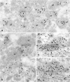
Immunogold labeling (10 nm gold) for delta 1/2 in the adult (a) and P21 (b,c) cerebellum. Note the abundant labeling of parallel fiber (pf) synapses and the low level or absence at climbing fiber (cf) synapses.Arrowheads indicate the postsynaptic densities/membranes (found on the heads of spines). P, Purkinje cell dendrite. Scale bar, 0.5 μm.
Fig. 2.
Immunogold labeling for delta 1/2 at P14 (a, b), P10 (c,d), and P2 (e). Both parallel fiber (pf) and climbing fiber (cf) synapses show abundant gold labeling at P10 and P14 in contrast to that seen at P21 and in the adult.Arrowheads indicate the postsynaptic densities/membranes. P, Purkinje cell dendrite. Scale bar, 0.5 μm.
Fig. 3.
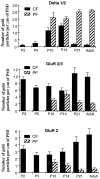
Histograms illustrating the changes in immunogold labeling for delta 1/2 (top), GluR2/3 (middle), and GluR2 (bottom) in the postsynaptic density/membrane (PSD) of parallel fiber (PF) and climbing fiber (CF) synapses on Purkinje cells during development of the cerebellum. Note especially the large differences in the pattern of immunolabeling between delta 1/2 and GluR2/3 and GluR2 at P2–P5 and P21–adult. Lower levels of immunogold labeling for_CF_ versus PF (delta 1/2) or for_PF_ versus CF (GluR2/3; GluR2) were statistically significant (p < 0.01) at P10 (GluR2/3 only), at P14 (GluR2 only), and at P21 and in adults for all three antibodies. For delta 1/2, statistical significance also was found for the following: P5 CF versus P10 CF, P14_CF_ versus either P21 or adult CF, and P14_PF_ versus P21 PF.
Fig. 4.
Colocalization of GABA (30 nm gold) neurotransmitter and delta 1/2 receptors (10 nm gold) at P10. Note the absence of GABA labeling in parallel fiber (pf; a, b) and climbing fiber (cf; b) terminals and the abundant labeling for GABA in Purkinje cell dendrites (P; b) and somata (P;c, d) and in pleomorphic vesicle-containing synaptic terminals (i). Delta receptor labeling is abundant in the postsynaptic density/membrane of parallel and climbing fiber synapses (arrowheads) but is absent from GABAergic synapses (arrows). Scale bar, 0.5 μm.
Fig. 5.
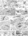
Immunogold labeling for GluR2/3 in the postsynaptic density/membrane (arrowheads) of parallel fiber (pf) and climbing fiber (cf) synapses on Purkinje cells (P; dendrites in c, f; somata in d, g) during development.a, c, Adult; b,d, P21; e, f, P14;g, h, P10; i, P5; and_j_, P2. Note the higher labeling of climbing fiber synapses compared with that of parallel fiber synapses. Scale bar, 0.5 μm.
Fig. 6.
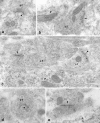
Immunogold labeling for GluR2 in the postsynaptic density/membrane (arrowheads) of parallel fiber (pf) and climbing fiber (cf) synapses on Purkinje cells during development. a, Adult; b, P21;c, d, P14; and e, P2. Note the higher labeling of climbing fiber synapses compared with that of parallel fiber synapses. P, Purkinje cell dendrite. Scale bar, 0.5 μm.
Fig. 7.
Immunogold labeling for GluR1 in the postsynaptic density/membrane (arrowheads) of parallel fiber (pf) and climbing fiber (cf) synapses on Purkinje cells during development. a, b, Adult;c, d, P10; and e, P2. Note the high level of staining at P10. Most synapses at P2 were not labeled as highly as those in the example shown. P, Purkinje cell dendrite (b) and somata (c,e). Scale bar, 0.5 μm.
Fig. 8.
Summary histogram (top) and diagrams (bottom) of development of glutamate receptors at parallel (P10–adult) and climbing (P2–adult) fiber synapses.Histogram, Note especially the peak in immunogold labeling of the delta receptors at P10–P14 in climbing fiber synapses (CF), the peaks of the AMPA receptors at P2–P5, and the inverse patterns of peaks for parallel fiber synapses (PF) and climbing fiber synapses in adults for AMPA versus delta receptors. Diagrams, Climbing fibers (cf) innervate the Purkinje cell (Pj) body up to approximately the end of the second postnatal week. By P21, climbing fiber innervation is reduced to a single fiber per Purkinje cell. Climbing fiber synapses on Purkinje cell bodies (early postnatal ages) have many postsynaptic AMPA receptors (based on labeling for GluR2/3 and GluR2) and few delta receptors. Climbing fiber synapses on Purkinje cell dendrites (later postnatal ages to adult) have many AMPA receptors; they have many delta receptors in the second postnatal week but very few from P21 to adult. Immunogold labeling for delta receptors at parallel fiber (pf) synapses is always abundant, but less immunogold labeling is seen for AMPA receptors. Labeled terminals are illustrated diagrammatically as postsynaptic spine heads and necks, with the receptors arranged along the surface of the spine head. The number of receptors shown is based approximately on the values of mean number of gold particles per synapse in Tables 1 and 2 and in the Results; it is intended only to show the relative amounts and not to represent actual numbers. The asterisk denotes a level of less than one-half of a receptor per synapse.
Similar articles
- Differential localization of delta glutamate receptors in the rat cerebellum: coexpression with AMPA receptors in parallel fiber-spine synapses and absence from climbing fiber-spine synapses.
Landsend AS, Amiry-Moghaddam M, Matsubara A, Bergersen L, Usami S, Wenthold RJ, Ottersen OP. Landsend AS, et al. J Neurosci. 1997 Jan 15;17(2):834-42. doi: 10.1523/JNEUROSCI.17-02-00834.1997. J Neurosci. 1997. PMID: 8987804 Free PMC article. - Delta-glutamate receptors are differentially distributed at parallel and climbing fiber synapses on Purkinje cells.
Zhao HM, Wenthold RJ, Wang YX, Petralia RS. Zhao HM, et al. J Neurochem. 1997 Mar;68(3):1041-52. doi: 10.1046/j.1471-4159.1997.68031041.x. J Neurochem. 1997. PMID: 9048749 - Differential neuronal and glial expression of GluR1 AMPA receptor subunit and the scaffolding proteins SAP97 and 4.1N during rat cerebellar development.
Douyard J, Shen L, Huganir RL, Rubio ME. Douyard J, et al. J Comp Neurol. 2007 May 1;502(1):141-56. doi: 10.1002/cne.21294. J Comp Neurol. 2007. PMID: 17335044 - Activity-dependent plasticity of developing climbing fiber-Purkinje cell synapses.
Bosman LW, Konnerth A. Bosman LW, et al. Neuroscience. 2009 Sep 1;162(3):612-23. doi: 10.1016/j.neuroscience.2009.01.032. Epub 2009 Jan 23. Neuroscience. 2009. PMID: 19302832 Review. - Influence of parallel fiber-Purkinje cell synapse formation on postnatal development of climbing fiber-Purkinje cell synapses in the cerebellum.
Hashimoto K, Yoshida T, Sakimura K, Mishina M, Watanabe M, Kano M. Hashimoto K, et al. Neuroscience. 2009 Sep 1;162(3):601-11. doi: 10.1016/j.neuroscience.2008.12.037. Epub 2008 Dec 31. Neuroscience. 2009. PMID: 19166909 Review.
Cited by
- Number and density of AMPA receptors in individual synapses in the rat cerebellum as revealed by SDS-digested freeze-fracture replica labeling.
Masugi-Tokita M, Tarusawa E, Watanabe M, Molnár E, Fujimoto K, Shigemoto R. Masugi-Tokita M, et al. J Neurosci. 2007 Feb 21;27(8):2135-44. doi: 10.1523/JNEUROSCI.2861-06.2007. J Neurosci. 2007. PMID: 17314308 Free PMC article. - Regulation of AMPA receptors by phosphorylation.
Carvalho AL, Duarte CB, Carvalho AP. Carvalho AL, et al. Neurochem Res. 2000 Oct;25(9-10):1245-55. doi: 10.1023/a:1007644128886. Neurochem Res. 2000. PMID: 11059799 Review. - Dynamics of trace element concentration during development and excitotoxic cell death in the cerebellum of Lurcher mutant mice.
Bäurle J, Kucera J, Frischmuth S, Lambertz M, Kranda K. Bäurle J, et al. Brain Pathol. 2009 Oct;19(4):586-95. doi: 10.1111/j.1750-3639.2008.00200.x. Epub 2008 Aug 13. Brain Pathol. 2009. PMID: 18702639 Free PMC article. - Ultrastructure of synapses in the mammalian brain.
Harris KM, Weinberg RJ. Harris KM, et al. Cold Spring Harb Perspect Biol. 2012 May 1;4(5):a005587. doi: 10.1101/cshperspect.a005587. Cold Spring Harb Perspect Biol. 2012. PMID: 22357909 Free PMC article. Review. - Distal extension of climbing fiber territory and multiple innervation caused by aberrant wiring to adjacent spiny branchlets in cerebellar Purkinje cells lacking glutamate receptor delta 2.
Ichikawa R, Miyazaki T, Kano M, Hashikawa T, Tatsumi H, Sakimura K, Mishina M, Inoue Y, Watanabe M. Ichikawa R, et al. J Neurosci. 2002 Oct 1;22(19):8487-503. doi: 10.1523/JNEUROSCI.22-19-08487.2002. J Neurosci. 2002. PMID: 12351723 Free PMC article.
References
- Altman J, Bayer SA. Development of the cerebellar system in relation to its evolution, structure, and functions. CRC; New York: 1997.
- Araki K, Meguro H, Kushiya E, Takayama C, Inoue Y, Mishina M. Selective expression of the glutamate receptor channel δ2 subunit in cerebellar Purkinje cells. Biochem Biophys Res Commun. 1993;197:1267–1276. - PubMed
- Bergmann M, Fox PA, Grabs D, Post A, Schilling K. Expression and subcellular distribution of glutamate receptor subunits 2/3 in the developing cerebellar cortex. J Neurosci Res. 1996;43:78–86. - PubMed
Publication types
MeSH terms
Substances
LinkOut - more resources
Full Text Sources
Research Materials
Miscellaneous


