Repertoire of chemokine receptor expression in the female genital tract: implications for human immunodeficiency virus transmission - PubMed (original) (raw)
Repertoire of chemokine receptor expression in the female genital tract: implications for human immunodeficiency virus transmission
B K Patterson et al. Am J Pathol. 1998 Aug.
Abstract
Sexually transmitted diseases, genital ulcer disease, and progesterone therapy increase susceptibility to lentivirus transmission. Infection of cells by human immunodeficiency virus (HIV) is dependent on expression of specific chemokine receptors known to function as HIV co-receptors. Quantitative kinetic reverse transcription-polymerase chain reaction was developed to determine the in vivo expression levels of CCR5, CXCR4, CCR3, CCR2b, and the cytomegalovirus-encoded US28 in peripheral blood mononuclear cells and cervical biopsies from 12 women with and without sexually transmitted diseases, genital ulcer disease, and progesterone-predominant conditions. Our data indicate that CCR5 is the major HIV co-receptor expressed in the female genital tract, and CXCR4 is the predominantly expressed HIV co-receptor in peripheral blood. CCR5 mRNA expression in the ectocervix was 10-fold greater than CXCR4, 20-fold greater than CCR2b, and 100-fold greater than CCR3. In peripheral blood, CXCR4 expression was 1.5-fold greater than CCR5, 10-fold greater than CCR2b, and 15-fold greater than CCR3. US28 was not expressed in cervical tissue despite expression in peripheral blood mononuclear cells from five individuals. CCR5 was significantly increased (p < 0.02) in biopsies from women with sexually transmitted diseases and others who were progesterone predominant. In vitro studies demonstrate that progesterone increases CCR5, CXCR4, and CCR3 expression and decreases CCR2b expression in lymphocytes and monocytes/macrophages. Characterization of chemokine receptors at the tissue level provides important information in identifying host determinants of HIV-1 transmission.
Figures
Figure 1.
Chemokine receptor mRNA quantification is linear over a range of at least 105 copies. Threshold cycle number (Ct) refers to the cycle number at which the reporter signal exceeds background. Results were based on triplicate determinations. The correlation coefficient for all curves was 0.99.
Figure 2.
Comparison of chemokine receptor expression in PBMCs and uterine cervix. CCR5 mRNA expression in the cervix was 10-fold greater than CXCR4, 20-fold greater than CCR2b, and 100-fold greater than CCR3. In PBMCs, CXCR4 expression was 1.5-fold greater than CCR5, 10-fold greater than CCR2b, and 15-fold greater than CCR3. US28 was not expressed in any of the cervical biopsies despite expression in PBMCs from five individuals.
Figure 3.
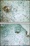
Digital microscopic images of chemokine receptor expression in an immunohistochemically stained cervical biopsy from HIV-1-seropositive patient 12 (Tables 1 and 2) ▶ ▶ . Chemokine receptor-expressing cells appear brown (arrows) in sections counterstained with hematoxylin (blue). A: CCR5-expressing cells are clustered beneath the surface epithelium. B: CXCR4-expressing cells from the same biopsy have a similar localization as the CCR5-expressing cells. Increased numbers of CCR5-expressing cells were found compared with CXCR4-expressing cells. Magnification, ×350.
Figure 4.
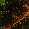
Localization of cells co-expressing CD4 and CCR5 (yellow) using double-label immunofluorescence and laser confocal image analysis (patient 6). Cells expressing only CD4 appeared red (arrowheads), and cells expressing only CCR5 were not identified. The epithelium-submucosa junction is denoted by arrows.
Figure 5.
Immunophenotypic characterization of cell types known to express CCR5 in representative cervical biopsies from patients listed in Table 1 ▶ . Activated/memory T lymphocytes (CD45RO), macrophages (CD68), and Langerhans’ cells (S-100) were visualized and quantified using image analysis. Positive cells appear brown (arrowheads) in sections counterstained with hematoxylin (blue). A lymphoid follicle was identified (arrow) in one of the biopsies. Magnification, ×200.
Figure 6.
Assisted computerized image analysis photomicrograph of cervical biopsies from patients 8 (A) and 9 (B) stained by immunohistochemistry for IL-2. IL-2-producing cells were identified by a juxtanuclear focal staining pattern (arrows) surrounded by extracellular immune reactivity caused by adherence of cytokines to matrix proteins. Tissue sections were counterstained with hematoxylin (blue). Magnification, ×400.
Figure 7.
A: Quantification of progesterone-induced chemokine receptor mRNA up-regulation after 3 days of PBMC culture in the presence of 50 ng/ml progesterone. CCR5, CXCR4, and CCR3 mRNA were significantly increased after progesterone treatment, whereas CCR2b mRNA decreased after treatment. B: Representative histograms from immunophenotyping/flow cytometry experiments. Untreated (top) and progesterone-treated (bottom) cells were double labeled with CD14-fluorescein isothiocyanate and CCR5-PE and gated based on CD14 expression and side scatter. Progesterone up-regulated CCR5 protein expression fivefold in CD14+ monocytes. The increase in CCR5/CXCR4 expression peaked at 3 days and remained elevated or slightly diminished by day 6.
Similar articles
- Influence of the CCR2-V64I polymorphism on human immunodeficiency virus type 1 coreceptor activity and on chemokine receptor function of CCR2b, CCR3, CCR5, and CXCR4.
Lee B, Doranz BJ, Rana S, Yi Y, Mellado M, Frade JM, Martinez-A C, O'Brien SJ, Dean M, Collman RG, Doms RW. Lee B, et al. J Virol. 1998 Sep;72(9):7450-8. doi: 10.1128/JVI.72.9.7450-7458.1998. J Virol. 1998. PMID: 9696841 Free PMC article. - In vivo evolution of HIV-1 co-receptor usage and sensitivity to chemokine-mediated suppression.
Scarlatti G, Tresoldi E, Björndal A, Fredriksson R, Colognesi C, Deng HK, Malnati MS, Plebani A, Siccardi AG, Littman DR, Fenyö EM, Lusso P. Scarlatti G, et al. Nat Med. 1997 Nov;3(11):1259-65. doi: 10.1038/nm1197-1259. Nat Med. 1997. PMID: 9359702 - HIV coreceptor and chemokine ligand gene expression in the male urethra and female cervix.
McClure CP, Tighe PJ, Robins RA, Bansal D, Bowman CA, Kingston M, Ball JK. McClure CP, et al. AIDS. 2005 Aug 12;19(12):1257-65. doi: 10.1097/01.aids.0000180096.50393.96. AIDS. 2005. PMID: 16052080 - [Deep lung--cellular reaction to HIV].
Tavares Marques MA, Alves V, Duque V, Botelho MF. Tavares Marques MA, et al. Rev Port Pneumol. 2007 Mar-Apr;13(2):175-212. Rev Port Pneumol. 2007. PMID: 17492233 Review. Portuguese. - Co-receptor use by HIV and inhibition of HIV infection by chemokine receptor ligands.
Simmons G, Reeves JD, Hibbitts S, Stine JT, Gray PW, Proudfoot AE, Clapham PR. Simmons G, et al. Immunol Rev. 2000 Oct;177:112-26. doi: 10.1034/j.1600-065x.2000.17719.x. Immunol Rev. 2000. PMID: 11138769 Review.
Cited by
- Translational Models to Predict Target Concentrations for Pre-Exposure Prophylaxis in Women.
Lantz AM, Nicol MR. Lantz AM, et al. AIDS Res Hum Retroviruses. 2022 Dec;38(12):909-923. doi: 10.1089/AID.2022.0057. Epub 2022 Oct 25. AIDS Res Hum Retroviruses. 2022. PMID: 36097755 Free PMC article. Review. - Expression of CCR5, CXCR4 and DC-SIGN in Cervix of HIV-1 Heterosexually Infected Mexican Women.
Rivera-Morales LG, Lopez-Guillen P, Vazquez-Guillen JM, Palacios-Saucedo GC, Rosas-Taraco AG, Ramirez-Pineda A, Amaya-Garcia PI, Rodriguez-Padilla C. Rivera-Morales LG, et al. Open AIDS J. 2012;6:239-44. doi: 10.2174/1874613601206010239. Epub 2012 Oct 5. Open AIDS J. 2012. PMID: 23115608 Free PMC article. - Phosphorothioate 2' deoxyribose oligomers as microbicides that inhibit human immunodeficiency virus type 1 (HIV-1) infection and block Toll-like receptor 7 (TLR7) and TLR9 triggering by HIV-1.
Fraietta JA, Mueller YM, Do DH, Holmes VM, Howett MK, Lewis MG, Boesteanu AC, Alkan SS, Katsikis PD. Fraietta JA, et al. Antimicrob Agents Chemother. 2010 Oct;54(10):4064-73. doi: 10.1128/AAC.00367-10. Epub 2010 Jul 12. Antimicrob Agents Chemother. 2010. PMID: 20625151 Free PMC article. - Proximity-dependent mapping of the HCMV US28 interactome identifies RhoGEF signaling as a requirement for efficient viral reactivation.
Medica S, Crawford LB, Denton M, Min CK, Jones TA, Alexander T, Parkins CJ, Diggins NL, Streblow GJ, Mayo AT, Kreklywich CN, Smith P, Jeng S, McWeeney S, Hancock MH, Yurochko A, Cohen MS, Caposio P, Streblow DN. Medica S, et al. PLoS Pathog. 2023 Oct 2;19(10):e1011682. doi: 10.1371/journal.ppat.1011682. eCollection 2023 Oct. PLoS Pathog. 2023. PMID: 37782657 Free PMC article. - Differential ligand binding to a human cytomegalovirus chemokine receptor determines cell type-specific motility.
Vomaske J, Melnychuk RM, Smith PP, Powell J, Hall L, DeFilippis V, Früh K, Smit M, Schlaepfer DD, Nelson JA, Streblow DN. Vomaske J, et al. PLoS Pathog. 2009 Feb;5(2):e1000304. doi: 10.1371/journal.ppat.1000304. Epub 2009 Feb 20. PLoS Pathog. 2009. PMID: 19229316 Free PMC article.
References
- Stakewski S, Schieck E, Rehmet S, Helm EB, Stille W: HIV transmission from a male after only two sexual contacts. Lancet 1987, 2:628-630 - PubMed
- Liu R, Paxton WA, Choe S, Ceridini D, Martin SR, Horuk R, MacDonald ME, Stuhlmann H, Koup RA, Landau NR: Homozygous defect in HIV-1 coreceptor accounts for resistance of some multiply exposed individuals to HIV-1 infection. Cell 1996, 86:367-377 - PubMed
- Padian NS, Shiboski SC, Jewell NP: Female to male transmission of human immunodeficiency virus. JAMA 1991, 266:1664-1667 - PubMed
- Plummer FA, Simonsen JN, Cameron DW, Ndinya-Achola JO, Kreiss JK, Gakinya MN, Waiyaki P, Cheang M, Piot P, Ronald AR, Ngugi EN: Cofactors in male-female sexual transmission of human immunodeficiency virus type 1. J Infect Dis 1991, 163:233-238 - PubMed
- Laga M, Diallo OM, Buve: Inter-relationship of sexually transmitted disease and HIV: where are we now? AIDS 1994, 8:S119-S124
Publication types
MeSH terms
Substances
LinkOut - more resources
Full Text Sources
Medical


