Increased anxiety of mice lacking the serotonin1A receptor - PubMed (original) (raw)
Increased anxiety of mice lacking the serotonin1A receptor
C L Parks et al. Proc Natl Acad Sci U S A. 1998.
Abstract
Brain serotonin (5-HT) has been implicated in a number of physiological processes and pathological conditions. These effects are mediated by at least 14 different 5-HT receptors. We have inactivated the gene encoding the 5-HT1A receptor in mice and found that receptor-deficient animals have an increased tendency to avoid a novel and fearful environment and to escape a stressful situation, behaviors consistent with an increased anxiety and stress response. Based on the role of the 5-HT1A receptor in the feedback regulation of the 5-HT system, we hypothesize that an increased serotonergic neurotransmission is responsible for the anxiety-like behavior of receptor-deficient animals. This view is consistent with earlier studies showing that pharmacological activation of the 5-HT system is anxiogenic in animal models and also in humans.
Figures
Figure 1
Genetic inactivation of the 5-HT1AR gene in ES cells and mice. (A) Genomic structure and restriction map of the 5-HT1AR locus (WT Genomic) and the targeting vector (KO Genomic). Primers used for PCR (wild-type 1–4 and KO 1, 2) and a probe employed in Southern experiments are depicted by arrowheads and a bar, respectively. (B) PCR screening of progeny derived from matings of heterozygote animals. (Upper) The presence of an amplified product derived from the substituted allele in heterozygote (+/−) and homozygote (−/−) mice. (Lower) PCR products derived from the wild-type allele in wild-type (+/+) and heterozygote animals. Size markers are in lane M. (C) Southern blot analysis of the wild-type receptor-specific _Xba_I fragment. The size of this fragment is increased from 1.88 to 3.0 kb by the substitution mutation.
Figure 2
5-HT1AR density of wild-type (+/+), heterozygote (+/−), and homozygote (−/−) animals in coronal sections of the hippocampus and cortex (A) and sections of the dorsal raphe nuclei (B). Receptors were labeled by the binding of [3H]-8-OH-DPAT. Nonspecific binding was determined in the presence of 5-HT. Three mice from each group were studied in this experiment, and sections from one is displayed.
Figure 3
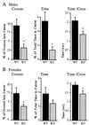
Open field test of wild-type and 5-HT1AR− males (A) and females (B). Fifteen wild-type and homozygotic males, 10 wild-type females, and 12 heterozygotic females were studied. The number of crosses into the center was normalized to locomotor activity and expressed as the percent of total crosses. Time spent in the center of the test apparatus is expressed as the percent of total time (10 min). Time per cross indicates the average time spent in the center of the open field for each entry. Asterisks designate statistically significant differences (P < 0.05) for the mutant as compared with the wild-type group.
Figure 4
Mobility in the forced swim test of wild-type and 5-HT1AR− mice. Each group consisted of 8–10 mice. The mobility of animals was measured in seconds between 0–2 min (1st block), 2–4 min (2nd block), and 4–6 min (3rd block) of the test. (□), Wild-type animals; (◊), 5-HT1AR− mice. Asterisks designate statistically significant differences (∗, P < 0.05 and ∗∗, P < 0.005) for the mutant as compared with wild-type group.
Similar articles
- 5-HT1A receptor knockout mouse as a genetic model of anxiety.
Toth M. Toth M. Eur J Pharmacol. 2003 Feb 28;463(1-3):177-84. doi: 10.1016/s0014-2999(03)01280-9. Eur J Pharmacol. 2003. PMID: 12600709 Review. - Serotonin1A receptor acts during development to establish normal anxiety-like behaviour in the adult.
Gross C, Zhuang X, Stark K, Ramboz S, Oosting R, Kirby L, Santarelli L, Beck S, Hen R. Gross C, et al. Nature. 2002 Mar 28;416(6879):396-400. doi: 10.1038/416396a. Nature. 2002. PMID: 11919622 - Involvement of 5-HT1A receptors in animal tests of anxiety and depression: evidence from genetic models.
Overstreet DH, Commissaris RC, De La Garza R 2nd, File SE, Knapp DJ, Seiden LS. Overstreet DH, et al. Stress. 2003 Jun;6(2):101-10. doi: 10.1080/1025389031000111311. Stress. 2003. PMID: 12775329 Review. - The 5-HT(1A) receptor knockout mouse and anxiety.
Olivier B, Pattij T, Wood SJ, Oosting R, Sarnyai Z, Toth M. Olivier B, et al. Behav Pharmacol. 2001 Nov;12(6-7):439-50. doi: 10.1097/00008877-200111000-00004. Behav Pharmacol. 2001. PMID: 11742137 Review. - Serotonin receptor 1A knockout: an animal model of anxiety-related disorder.
Ramboz S, Oosting R, Amara DA, Kung HF, Blier P, Mendelsohn M, Mann JJ, Brunner D, Hen R. Ramboz S, et al. Proc Natl Acad Sci U S A. 1998 Nov 24;95(24):14476-81. doi: 10.1073/pnas.95.24.14476. Proc Natl Acad Sci U S A. 1998. PMID: 9826725 Free PMC article.
Cited by
- Phytocannabinoids restore seizure-induced alterations in emotional behaviour in male rats.
Gom RC, Wickramarachchi P, George AG, Lightfoot SHM, Newton-Gunderson D, Hill MN, Teskey GC, Colangeli R. Gom RC, et al. Neuropsychopharmacology. 2024 Oct 21. doi: 10.1038/s41386-024-02005-y. Online ahead of print. Neuropsychopharmacology. 2024. PMID: 39433952 - Dissociating the therapeutic effects of environmental enrichment and exercise in a mouse model of anxiety with cognitive impairment.
Rogers J, Vo U, Buret LS, Pang TY, Meiklejohn H, Zeleznikow-Johnston A, Churilov L, van den Buuse M, Hannan AJ, Renoir T. Rogers J, et al. Transl Psychiatry. 2016 Apr 26;6(4):e794. doi: 10.1038/tp.2016.52. Transl Psychiatry. 2016. PMID: 27115125 Free PMC article. - The therapeutic role of 5-HT1A and 5-HT2A receptors in depression.
Celada P, Puig M, Amargós-Bosch M, Adell A, Artigas F. Celada P, et al. J Psychiatry Neurosci. 2004 Jul;29(4):252-65. J Psychiatry Neurosci. 2004. PMID: 15309042 Free PMC article. Review. - The recombinant 5-HT1A receptor: G protein coupling and signalling pathways.
Raymond JR, Mukhin YV, Gettys TW, Garnovskaya MN. Raymond JR, et al. Br J Pharmacol. 1999 Aug;127(8):1751-64. doi: 10.1038/sj.bjp.0702723. Br J Pharmacol. 1999. PMID: 10482904 Free PMC article. Review. - Freud-1: A neuronal calcium-regulated repressor of the 5-HT1A receptor gene.
Ou XM, Lemonde S, Jafar-Nejad H, Bown CD, Goto A, Rogaeva A, Albert PR. Ou XM, et al. J Neurosci. 2003 Aug 13;23(19):7415-25. doi: 10.1523/JNEUROSCI.23-19-07415.2003. J Neurosci. 2003. PMID: 12917378 Free PMC article.
References
- Jacobs B L, Wilkinson L O, Fornal C A. Neuropsychopharmacology. 1990;3:473–479. - PubMed
- Leysen J E. Neuropsychopharmacology. 1990;3:361–369. - PubMed
- Fuller R W. Neuropsychopharmacology. 1990;3:495–502. - PubMed
- Buhot M C. Curr Opin Neurobiol. 1997;7:243–254. - PubMed
- Weiger W A. Biol Rev Camb Philos Soc. 1997;72:61–95. - PubMed
Publication types
MeSH terms
Substances
LinkOut - more resources
Full Text Sources
Other Literature Sources
Medical
Molecular Biology Databases


