Expression of the Epstein-Barr virus latent membrane protein 1 induces B cell lymphoma in transgenic mice - PubMed (original) (raw)
Expression of the Epstein-Barr virus latent membrane protein 1 induces B cell lymphoma in transgenic mice
W Kulwichit et al. Proc Natl Acad Sci U S A. 1998.
Abstract
The latent membrane protein 1 (LMP1) of the Epstein-Barr virus has transforming properties in rodent fibroblasts and is expressed in most of the cancers associated with Epstein-Barr virus (EBV) infection including posttransplant lymphomas, Hodgkin's disease, nasopharyngeal carcinoma, and AIDS-related lymphomas. In this study, three lineages of LMP1 transgenic mice were established with LMP1 expressed under the control of the Ig heavy chain promoter and enhancer. Lymphoma developed in all three lineages, and the incidence of lymphoma increased significantly with age with lymphomas developing in 42% of transgenic mice over 18 months. The expression of LMP1 was detected at high levels in the lymphoma tissues but only at trace levels in normal lymphoid tissues. Gene rearrangement of the Ig heavy chain indicated monoclonality or oligoclonality in all lymphomas, some of the lymphoid hyperplastic spleens, and some histologically normal spleens. These data reveal that LMP1, without the expression of other EBV genes, is oncogenic in vivo and indicate that LMP1 is a major contributing factor to the development of EBV-associated lymphomas.
Figures
Figure 1
Histopathology and immunohistochemistry for LMP1 in lymphoma and lymphoid infiltrate. (A) Follicular center cell lymphoma (lineage 3), large cell type. The normal architecture of the spleen is effaced by sheets of large neoplastic cells with abundant cytoplasm. The nuclei are irregularly shaped, with clumped or marginated chromatin and sometimes prominent nucleoli. Mitotic figures are frequently present. (Hematoxylin and eosin; ×600.) (B) Lymphoid infiltrate in the lung. An aggressive lymphoid-cell infiltrate (lineage 6) has invaded the adjacent alveolar tissue. Analysis of DNA extracted from this tissue demonstrated monoclonal heavy chain rearrangement on Southern blot. A high level of LMP1 was detected by immunoblot analysis of a protein lysate prepared from this tissue. (Hematoxylin and eosin; ×200.) (C) Expression of LMP1 in the lymphoid infiltrate shown in B. The tissue was stained with rabbit polyclonal anti-LMP1 and detected with an immunoperoxidase-tagged goat anti-rabbit serum. LMP1 is detected as dark brown staining in the cell membrane, with some cells showing the characteristic capping. (×1,000.) (D and_E_) Expression of LMP1 in lymphoma tissue and negative-staining control (lineage 3). The splenic lymphoma tissue was stained with either rabbit polyclonal anti-LMP1 (D) or normal rabbit serum at same dilution (E). The staining was detected with fluorescein-conjugated goat anti-rabbit antibody. LMP1 is detected as green fluorescence in the plasma membrane, with some cells showing the characteristic capping. (×1,000.) (F) Presence of cytoplasmic Ig in the lymphoma. The same lymphoma tissue depicted in_D_ and E was stained with tetramethylrhodamine B isothiocyanate -conjugated goat anti-mouse IgG. IgG is detected as red fluorescence in the cytoplasm of most cells. This lymphoma also expresses a high level of LMP1 and was shown to be monoclonal by IgH gene rearrangement (see also Fig. 4_A_and B). (×1,000.)
Figure 2
Levels of the expression of LMP1 correlate with lymphoid pathologies. Immunoblots were prepared with total protein tissue lysate (200 μg) and 50 μg of protein lysate from the B95–8 EBV-positive lymphoid cell line (lane 1) and reacted with the CS1–4 mouse monoclonal antibody. A spontaneous lymphoma was obtained from a 2-year-old LMP1-negative littermate (lane 2). Lanes 3–5 are lymphoma tissues; lanes 6–7 are lymphoid hyperplasia; lanes 8–10 are normal lymphoid tissues. Lanes 3–10 are transgenic splenic tissues from lineage 3.
Figure 3
Transgenic lymphomas and lymphoid hyperplasia are clonal. Southern blots were prepared with DNA from samples of lymphoma, hyperplasia, or normal spleens and hybridized to a PJ11 probe representing the JH region of the heavy chain locus. “Normal spleen” is a nontransgenic control spleen with the genomic 6.6-kb _Eco_RI fragment. The upper genomic band (23 kb) in mice of lineage 3 is caused by the integration of the transgene into the heavy chain locus in this lineage (lanes 1, 2, 3, 6, 9, and 10). Lanes 7, 8, 11, and 13 are from lineage 6. Lanes 4, 5, and 12 are from lineage 9.
Figure 4
Expression of LMP1 correlates with IgH gene rearrangement and lymphoma detection. (A) Immunoblots were prepared with protein lysates from lymphoma samples and control tissues from three mice and the B95–8 cell line. Mouse A had lymphoma in hilar LN (lymph nodes) and the spleen, but not the skin and kidney. Mouse B had lymphoma in the mesenteric LN but not in the spleen. Mouse C had lymphoma in the lungs but not the spleen. “Normal spleen” was a spleen from a nontransgenic control mouse. (B) IgH gene rearrangement. Southern blots were hybridized to the PJ11 probe and showed the correlation of the lymphoma phenotype in the organs described in A with clonal rearrangement. In mouse A, gene rearrangement was analyzed and detected only in the spleen. All three mice were from lineage 3, which contained two genomic bands. The upper band (23 kb) resulted from the integration of the transgene into the heavy chain locus.
Figure 5
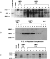
A20, Bcl-2, and c-Myc are expressed at elevated levels in transgenic lymphoma. Immunoblots were prepared with 200 μg total protein lysates from lymphomas that developed in nontransgenic littermates and transgenic lymphoma tissues and reacted with mouse monoclonal anti-A20, rabbit polyclonal anti-Bcl-2, and rabbit polyclonal anti-c-Myc.
References
- Raab-Traub N. Semin Virol. 1996;7:315–323.
- Kieff E. In: Fields Virology. Fields B N, Knipe D M, Howley P M, Chanock R M, Melnick J L, Monath T P, Roizman B, Straus S E, editors. Philadelphia: Lippincott–Raven; 1996. pp. 2343–2396.
- Wang D, Liebowitz D, Kieff E. Cell. 1985;43:831–840. - PubMed
- Henderson S, Rowe M, Gregory C, Croom-Carter D, Wang F, Longnecker R, Kieff E, Rickinson A. Cell. 1991;65:1107–1115. - PubMed
- Laherty C D, Hu H M, Opipari A W, Wang F, Dixit V M. J Biol Chem. 1992;267:24157–24160. - PubMed
Publication types
MeSH terms
Substances
LinkOut - more resources
Full Text Sources
Other Literature Sources
Molecular Biology Databases
Research Materials


