Complex gangliosides are essential in spermatogenesis of mice: possible roles in the transport of testosterone - PubMed (original) (raw)
. 1998 Oct 13;95(21):12147-52.
doi: 10.1073/pnas.95.21.12147.
A Yamamoto, K Furukawa, J Zhao, S Fukumoto, S Yamashiro, M Okada, M Haraguchi, M Shin, M Kishikawa, H Shiku, S Aizawa, K Furukawa
Affiliations
- PMID: 9770454
- PMCID: PMC22799
- DOI: 10.1073/pnas.95.21.12147
Complex gangliosides are essential in spermatogenesis of mice: possible roles in the transport of testosterone
K Takamiya et al. Proc Natl Acad Sci U S A. 1998.
Abstract
Mice, homozygous for disrupted ganglioside GM2/GD2 synthase (EC 2.4. 1.94) gene and lacking all complex gangliosides, do not display any major neurologic abnormalities. Further examination of these mutant mice, however, revealed that the males were sterile and aspermatogenic. In the seminiferous tubules of the mutant mice, a number of multinuclear giant cells and vacuolated Sertoli cells were observed. The levels of testosterone in the serum of these mice were very low, although testosterone production equaled that produced in wild-type mice. Testosterone was found to be accumulated in interstitial Leydig cells, and intratesticularly injected testosterone was poorly drained in seminiferous fluid in the mutant mice. These results suggested that complex gangliosides are essential in the transport of testosterone to the seminiferous tubules and bloodstream from Leydig cells. Our results provide insights into roles of gangliosides in vivo.
Figures
Figure 1
Proposed pathway of ganglioside synthesis. The box encloses gangliosides, whose biosynthesis is blocked in β1,4GalNAc-T-deficient mice. LacCer, lactosylceramide .
Figure 2
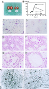
Male sterility in β1,4GalNAc-T- deficient mice. Morphology and growth of the testis of wild-type and mutant mice. (A) Eight-week-old wild-type (+/+) and mutant (−/−) testes. (B) Changes in testicular weight in wild-type and mutant mice. (C and D) Smear of seminiferous fluid from wild-type (C) and mutant (D) mice. (Hematoxylin/eosin; ×280.) (E and F) Histopathology of testis from 10-week-old wild-type (E) and mutant (F) mice. (Hematoxylin/eosin; ×140.) (G and H) High magnification of mutant mice testis. (Hematoxylin/eosin; ×280.) Note the diffuse vacuoles in Sertoli cells (in H and at asterisks in F). (I and J) Electron micrographs of multinuclear giant cells. A giant cell (I) and an unseparated prematurely opened intercellular bridge (arrows) (J) are shown. (Bars indicate 5 μm).
Figure 3
Expression of β1,4GalNAc-T gene in testis. (A–C) In situ hybridization of β1,4GalNAc-T gene in wild-type testis. (A) Hematoxylin/eosin staining of the testis from a wild-type mouse ×140. (B) With digoxigenin-labeled RNA probe, in situ hybridization was performed as described in Materials and Methods. (×140. (C) High magnification (×560) of B. (D) Total RNA was extracted from testes of ≈2- to 8-week-old wild-type mice and 20 μg each was applied for Northern blotting. Glyceraldehyde-3-phosphate dehydrogenase (GAPDH) cDNA was used to hybridize the same membrane as a control.
Figure 4
Expression of gangliosides in testis. (A) TLC of acidic glycosphingolipids from testis. St., bovine brain ganglioside mixture as a standard. Acidic glycolipids were separated by TLC with a solvent (chloroform/methanol/0.2% CaCl2, 55:45:10). Resorcinol spray was used for detection. (B) Profiles of acidic glycosphingolipids in nongerm cells. Nongerm cells from testis were cultured primarily after elimination of germ cells, then metabolically labeled with [14C]glucosamine and analyzed for acidic glycolipids by TLC. Solvent for TLC was as in A.
Figure 5
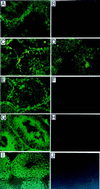
Distribution of gangliosides in testis. (A–C) Cholera toxin staining. (A) Staining in wild-type testis. (B) No staining of mutant testis. (C) Staining after neuraminidase treatment of wild-type testis for detection of the complex gangliosides. (D) GD3 expression in mutant testis revealed by anti-GD3 mAb R24. Testis of wild-type mice did not show definite staining (data not shown). (E–J) Immunohistostaining with mAbs specific for GD1a (E, F), GT1b (G, H), and GD1b (I, J). Samples from wild-type (E, G, I) and mutant testis (F, H, J) were compared.
Figure 6
Hormonal changes and their effects in β1,4GalNAc-T mutant mice. Black bars represent wild type and white bars homozygous mutant (A–F). Ten- to 12-week-old mice were analyzed. (A) Testosterone levels in serum. Note marked reduction in serum testosterone level in the mutant (21.0 ± 4.10 ng/dl) in contrast to the wild type (386.63 ± 111.90 ng/dl). ∗, P < 0.005. (B) Testosterone in testis. In testis, the amounts of total testosterone are comparable, and the amount per weight is somewhat higher in the mutant. ∗∗, P < 0.05. (C) Production of testosterone in cultured testicular cells with or without human chorionic gonadotrophin stimulation in vitro. (D) Serum FSH and LH levels in wild-type and mutant mice. (E) Comparison of muscle size. Circumferences of the gastrocnemius muscles and weights of parts between the knee joints and Achilles tendon were measured. ∗∗, P <0.05. (F) Neuron density at the preoptic area was examined by serial cross-sections, as described (34). Ordinate is unit of pixels. ∗, <0.005.
Figure 7
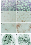
Testosterone production in the interstitial cells of testis. (A and B) Hematoxylin/eosin staining of testis from wild-type (A) and mutant (B) mice. (C–F) Immunohistochemistry for testosterone with polyclonal antibody (Zymed) in wild-type (C and E) and mutant (D and F) testis. (×140 for C and D, ×280 for E and F.) (G and H) Electron micrograph of Leydig cells of wild-type (G) and mutant (H) mice. (Bars indicate 2 μm.)
Figure 8
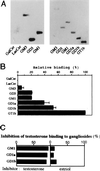
Ganglioside–testosterone interaction. [14C]testosterone was applied to a poly(vinylidene difluoride) membrane on which individual glycolipids had been blotted. Binding and washing were performed as described in Materials and Methods. (A) Each lane contained 1.6 (left) and 0.4 (right) nmol of glycolipids. (B) Relative binding of testosterone to 0.4 nmol of each ganglioside is given as a percentage of the GT1b value (an average of three experiments). (C) Specific inhibition of testosterone binding to gangliosides by unlabeled testosterone. [14C]testosterone was applied to a poly(vinylidene difluoride) membrane, as in A, with individual inhibitors. Similar experiments with wide range of gangliosides were repeated 3 times, and a representative result only for testosterone and estriol is presented as percent inhibition.
Figure 9
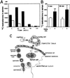
(A) Transport of intratesticularly injected [14C]testosterone into semen. The radioactivity in semen after injection was measured at the time points indicated. These experiments were repeated at least three times and showed essentially the same results. A representative result is given. Black bars, wild type; white bars, mutant. (B) Improvement of [14C]testosterone excretion in the presence of gangliosides in the mutant mice. [14C]testosterone was injected as in A, with or without gangliosides (10 μg), then cpm in semen was examined at 5 and 30 min after injection. ∗∗, P < 0.05. (C) A scheme to show the essential role of complex gangliosides in testosterone transport from Leydig cells in testis. T, testosterone. An X superimposed on an arrow represents disrupted transport.
Similar articles
- Disordered testosterone transport in mice lacking the ganglioside GM2/GD2 synthase gene.
Furukawa K, Takamiya K, Ohmi Y, Bhuiyan RH, Tajima O, Furukawa K. Furukawa K, et al. FEBS Open Bio. 2023 Sep;13(9):1615-1624. doi: 10.1002/2211-5463.13603. Epub 2023 Apr 6. FEBS Open Bio. 2023. PMID: 36999634 Free PMC article. Review. - Beta1,4-N-acetylgalactosaminyltransferase--GM2/GD2 synthase: a key enzyme to control the synthesis of brain-enriched complex gangliosides.
Furukawa K, Takamiya K, Furukawa K. Furukawa K, et al. Biochim Biophys Acta. 2002 Dec 19;1573(3):356-62. doi: 10.1016/s0304-4165(02)00403-8. Biochim Biophys Acta. 2002. PMID: 12417418 Review. - Mice with disrupted GM2/GD2 synthase gene lack complex gangliosides but exhibit only subtle defects in their nervous system.
Takamiya K, Yamamoto A, Furukawa K, Yamashiro S, Shin M, Okada M, Fukumoto S, Haraguchi M, Takeda N, Fujimura K, Sakae M, Kishikawa M, Shiku H, Furukawa K, Aizawa S. Takamiya K, et al. Proc Natl Acad Sci U S A. 1996 Oct 1;93(20):10662-7. doi: 10.1073/pnas.93.20.10662. Proc Natl Acad Sci U S A. 1996. PMID: 8855236 Free PMC article. - Wt1 is involved in leydig cell steroid hormone biosynthesis by regulating paracrine factor expression in mice.
Chen M, Wang X, Wang Y, Zhang L, Xu B, Lv L, Cui X, Li W, Gao F. Chen M, et al. Biol Reprod. 2014 Apr 3;90(4):71. doi: 10.1095/biolreprod.113.114702. Print 2014 Apr. Biol Reprod. 2014. PMID: 24571983 - Luteinizing hormone receptor-mediated effects on initiation of spermatogenesis in gonadotropin-deficient (hpg) mice are replicated by testosterone.
Spaliviero JA, Jimenez M, Allan CM, Handelsman DJ. Spaliviero JA, et al. Biol Reprod. 2004 Jan;70(1):32-8. doi: 10.1095/biolreprod.103.019398. Epub 2003 Sep 3. Biol Reprod. 2004. PMID: 12954730
Cited by
- Simplifying complexity: genetically resculpting glycosphingolipid synthesis pathways in mice to reveal function.
Allende ML, Proia RL. Allende ML, et al. Glycoconj J. 2014 Dec;31(9):613-22. doi: 10.1007/s10719-014-9563-5. Epub 2014 Oct 29. Glycoconj J. 2014. PMID: 25351657 Free PMC article. Review. - Role of sphingolipid metabolites in the homeostasis of steroid hormones and the maintenance of testicular functions.
Wang D, Tang Y, Wang Z. Wang D, et al. Front Endocrinol (Lausanne). 2023 Mar 17;14:1170023. doi: 10.3389/fendo.2023.1170023. eCollection 2023. Front Endocrinol (Lausanne). 2023. PMID: 37008929 Free PMC article. Review. - Novel Molecular Mechanisms of Gangliosides in the Nervous System Elucidated by Genetic Engineering.
Furukawa K, Ohmi Y, Yesmin F, Tajima O, Kondo Y, Zhang P, Hashimoto N, Ohkawa Y, Bhuiyan RH, Furukawa K. Furukawa K, et al. Int J Mol Sci. 2020 Mar 11;21(6):1906. doi: 10.3390/ijms21061906. Int J Mol Sci. 2020. PMID: 32168753 Free PMC article. Review. - The regulation of spermatogenesis by androgens.
Smith LB, Walker WH. Smith LB, et al. Semin Cell Dev Biol. 2014 Jun;30:2-13. doi: 10.1016/j.semcdb.2014.02.012. Epub 2014 Mar 2. Semin Cell Dev Biol. 2014. PMID: 24598768 Free PMC article. Review. - Roles for Golgi Glycans in Oogenesis and Spermatogenesis.
Akintayo A, Stanley P. Akintayo A, et al. Front Cell Dev Biol. 2019 Jun 7;7:98. doi: 10.3389/fcell.2019.00098. eCollection 2019. Front Cell Dev Biol. 2019. PMID: 31231650 Free PMC article. Review.
References
- Suzuki K. J Neurochem. 1965;12:969–979. - PubMed
- Ledeen R, Yu R K. Methods Enzymol. 1982;83:139–191. - PubMed
- Wiegandt H. In: Glycolipids. Wiegandt H, editor. Amsterdam: Elsevier; 1985. pp. 199–260.
- Hakomori S. J Biol Chem. 1990;265:18713–18716. - PubMed
- Schengrund C L. Brain Res Bull. 1990;24:131–141. - PubMed
Publication types
MeSH terms
Substances
LinkOut - more resources
Full Text Sources
Molecular Biology Databases

