Derivation of pluripotent stem cells from cultured human primordial germ cells - PubMed (original) (raw)
Derivation of pluripotent stem cells from cultured human primordial germ cells
M J Shamblott et al. Proc Natl Acad Sci U S A. 1998.
Erratum in
- Proc Natl Acad Sci U S A 1999 Feb 2;96(3):1162
Abstract
Human pluripotent stem cells would be invaluable for in vitro studies of aspects of human embryogenesis. With the goal of establishing pluripotent stem cell lines, gonadal ridges and mesenteries containing primordial germ cells (PGCs, 5-9 weeks postfertilization) were cultured on mouse STO fibroblast feeder layers in the presence of human recombinant leukemia inhibitory factor, human recombinant basic fibroblast growth factor, and forskolin. Initially, single PGCs in culture were visualized by alkaline phosphatase activity staining. Over a period of 7-21 days, PGCs gave rise to large multicellular colonies resembling those of mouse pluripotent stem cells termed embryonic stem and embryonic germ (EG) cells. Throughout the culture period most cells within the colonies continued to be alkaline phosphatase-positive and tested positive against a panel of five immunological markers (SSEA-1, SSEA-3, SSEA-4, TRA-1-60, and TRA-1-81) that have been used routinely to characterize embryonic stem and EG cells. The cultured cells have been continuously passaged and found to be karyotypically normal and stable. Both XX and XY cell cultures have been obtained. Immunohistochemical analysis of embryoid bodies collected from these cultures revealed a wide variety of differentiated cell types, including derivatives of all three embryonic germ layers. Based on their origin and demonstrated properties, these human PGC-derived cultures meet the criteria for pluripotent stem cells and most closely resemble EG cells.
Figures
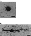
Figure 1
AP activity of individual human PGCs in culture. (A) Stationary and (B) migratory PGCs in a primary culture, growing on a feeder layer of mitotically inactivated mouse STO fibroblasts. (Bars represent 10 μm.)
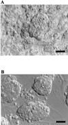
Figure 2
Colony morphology. (A) Human PGC-derived cell colony growing on a feeder layer of mitotically inactivated mouse STO fibroblasts. (B) Mouse ES colony growing on a feeder layer of mitotically inactivated mouse fibroblasts. Hoffman modulation optics. (Bars represent 100 μm.)
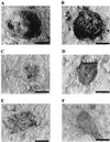
Figure 3
Expression of cell surface markers by human PGC-derived cell colonies. (A) AP. (B) SSEA-1. (C) SSEA-3. (D) SSEA-4. (E) TRA-1–60. (F) TRA-1–81. (Bars represent 100 μm.)
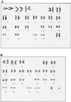
Figure 4
Karyotype of human PGC-derived cell cultures. (A) XX, eight passages. (B) XY, 10 passages.
Figure 5
Immunohistological analysis of human EB sections. EB culture designation, antibody epitope, and objective power are as follows: (A) BF1, muscle-specific actin, ×100. Arrow indicates a cell with eccentric nuclei and cytoplasmic filaments. (B) RI5, desmin, ×60. (C) BF1, CD34, ×60. (D) BF1, S-100, ×100. (E) RI5, S-100, ×60. (F) RI, pan-neurofilament, ×100. (G) BF1, α-1-fetoprotein, ×60. (H) BF1, pan-cytokeratin, ×60. (I) RI5, cytokeratin 7, ×60. (Bars represent 20 μm.)
Similar articles
- Fibroblast-like cells derived from the gonadal ridges and dorsal mesenteries of human embryos as feeder cells for the culture of human embryonic germ cells.
He J, Wang Y, Li YL. He J, et al. J Biomed Sci. 2007 Sep;14(5):617-28. doi: 10.1007/s11373-007-9185-z. Epub 2007 Jun 14. J Biomed Sci. 2007. PMID: 17566873 - [Isolation and culture of human pluripotent embryonic germ cells].
Li XH, Cong HC, Wang Z, Wu CF, Cao YL. Li XH, et al. Shi Yan Sheng Wu Xue Bao. 2002 Jun;35(2):142-6. Shi Yan Sheng Wu Xue Bao. 2002. PMID: 15344333 Chinese. - Establishment of a human embryonic germ cell line and comparison with mouse and human embryonic stem cells.
Park JH, Kim SJ, Lee JB, Song JM, Kim CG, Roh S 2nd, Yoon HS. Park JH, et al. Mol Cells. 2004 Apr 30;17(2):309-15. Mol Cells. 2004. PMID: 15179047 Retracted. - Embryonic germ cell lines and their derivation from mouse primordial germ cells.
Labosky PA, Barlow DP, Hogan BL. Labosky PA, et al. Ciba Found Symp. 1994;182:157-68; discussion 168-78. doi: 10.1002/9780470514573.ch9. Ciba Found Symp. 1994. PMID: 7835148 Review. - Germ cells and pluripotent stem cells in the mouse.
McLaren A, Durcova-Hills G. McLaren A, et al. Reprod Fertil Dev. 2001;13(7-8):661-4. doi: 10.1071/rd01080. Reprod Fertil Dev. 2001. PMID: 11999318 Review.
Cited by
- The reciprocal relationship between primordial germ cells and pluripotent stem cells.
Pirouz M, Klimke A, Kessel M. Pirouz M, et al. J Mol Med (Berl). 2012 Jul;90(7):753-61. doi: 10.1007/s00109-012-0912-1. Epub 2012 May 15. J Mol Med (Berl). 2012. PMID: 22584374 Review. - Human post-implantation blastocyst-like characteristics of Muse cells isolated from human umbilical cord.
Kushida Y, Oguma Y, Abe K, Deguchi T, Barbera FG, Nishimura N, Fujioka K, Iwatani S, Dezawa M. Kushida Y, et al. Cell Mol Life Sci. 2024 Jul 11;81(1):297. doi: 10.1007/s00018-024-05339-4. Cell Mol Life Sci. 2024. PMID: 38992309 Free PMC article. - Stem cells to replace or regenerate the diabetic pancreas: Huge potential & existing hurdles.
Bhartiya D. Bhartiya D. Indian J Med Res. 2016 Mar;143(3):267-74. doi: 10.4103/0971-5916.182615. Indian J Med Res. 2016. PMID: 27241638 Free PMC article. Review. - Exploiting CRISPR Cas9 in Three-Dimensional Stem Cell Cultures to Model Disease.
Gopal S, Rodrigues AL, Dordick JS. Gopal S, et al. Front Bioeng Biotechnol. 2020 Jun 24;8:692. doi: 10.3389/fbioe.2020.00692. eCollection 2020. Front Bioeng Biotechnol. 2020. PMID: 32671050 Free PMC article. Review. - Ambiguous cells: the emergence of the stem cell concept in the nineteenth and twentieth centuries.
Maehle AH. Maehle AH. Notes Rec R Soc Lond. 2011 Dec 20;65(4):359-78. doi: 10.1098/rsnr.2011.0023. Notes Rec R Soc Lond. 2011. PMID: 22332468 Free PMC article.
References
- Evans M J, Kaufman M H. Nature (London) 1981;292:154–156. - PubMed
- Matsui Y, Toksoz D, Nishikawa S, Nishikawa S, Williams D, Zsebo K, Hogan B L. Nature (London) 1991;353:750–752. - PubMed
- Resnick J L, Bixler L S, Cheng L, Donovan P J. Nature (London) 1992;359:550–551. - PubMed
- Stewart C, Gadi I, Bhatt H. Dev Biol. 1994;161:626–628. - PubMed
Publication types
MeSH terms
Substances
LinkOut - more resources
Full Text Sources
Other Literature Sources
Medical
Miscellaneous


