Expression of plasminogen activator pla of Yersinia pestis enhances bacterial attachment to the mammalian extracellular matrix - PubMed (original) (raw)
Expression of plasminogen activator pla of Yersinia pestis enhances bacterial attachment to the mammalian extracellular matrix
K Lähteenmäki et al. Infect Immun. 1998 Dec.
Abstract
The effect of the plasminogen activator Pla of Yersinia pestis on the adhesiveness of bacteria to the mammalian extracellular matrix was determined. Y. pestis KIM D27 harbors the 9.5-kb plasmid pPCP1, encoding Pla and pesticin; the strain efficiently adhered to the reconstituted basement membrane preparation Matrigel, to the extracellular matrix prepared from human lung NCI-H292 epithelial cells, as well as to immobilized laminin. The isogenic strain Y. pestis KIM D34 lacking pPCP1 exhibited lower adhesiveness to both matrix preparations and to laminin. Both strains showed weak adherence to type I, IV, and V collagens as well as to human plasma and cellular fibronectin. The Pla-expressing recombinant Escherichia coli LE392(pC4006) exhibited specific adhesiveness to both extracellular matrix preparations as well as to laminin. The Pla-expressing strains showed a low-affinity adherence to another basement membrane component, heparan sulfate proteoglycan, but not to chondroitin sulfate proteoglycan. The degradation of radiolabeled laminin, heparan sulfate proteoglycan, or human lung extracellular matrix by the Pla-expressing recombinant E. coli required the presence of plasminogen, and degradation was inhibited by the plasmin inhibitors aprotinin and alpha2-antiplasmin. Our results indicate a function of Pla in enhancing bacterial adhesion to extracellular matrices. Y. pestis also exhibits a low level of Pla-independent adhesiveness to extracellular matrices.
Figures
FIG. 1
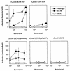
Adherence of Pla+ Y. pestis KIM D27, Pla− Y. pestis KIM D34, Pla+ E. coli LE392(pC4006), and Pla− E. coli LE392(pC4007) to the reconstituted mouse BM preparation Matrigel and to the ECM prepared by detergent extraction from human lung NCI-H292 epithelial cells. The control surface was coated with BSA. The bacteria were tested at a concentration of 109 cells/ml.
FIG. 2
Adherence of Pla+ Y. pestis KIM D27 and E. coli LE392(pC4006) as well as of Pla− Y. pestis KIM D34, E. coli LE392(pC4007), and E. coli LE392 to the BM preparation Matrigel reconstituted on glass slides as well as to the ECM prepared by detergent extraction from human lung NCI-H292 cells. The bacteria were tested at four different concentrations as indicated, and the data shown are means ± SDs for 20 randomly chosen microscopic fields of 1.6 × 104 μm2. The control surface was coated with BSA.
FIG. 3
Adherence of Pla+ Y. pestis KIM D27 and E. coli LE392(pC4006) as well as of Pla− Y. pestis KIM D34, E. coli LE392(pC4007), and E. coli LE392 to immobilized proteins of the ECM. Bacterial concentration varied from 108 to 5 × 10⁹ cells/ml, and the surface concentration of the target proteins was 2.5 pmol. The control surfaces were coated with the highly glycosylated fetuin and with the nonglycosylated BSA from a solution of 25 μg/ml. Data are means ± SDs for 20 randomly chosen microscopic fields of 1.6 × 104 μm2.
FIG. 4
Adherence of Pla+ Y. pestis KIMD27 and E. coli LE392(pC4006) as well as of Pla− Y. pestis KIM D34, E. coli LE392(pC4007), and E. coli LE392 to immobilized proteoglycans. For comparison, bacterial adhesiveness to laminin and BSA is also shown. All target proteins were coated from a solution of 50 μg/ml, and the bacteria were tested at the concentrations of 108 cells/ml (A) and 109 cells/ml (B). Data are means ± SDs for 20 randomly chosen fields of 1.6 × 104 μm2.
FIG. 5
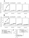
Degradation of 125I-labeled laminin (A) and 35S-labeled extracellular matrix prepared from human lung NCI-H292 cells (B) by Pla+ or Pla− recombinant E. coli in the presence of plasminogen and inhibitors of plasmin activity. (C) Plasmin activity associated with the E. coli cells or in PBS without added bacteria, measured with the chromogenic substrate. Bacteria were used at a concentration of 2 × 10⁹ cells/ml.
Similar articles
- Adhesive properties of the purified plasminogen activator Pla of Yersinia pestis.
Lobo LA. Lobo LA. FEMS Microbiol Lett. 2006 Sep;262(2):158-62. doi: 10.1111/j.1574-6968.2006.00382.x. FEMS Microbiol Lett. 2006. PMID: 16923070 - The Pla surface protease/adhesin of Yersinia pestis mediates bacterial invasion into human endothelial cells.
Lähteenmäki K, Kukkonen M, Korhonen TK. Lähteenmäki K, et al. FEBS Lett. 2001 Aug 24;504(1-2):69-72. doi: 10.1016/s0014-5793(01)02775-2. FEBS Lett. 2001. PMID: 11522299 - Protein regions important for plasminogen activation and inactivation of alpha2-antiplasmin in the surface protease Pla of Yersinia pestis.
Kukkonen M, Lähteenmäki K, Suomalainen M, Kalkkinen N, Emödy L, Lång H, Korhonen TK. Kukkonen M, et al. Mol Microbiol. 2001 Jun;40(5):1097-111. doi: 10.1046/j.1365-2958.2001.02451.x. Mol Microbiol. 2001. PMID: 11401715 - Using every trick in the book: the Pla surface protease of Yersinia pestis.
Suomalainen M, Haiko J, Ramu P, Lobo L, Kukkonen M, Westerlund-Wikström B, Virkola R, Lähteenmäki K, Korhonen TK. Suomalainen M, et al. Adv Exp Med Biol. 2007;603:268-78. doi: 10.1007/978-0-387-72124-8_24. Adv Exp Med Biol. 2007. PMID: 17966423 Review. - Plasminogen activation in degradation and penetration of extracellular matrices and basement membranes by invasive bacteria.
Lähteenmäki K, Kuusela P, Korhonen TK. Lähteenmäki K, et al. Methods. 2000 Jun;21(2):125-32. doi: 10.1006/meth.2000.0983. Methods. 2000. PMID: 10816373 Review.
Cited by
- Influence of Na(+), dicarboxylic amino acids, and pH in modulating the low-calcium response of Yersinia pestis.
Brubaker RR. Brubaker RR. Infect Immun. 2005 Aug;73(8):4743-52. doi: 10.1128/IAI.73.8.4743-4752.2005. Infect Immun. 2005. PMID: 16040987 Free PMC article. - Effects of Psa and F1 on the adhesive and invasive interactions of Yersinia pestis with human respiratory tract epithelial cells.
Liu F, Chen H, Galván EM, Lasaro MA, Schifferli DM. Liu F, et al. Infect Immun. 2006 Oct;74(10):5636-44. doi: 10.1128/IAI.00612-06. Infect Immun. 2006. PMID: 16988239 Free PMC article. - Plasminogen activator Pla of Yersinia pestis utilizes murine DEC-205 (CD205) as a receptor to promote dissemination.
Zhang SS, Park CG, Zhang P, Bartra SS, Plano GV, Klena JD, Skurnik M, Hinnebusch BJ, Chen T. Zhang SS, et al. J Biol Chem. 2008 Nov 14;283(46):31511-21. doi: 10.1074/jbc.M804646200. Epub 2008 Jul 23. J Biol Chem. 2008. PMID: 18650418 Free PMC article. - Plague Prevention and Therapy: Perspectives on Current and Future Strategies.
Rosario-Acevedo R, Biryukov SS, Bozue JA, Cote CK. Rosario-Acevedo R, et al. Biomedicines. 2021 Oct 9;9(10):1421. doi: 10.3390/biomedicines9101421. Biomedicines. 2021. PMID: 34680537 Free PMC article. Review. - Receptor mimicry as novel therapeutic treatment for biothreat agents.
Thomas RJ. Thomas RJ. Bioeng Bugs. 2010 Jan-Feb;1(1):17-30. doi: 10.4161/bbug.1.1.10049. Bioeng Bugs. 2010. PMID: 21327124 Free PMC article. Review.
References
- Boyle M D P, Lottenberg R. Plasminogen activation by invasive human pathogens. Thromb Haemost. 1997;77:1–10. - PubMed
- Branden C, Tooze J. Introduction to protein structure. New York, N.Y: Garland Publishing Inc.; 1991.
- Brubaker R R, Beesely E D, Surgalla M J. Pasteurella pestis: role of pesticin I and iron in experimental plague. Science. 1965;149:422–424. - PubMed
- Coleman J L, Gebbia J A, Piesman J, Degen J L, Buggs T H, Benach J L. Plasminogen is required for efficient dissemination of B. burgdorferi in ticks and for enhancement of spirochetemia in mice. Cell. 1997;89:1111–1119. - PubMed
Publication types
MeSH terms
Substances
LinkOut - more resources
Full Text Sources
Other Literature Sources


