Phosphatidylcholine-specific phospholipase C from Listeria monocytogenes is an important virulence factor in murine cerebral listeriosis - PubMed (original) (raw)
Phosphatidylcholine-specific phospholipase C from Listeria monocytogenes is an important virulence factor in murine cerebral listeriosis
D Schlüter et al. Infect Immun. 1998 Dec.
Abstract
Meningoencephalitis is a serious and often fatal complication of Listeria monocytogenes infection. The aim of the present study was to analyze the role of internalin A (InlA) and B, which are involved in the invasion of L. monocytogenes into cultivated host tissue cells, and that of phosphatidylcholine-specific phospholipase C (PlcB), which mainly promotes the direct cell-to-cell spread of L. monocytogenes, in murine cerebral listeriosis by use of an InlA/B (DeltainlAB2)- and a PlcB (DeltaplcB2)-deficient isogenic deletion mutant strain and the wild-type (WT) L. monocytogenes EGD. Listeria strains were directly applied to the brain, a technique which has been employed previously to study the pathogenesis of cerebral listeriosis (D. Schlüter, S. B. Oprisiu, S. Chahoud, D. Weiner, O. D. Wiestler, H. Hof, and M. Deckert-Schlüter, Eur. J. Immunol. 25:2384-2391, 1995). We demonstrated that PlcB, but not InlA or InlB, is an important virulence factor in cerebral listeriosis. Nonimmunized mice infected intracerebrally with the DeltaplcB2 strain survived significantly longer and had a reduced intracerebral bacterial load compared to mice infected with the DeltainlAB2 strain or WT bacteria. In addition, immunization with the WT prior to intracerebral infection significantly increased the survival rate of mice challenged intracerebrally with the DeltaplcB2 strain compared to that of mice infected with the WT or DeltainlAB2 strain. Histopathology revealed that the major difference between the various experimental groups was a significantly delayed intracerebral spread of the DeltaplcB2 mutant strain, indicating that cell-to-cell spread is an important pathogenic feature of cerebral listeriosis. Interestingly, irrespective of the Listeria mutant used, the apoptosis of hippocampal and cerebellar neurons and an internal hydrocephalus developed in surviving mice, indicating that these complications are not dependent on the virulence factors InlA/B and PlcB. In conclusion, this study points to PlcB as a virulence factor important for the intracerebral pathogenesis of murine L. monocytogenes meningoencephalitis.
Figures
FIG. 1
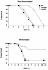
Survival rates of nonimmunized (upper panel) and immunized (lower panel) mice infected i.c. with L. monocytogenes WT, Δ_inlAB2_, and Δ_plcB2_. Nonimmunized mice infected i.c. with Δ_plcB2_ survived significantly longer than mice infected with WT or Δ_inlAB2_ (P < 0.05). Immunized mice infected i.c. with Δ_plcB2_ had a significantly increased survival rate compared to that of mice infected with WT or Δ_inlAB2 L. monocytogenes_ (P < 0.05). Data represent survival rates of 10 mice per experimental group. The results of one of two experiments which gave comparable results are shown.
FIG. 2
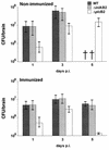
Parasitic load of nonimmunized (upper panel) and immunized (lower panel) mice i.c. infected with L. monocytogenes WT, Δ_inlAB2_, and Δ_plcB2_. At each time point after infection, the i.c. bacterial load of mice infected with Δ_plcB2_ was significantly lower than that of mice infected with WT or Δ_inlAB2_ (P < 0.05). †, mice were already deceased. Five mice per group were analyzed, and the median ± the highest and lowest values of each group are shown.
FIG. 3
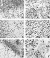
CNS pathology of nonimmunized mice i.c. infected with L. monocytogenes WT (a and b), Δ_inlAB2_ (c and d), and Δ_plcB2_ (e and f) at day 3 p.i. (a) Severe empyema of the ventricle (area above the arrows). The ependymal wall is largely destroyed and barely discernible, and the periventricular brain stem is infiltrated by numerous neutrophils and macrophages. (b) L. monocytogenes WT cluster in the largely destroyed plexus. The arrow points to a group of remarkably elongated WT bacteria. (c) Δ_inlAB2_ has also largely destroyed the ependyma of the ventricular wall (area left of the arrowheads) and has invaded the adjacent brain parenchyma. Brain stem neurons are surrounded by inflammatory leukocytes (arrow). (d) Δ_inlAB2_ cluster in the partially necrotic choroid plexus. Note that the elongated shape of the Δ_inlAB2_ strain (arrow) is identical to that of the WT in panel b. (e) Δ_plcB2_ has also infected the fourth ventricle, and the bacteria are accompanied by intraventricular neutrophils and macrophages. In contrast to WT (a) and Δ_inlAB2_ (c), the ependymal lining is largely intact (arrows). Very few bacteria are detectable in the periventricular tissue (arrowhead). (f) Some Δ_plcB2_ are detectable either as single or small groups of coccoid bacteria (arrowheads) in the choroid plexus. The choroid plexus is infiltrated by neutrophils and macrophages, but in contrast to the images shown in panels b and d, its structure is largely preserved. Specimens in panels a to f were stained with cresyl violet. Magnification is as follows: for panels a and e, ×260; b and d, ×1,250; c, ×520; f, ×1,470.
FIG. 4
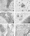
CNS pathology of immunized mice i.c. infected with L. monocytogenes WT (a and b), Δ_inlAB2_ (c and d), and Δ_plcB2_ (e and f) at days 3 (a, c, and e) and 5 (b, d, and f) p.i. (a) Inflammation of the lateral ventricle (V) in a mouse infected with L. monocytogenes WT. Additional infiltrates are present in the adjacent brain parenchyma. (b) Small, circumscribed infiltrates are present in the brain stem in the vicinity of neurons. (c) In Δ_inlAB2_-infected mice, inflammation is also largely confined to the lateral ventricle, and small parts of the choroid plexus are preserved (asterisk). A significant part of the ependyma is still intact (arrows). (d) From days 3 to 5 p.i., disease progressed in Δ_inlAB2_-infected mice. Purulent ventriculitis and focal brain stem encephalitis (arrows) developed. (e) Only discrete infiltrates were detectable in the largely normal fourth ventricle in a Δ_plcB2_-infected mouse at day 3 p.i. The choroid plexus was largely preserved (arrowhead), and ventricular empyema was absent. The ependyma was only focally destroyed, and small, single infiltrates were present in the periventricular tissue (arrow). (f) At day 5 p.i., the inflammation was largely resolved, and only small residual infiltrates were present (arrow) in the wall of the fourth ventricle. Specimens in panels a to f were stained with cresyl violet. Magnification, ×260.
FIG. 5
CNS complications in L. monocytogenes meningoencephalitis at day 10 p.i. (a and b) A severe obstructive hydrocephalus developed at day 10 p.i. Note the massive enlargement of the third ventricle (a) compared to the normal size of the third ventricle of an immunized mouse i.c. infected with Δ_plcB2_ at day 1 p.i. (b). (c) Postinflammatory scarring of the ventricular wall of the fourth ventricle. Subependymal formation of a membranous gliotic tissue (large arrow). In this area, the ependyma is largely intact (arrowheads). In addition, some Purkinje cells in the adjacent cerebellum have small, pyknotic nuclei with condensed chromatin (small arrows), which is characteristic of apoptotic cells. (d) Apoptotic, TUNEL-positive neurons with small, pyknotic nuclei in the CA1 segment of the hippocampus (large arrow). Note the relatively sharp demarcation from the adjacent hippocampal segments (small arrows). Specimens in panels a to c were stained with cresyl violet, and panel d shows TUNEL staining. Magnification for panels a to d, ×260. Images shown in panels a, c, and d are from immunized mice i.c. infected with Δ_plcB2_ at day 10 p.i. Similar observations were made about the brains of surviving mice infected with L. monocytogenes WT and Δ_inlAB2_ at day 10 p.i.
Similar articles
- Listeria pathogenesis and molecular virulence determinants.
Vázquez-Boland JA, Kuhn M, Berche P, Chakraborty T, Domínguez-Bernal G, Goebel W, González-Zorn B, Wehland J, Kreft J. Vázquez-Boland JA, et al. Clin Microbiol Rev. 2001 Jul;14(3):584-640. doi: 10.1128/CMR.14.3.584-640.2001. Clin Microbiol Rev. 2001. PMID: 11432815 Free PMC article. Review. - Directed evolution and targeted mutagenesis to murinize Listeria monocytogenes internalin A for enhanced infectivity in the murine oral infection model.
Monk IR, Casey PG, Hill C, Gahan CG. Monk IR, et al. BMC Microbiol. 2010 Dec 13;10:318. doi: 10.1186/1471-2180-10-318. BMC Microbiol. 2010. PMID: 21144051 Free PMC article. - Relative Roles of Listeriolysin O, InlA, and InlB in Listeria monocytogenes Uptake by Host Cells.
Phelps CC, Vadia S, Arnett E, Tan Y, Zhang X, Pathak-Sharma S, Gavrilin MA, Seveau S. Phelps CC, et al. Infect Immun. 2018 Sep 21;86(10):e00555-18. doi: 10.1128/IAI.00555-18. Print 2018 Oct. Infect Immun. 2018. PMID: 30061379 Free PMC article. - Influence of internalin A murinisation on host resistance to orally acquired listeriosis in mice.
Bergmann S, Beard PM, Pasche B, Lienenklaus S, Weiss S, Gahan CG, Schughart K, Lengeling A. Bergmann S, et al. BMC Microbiol. 2013 Apr 23;13:90. doi: 10.1186/1471-2180-13-90. BMC Microbiol. 2013. PMID: 23617550 Free PMC article. - Role of internalin proteins in the pathogenesis of Listeria monocytogenes.
Ireton K, Mortuza R, Gyanwali GC, Gianfelice A, Hussain M. Ireton K, et al. Mol Microbiol. 2021 Dec;116(6):1407-1419. doi: 10.1111/mmi.14836. Epub 2021 Nov 7. Mol Microbiol. 2021. PMID: 34704304 Review.
Cited by
- Biological effects of listeriolysin O: implications for vaccination.
Hernández-Flores KG, Vivanco-Cid H. Hernández-Flores KG, et al. Biomed Res Int. 2015;2015:360741. doi: 10.1155/2015/360741. Epub 2015 Mar 22. Biomed Res Int. 2015. PMID: 25874208 Free PMC article. Review. - Listeria pathogenesis and molecular virulence determinants.
Vázquez-Boland JA, Kuhn M, Berche P, Chakraborty T, Domínguez-Bernal G, Goebel W, González-Zorn B, Wehland J, Kreft J. Vázquez-Boland JA, et al. Clin Microbiol Rev. 2001 Jul;14(3):584-640. doi: 10.1128/CMR.14.3.584-640.2001. Clin Microbiol Rev. 2001. PMID: 11432815 Free PMC article. Review. - Granzymes drive a rapid listeriolysin O-induced T cell apoptosis.
Carrero JA, Vivanco-Cid H, Unanue ER. Carrero JA, et al. J Immunol. 2008 Jul 15;181(2):1365-74. doi: 10.4049/jimmunol.181.2.1365. J Immunol. 2008. PMID: 18606691 Free PMC article. - Contribution of membrane-damaging toxins to Bacillus endophthalmitis pathogenesis.
Callegan MC, Cochran DC, Kane ST, Gilmore MS, Gominet M, Lereclus D. Callegan MC, et al. Infect Immun. 2002 Oct;70(10):5381-9. doi: 10.1128/IAI.70.10.5381-5389.2002. Infect Immun. 2002. PMID: 12228262 Free PMC article. - Mycobacterium marinum Degrades Both Triacylglycerols and Phospholipids from Its Dictyostelium Host to Synthesise Its Own Triacylglycerols and Generate Lipid Inclusions.
Barisch C, Soldati T. Barisch C, et al. PLoS Pathog. 2017 Jan 19;13(1):e1006095. doi: 10.1371/journal.ppat.1006095. eCollection 2017 Jan. PLoS Pathog. 2017. PMID: 28103313 Free PMC article.
References
- Berche P. Bacteremia is required for invasion of the murine central nervous system by Listeria monocytogenes. Microb Pathog. 1995;18:323–336. - PubMed
- Bogdan I, Leib S L, Bergeron M, Chow L, Täubner M G. Tumor necrosis factor-alpha contributes to apoptosis in hippocampal neurons during experimental group B streptococcal meningitis. J Infect Dis. 1997;176:693–697. - PubMed
- Braun L, Dramsi S, Dehoux P, Bierne H, Lindahl G, Cossart P. InlB: an invasion protein of Listeria monocytogenes with a novel type of surface association. Mol Microbiol. 1997;25:285–294. - PubMed
- Chakraborty T, Wehland J. The host cell infected with Listeria monocytogenes. In: Kaufmann S H E, editor. Host response to intracellular pathogens. R. G. Austin, Tex: Landes Company; 1997. pp. 271–290.
- Domann E, Zechel S, Lingnau A, Hain T, Darji A, Nichterlein T, Wehland J, Chakraborty T. Identification and characterization of a novel PrfA-regulated gene in Listeria monocytogenes whose product, IrpA, is highly homologous to internalin proteins, which contain leucine-rich repeats. Infect Immun. 1997;65:101–109. - PMC - PubMed
Publication types
MeSH terms
Substances
LinkOut - more resources
Full Text Sources
Other Literature Sources


