Chronic experimental colitis induced by dextran sulphate sodium (DSS) is characterized by Th1 and Th2 cytokines - PubMed (original) (raw)
Chronic experimental colitis induced by dextran sulphate sodium (DSS) is characterized by Th1 and Th2 cytokines
L A Dieleman et al. Clin Exp Immunol. 1998 Dec.
Abstract
Oral administration of DSS has been reported to induce an acute and chronic colitis in mice. The aim of our study was to evaluate if the chronic phase of DSS-induced colitis was characterized by a Th1/Th2 response and how this would relate to mucosal regeneration. Swiss Webster mice were fed 5% DSS in their drinking water for 7 days, followed by 2-5 weeks consumption of water. Control mice received only water. The animals were killed at 3 and 6 weeks after induction. Their colons were isolated for histology and immunohistochemistry, using specific MoAbs for T and B cells, macrophages, interferon-gamma (IFN-gamma), IL-4 and IL-5. Colons were scored for inflammation, damage and regeneration. Two weeks after stopping DSS the colonic epithelium had only partially healed. Total colitis scores were still increased, especially in the distal colon, which was due to more inflammation, damage and less regeneration. In areas of incomplete colonic healing the basal parts of the lamina propria contained macrophages and CD4+ T cells. These CD4+ T cells showed a focal increase of IFN-gamma and IL-4 staining compared with control animals. These findings were still observed 5 weeks after stopping DSS in some mice, albeit less extensive. Chronic DSS-induced colitis is characterized by focal epithelial regeneration and a Th1 as well as Th2 cytokine profile. We postulate that chronic immune activation mediated by both populations of Th cells can interfere with colonic healing and can play a role in the pathogenesis of chronic colitis.
Figures
Fig. 1
Histological grading of colitis. (a) Sum of the scores of b, c, and d. (b) Inflammation score. (c) Extent of inflammation. (d) Crypt damage/regeneration score, as indicated in Table 1. Hatched bars represent mice at day 7 of DSS feeding, cross-hatched bars represent mice killed 14 days after stopping DSS, open bars depict mice 35 days after stopping DSS.
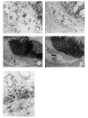
Fig. 2
(A) DSS 7 days. Multiple MOMA-2+ macrophages in submocosal oedema. (B) Two weeks after stopping DSS, the submucosa still contains some MOMA-2+ macrophages. Large lymphoid aggregates in the lamina propria, consisting mainly of B cells (C), but also T cells in the periphery (D), are shown. (E) Two weeks after stopping DSS, below an incompletely regenerated epithelium the base of the lamina propria contains bands of MT4+ cells.
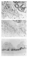
Fig. 3
Colonic tissue 2 weeks after stopping DSS (same area as in Fig. 2E). (A) IFN-γ-producing cells (arrows). (B) IL-5-producing cells are not present. (C) IL-4-producing cells at the base of the lamina propria.
Fig. 4
IFN-γ production in organ cultures from colons of indicated time points during DSS-induced colitis and control animals; solid bars represent healthy animals, hatched bars represent mice at day 7 of DSS feeding, cross-hatched bars represent mice at day 14 after stopping DSS. *Significantly different compared with healthy animals, P < 0.05.
Fig. 5
Number of IFN-γ-, IL-5- and IL-4-positive cells in 5–10 representative areas per spleen section, expressed as number of cells/mm2; solid bars represent healthy animals, hatched bars show mice at day 7 of DSS feeding, cross-hatched bars represent mice at day 14 after stopping DSS, open bars indicate mice at day 35 after stopping DSS. *Significantly different compared with healthy controls, P < 0.05.
Similar articles
- Oral administration of bovine milk from cows hyperimmunized with intestinal bacterin stimulates lamina propria T lymphocytes to produce Th1-biased cytokines in mice.
Wang Y, Lin L, Yin C, Othtani S, Aoyama K, Lu C, Sun X, Yoshikai Y. Wang Y, et al. Int J Mol Sci. 2014 Mar 28;15(4):5458-71. doi: 10.3390/ijms15045458. Int J Mol Sci. 2014. PMID: 24686517 Free PMC article. - Interleukin-19 protects mice from innate-mediated colonic inflammation.
Azuma YT, Matsuo Y, Kuwamura M, Yancopoulos GD, Valenzuela DM, Murphy AJ, Nakajima H, Karow M, Takeuchi T. Azuma YT, et al. Inflamm Bowel Dis. 2010 Jun;16(6):1017-28. doi: 10.1002/ibd.21151. Inflamm Bowel Dis. 2010. PMID: 19834971 - IL-33 attenuates development and perpetuation of chronic intestinal inflammation.
Groβ P, Doser K, Falk W, Obermeier F, Hofmann C. Groβ P, et al. Inflamm Bowel Dis. 2012 Oct;18(10):1900-9. doi: 10.1002/ibd.22900. Epub 2012 Apr 16. Inflamm Bowel Dis. 2012. PMID: 22508383 - IL-33 Aggravates DSS-Induced Acute Colitis in Mouse Colon Lamina Propria by Enhancing Th2 Cell Responses.
Zhu J, Yang F, Sang L, Zhai J, Zhang X, Yue D, Li S, Li Y, Lu C, Sun X. Zhu J, et al. Mediators Inflamm. 2015;2015:913041. doi: 10.1155/2015/913041. Epub 2015 May 28. Mediators Inflamm. 2015. PMID: 26161006 Free PMC article. - Cytokines in experimental colitis.
Garside P. Garside P. Clin Exp Immunol. 1999 Dec;118(3):337-9. doi: 10.1046/j.1365-2249.1999.01088.x. Clin Exp Immunol. 1999. PMID: 10594548 Free PMC article. Review. No abstract available.
Cited by
- The bone-liver interaction modulates immune and hematopoietic function through Pinch-Cxcl12-Mbl2 pathway.
He T, Zhou B, Sun G, Yan Q, Lin S, Ma G, Yao Q, Wu X, Zhong Y, Gan D, Huo S, Jin W, Chen D, Bai X, Cheng T, Cao H, Xiao G. He T, et al. Cell Death Differ. 2024 Jan;31(1):90-105. doi: 10.1038/s41418-023-01243-9. Epub 2023 Dec 7. Cell Death Differ. 2024. PMID: 38062244 Free PMC article. - MyD88 mediates the protective effects of probiotics against the arteriolar thrombosis and leukocyte recruitment associated with experimental colitis.
Souza DG, Senchenkova EY, Russell J, Granger DN. Souza DG, et al. Inflamm Bowel Dis. 2015 Apr;21(4):888-900. doi: 10.1097/MIB.0000000000000331. Inflamm Bowel Dis. 2015. PMID: 25738377 Free PMC article. - Osteopontin: participation in inflammation or mucosal protection in inflammatory bowel diseases?
Chen F, Liu H, Shen Q, Yuan S, Xu L, Cai X, Lian J, Chen SY. Chen F, et al. Dig Dis Sci. 2013 Jun;58(6):1569-80. doi: 10.1007/s10620-012-2556-y. Epub 2013 Jan 30. Dig Dis Sci. 2013. PMID: 23361573 - Dextran sulfate sodium leads to chronic colitis and pathological angiogenesis in Endoglin heterozygous mice.
Jerkic M, Peter M, Ardelean D, Fine M, Konerding MA, Letarte M. Jerkic M, et al. Inflamm Bowel Dis. 2010 Nov;16(11):1859-70. doi: 10.1002/ibd.21288. Inflamm Bowel Dis. 2010. PMID: 20848471 Free PMC article. - Effects of kefir fermented milk beverage on sodium dextran sulfate (DSS)-induced colitis in rats.
Nascimento da Silva K, Fávero AG, Ribeiro W, Ferreira CM, Sartorelli P, Cardili L, Bogsan CS, Bertaglia Pereira JN, de Cássia Sinigaglia R, Cristina de Moraes Malinverni A, Ribeiro Paiotti AP, Miszputen SJ, Ambrogini-Júnior O. Nascimento da Silva K, et al. Heliyon. 2022 Dec 29;9(1):e12707. doi: 10.1016/j.heliyon.2022.e12707. eCollection 2023 Jan. Heliyon. 2022. PMID: 36685418 Free PMC article.
References
- Okayasu I, Hatakeyama S, Ohkusa T, et al. A novel method in the induction of reliable experimental acute and chronic ulcerative colitis in mice. Gastroenterology. 1990;98:694–702. - PubMed
- Cooper H, Murthy SNS, Shah RS, et al. Clinicopathologic study of dextran sulfate sodium experimental murine colitis. Lab Invest. 1993;69:238–49. - PubMed
- Dieleman LA, Ridwan BU, Tennyson GS, et al. Dextran sulfate sodium (DSS)-induced colitis occurs in severe combined immunodeficient (SCID) mice. Gastroenterology. 1994;107:1643–52. - PubMed
- Graham RC, Karnovsky MC. The early stages of absorption of injected horseradish peroxidase in the proximal tubules of mouse kidney: ultrastructural cytochemistry by a new technique. J Histochem Cytochem. 1966;14:291–302. - PubMed
- Kraal G, Rep M, Janse M. Macrophages in T and B cell compartments and other tissue macrophages recognized by monoclonal antibody MOMA-2. Scan J Immunol. 1987;26:653–61. - PubMed
MeSH terms
Substances
LinkOut - more resources
Full Text Sources
Other Literature Sources
Research Materials


