Restricted high level expression of Tcf-4 protein in intestinal and mammary gland epithelium - PubMed (original) (raw)
Restricted high level expression of Tcf-4 protein in intestinal and mammary gland epithelium
N Barker et al. Am J Pathol. 1999 Jan.
Abstract
Tcf-4 is a member of the Tcf/Lef family of transcription factors that interact functionally with beta-catenin to mediate Wnt signaling in vertebrates. We have previously demonstrated that the tumor suppressor function of APC in the small intestine is mediated via regulation of Tcf-4/beta-catenin transcriptional activity. To gain further insight into the role of Tcf-4 in development and carcinogenesis we have generated several mouse monoclonal antibodies, one of which is specific for Tcf-4 and another of which recognizes both Tcf-3 and Tcf-4. Immunohistochemistry performed with the Tcf 4- specific monoclonal antibody revealed high levels of expression in normal intestinal and mammary epithelium and carcinomas derived therefrom. Additional sites of Tcf-3 expression, as revealed by staining with the Tcf-3/-4 antibody, occurred only within the stomach epithelium, hair follicles, and keratinocytes of the skin. A temporal Tcf-4 expression gradient was observed along the crypt-villus axis of human small intestinal epithelium: strong Tcf-4 expression was present within the crypts of early (week 16) human fetal small intestine, with the villi showing barely detectable Tcf-4 protein levels. Tcf-4 expression levels increased dramatically on the villi of more highly developed (week 22) fetal small intestine. We conclude that Tcf-4 exhibits a highly restricted expression pattern related to the developmental stage of the intestinal epithelium. The high levels of Tcf-4 expression in mammary epithelium and mammary carcinomas may also indicate a role in the development of this tissue and breast carcinoma.
Figures
Figure 1.
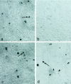
Immunohistochemical staining of hTcf-4 and mTcf-3 in COS cells using the 6H5 and 6F12 mAbs. a: Staining of COS cells expressing hTcf-4 (arrows) by the 6H5 mAb. b: Absence of staining on COS cells expressing mTcf-3 by the 6H5 mAb. Magnification, ×200. c,d: Staining of COS cells expressing hTcf-4 or mTcf-3 (arrows) by the 6F12 mAb. Magnification, ×200.
Figure 2.
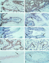
Immunohistochemical analysis of Tcf-4 and Tcf-3 expression in human and mouse tissues. a-h: Immunohistochemical stainings generated using the Tcf-4 specific mAb (6H5). i-j: Immunohistochemical stainings generated using the Tcf-3/−4 mAb (6F12). a,b: Tcf-4 is highly expressed at the tops of the crypts (arrows) of human adult colonic epithelium (a) and human colon carcinoma (b). Magnification, ×150. c,d: Tcf-4 is expressed at high levels in the fibrous tissue (open arrows) and crypts (shaded arrows) of human (c) and mouse (d) adult small intestinal epithelium. Magnification, ×100. e,f: Tcf-4 expression increases along the crypt-villus axis (arrows) during week 16 (e) and week 22 (f) of the development of human small intestinal epithelium. Magnification, ×50. g,h: Tcf-4 is expressed at high levels in the epithelium (shaded arrows) and fibrous tissue (open arrows) of human mammary gland (g) and mammary carcinoma (h). Magnification, ×100. i-j: Tcf-3 is expressed in hair follicles (i) and keratinocytes of skin (j). Magnification, ×100.
Figure 3.
a: Gel retardation of Tcf-4 complexes from HT-29 colon carcinoma cells. Tcf-4 complexed to an optimal Tcf binding site probe can be supershifted by addition of the Tcf-4-specific mAb 6H5. Lane 1: Nonspecific (NS) bands generated using a probe comprising a disrupted Tcf binding site. Lane 2 : Specific Tcf-4/probe complex. Lane 3: Supershift of the Tcf-4/probe complex by addition of 6H5 mAb. Lane 4: Supershift of the Tcf-4/probe complex on addition of 6F12 mAb. Lanes 5 and 6: No supershift of the Tcf-4/probe complex induced on addition of control antibodies (anti-APC and anti-Plakoglobin). b: Co-immunoprecipitation of Tcf-4 and β-catenin from SW620 nuclear extracts. Lane 1: A 92-kd band (arrow) visualized by Western blot using an anti-β-catenin mAb after Tcf-4 immunoprecipitation from SW620 nuclear extracts using the 6H5 mAb. H, antibody heavy chain; L, antibody light chain. Lane 2: Western blot analysis of total β-catenin (arrow) in SW620 total cell extracts.
Similar articles
- Sox17 and Sox4 differentially regulate beta-catenin/T-cell factor activity and proliferation of colon carcinoma cells.
Sinner D, Kordich JJ, Spence JR, Opoka R, Rankin S, Lin SC, Jonatan D, Zorn AM, Wells JM. Sinner D, et al. Mol Cell Biol. 2007 Nov;27(22):7802-15. doi: 10.1128/MCB.02179-06. Epub 2007 Sep 17. Mol Cell Biol. 2007. PMID: 17875931 Free PMC article. - Two members of the Tcf family implicated in Wnt/beta-catenin signaling during embryogenesis in the mouse.
Korinek V, Barker N, Willert K, Molenaar M, Roose J, Wagenaar G, Markman M, Lamers W, Destree O, Clevers H. Korinek V, et al. Mol Cell Biol. 1998 Mar;18(3):1248-56. doi: 10.1128/MCB.18.3.1248. Mol Cell Biol. 1998. PMID: 9488439 Free PMC article. - SOX9 is an intestine crypt transcription factor, is regulated by the Wnt pathway, and represses the CDX2 and MUC2 genes.
Blache P, van de Wetering M, Duluc I, Domon C, Berta P, Freund JN, Clevers H, Jay P. Blache P, et al. J Cell Biol. 2004 Jul 5;166(1):37-47. doi: 10.1083/jcb.200311021. J Cell Biol. 2004. PMID: 15240568 Free PMC article. - Dynamic expression of Lef/Tcf family members and beta-catenin during chick gastrulation, neurulation, and early limb development.
Schmidt M, Patterson M, Farrell E, Münsterberg A. Schmidt M, et al. Dev Dyn. 2004 Mar;229(3):703-7. doi: 10.1002/dvdy.20010. Dev Dyn. 2004. PMID: 14991726 - Mammary gland development requires syndecan-1 to create a beta-catenin/TCF-responsive mammary epithelial subpopulation.
Liu BY, Kim YC, Leatherberry V, Cowin P, Alexander CM. Liu BY, et al. Oncogene. 2003 Dec 18;22(58):9243-53. doi: 10.1038/sj.onc.1207217. Oncogene. 2003. PMID: 14681683
Cited by
- CFTR Cooperative _Cis_-Regulatory Elements in Intestinal Cells.
Collobert M, Bocher O, Le Nabec A, Génin E, Férec C, Moisan S. Collobert M, et al. Int J Mol Sci. 2021 Mar 5;22(5):2599. doi: 10.3390/ijms22052599. Int J Mol Sci. 2021. PMID: 33807548 Free PMC article. - Single-Cell Sequencing of Developing Human Gut Reveals Transcriptional Links to Childhood Crohn's Disease.
Elmentaite R, Ross ADB, Roberts K, James KR, Ortmann D, Gomes T, Nayak K, Tuck L, Pritchard S, Bayraktar OA, Heuschkel R, Vallier L, Teichmann SA, Zilbauer M. Elmentaite R, et al. Dev Cell. 2020 Dec 21;55(6):771-783.e5. doi: 10.1016/j.devcel.2020.11.010. Epub 2020 Dec 7. Dev Cell. 2020. PMID: 33290721 Free PMC article. - Regulation of epithelial-mesenchymal transition and organoid morphogenesis by a novel TGFβ-TCF7L2 isoform-specific signaling pathway.
Karve K, Netherton S, Deng L, Bonni A, Bonni S. Karve K, et al. Cell Death Dis. 2020 Aug 25;11(8):704. doi: 10.1038/s41419-020-02905-z. Cell Death Dis. 2020. PMID: 32843642 Free PMC article. - Roles of β-catenin, TCF-4, and survivin in nasopharyngeal carcinoma: correlation with clinicopathological features and prognostic significance.
Jin PY, Zheng ZH, Lu HJ, Yan J, Zheng GH, Zheng YL, Wu DM, Lu J. Jin PY, et al. Cancer Cell Int. 2019 Feb 28;19:48. doi: 10.1186/s12935-019-0764-7. eCollection 2019. Cancer Cell Int. 2019. PMID: 30867651 Free PMC article. - In Vitro Polarization of Colonoids to Create an Intestinal Stem Cell Compartment.
Attayek PJ, Ahmad AA, Wang Y, Williamson I, Sims CE, Magness ST, Allbritton NL. Attayek PJ, et al. PLoS One. 2016 Apr 21;11(4):e0153795. doi: 10.1371/journal.pone.0153795. eCollection 2016. PLoS One. 2016. PMID: 27100890 Free PMC article.
References
- Molenaar M, van de Wetering M, Oosterwegel M, Peterson-Maduro J, Godsave S, Korinek V, Roose J, Destree O, Clevers H: Xtcf-3 transcription factor mediates β-catenin-induced axis formation in Xenopus embryos. Cell 1996, 86:391-399 - PubMed
- Riese J, Yu X, Munnerlyn A, Eresh S, Hsu SC, Grosschedl R, Bienz M: LEF-1, a nuclear factor coordinating signaling inputs from wingless and decapentaplegic. Cell 1997, 88:777-787 - PubMed
- Brunner E, Peter O, Schweizer L, Basler K: Pangolin encodes a Lef-1 homologue that acts downstream of Armadillo to transduce the Wingless signal in Drosophila. Nature 1997, 385:829-833 - PubMed
- van de Wetering M, Cavallo R, Dooijes D, van Beest M, van Es J, Loureiro A, Ypma D, Hursh D, Jones T, Bejsovec A, Peifer M, Mortin M, Clevers H: Armadillo co-activates transcription driven by the product of the Drosophila segment polarity gene dTCF. Cell 1997, 88:789-799 - PubMed
- Behrens J, van Kries JP, Kuehl M, Bruhn D, Wedlich R, Grosschedl R, Birchmeier W: Functional interaction of β-catenin with the transcription factor LEF-1. Nature 1996, 382:638-642 - PubMed
MeSH terms
Substances
LinkOut - more resources
Full Text Sources
Molecular Biology Databases
Miscellaneous


