Enhancing versus suppressive effects of stress hormones on skin immune function - PubMed (original) (raw)
Enhancing versus suppressive effects of stress hormones on skin immune function
F S Dhabhar et al. Proc Natl Acad Sci U S A. 1999.
Abstract
Delayed-type hypersensitivity (DTH) reactions are antigen-specific cell-mediated immune responses that, depending on the antigen, mediate beneficial (e.g., resistance to viruses, bacteria, and fungi) or harmful (e.g., allergic dermatitis and autoimmunity) aspects of immune function. Contrary to the idea that stress suppresses immunity, we have reported that short-duration stressors significantly enhance skin DTH and that a stress-induced trafficking of leukocytes to the skin may mediate this immunoenhancement. Here, we identify the hormonal mediators of a stress-induced enhancement of skin immunity. Adrenalectomy, which eliminates the glucocorticoid and epinephrine stress response, eliminated the stress-induced enhancement of skin DTH. Low-dose corticosterone or epinephrine administration significantly enhanced skin DTH and produced a significant increase in the number of T cells in lymph nodes draining the site of the DTH reaction. In contrast, high-dose corticosterone, chronic corticosterone, or low-dose dexamethasone administration significantly suppressed skin DTH. These results suggest a role for adrenal stress hormones as endogenous immunoenhancing agents. These results also show that hormones released during an acute stress response may help prepare the immune system for potential challenges (e.g., wounding or infection) for which stress perception by the brain may serve as an early warning signal.
Figures
Figure 1
Adrenalectomy eliminates a stress-induced enhancement of skin DTH. A 6-day time course of changes in thickness of right pinnae of previously sensitized animals challenged with DNFB is shown. Stressed intact (A) and sham (B) animals showed a significant increase in the DTH response compared with unstressed animals. (C) ADX animals did not show a stress-induced increase skin DTH. Data are expressed as means ± SEM (n = 6 per treatment group). Statistically significant differences are indicated. (∗, P < 0.05; ∗∗, P < 0.005, independent t test.)
Figure 2
Acute administration of corticosterone enhances skin DTH. A time course of changes in the thickness of right pinnae of previously sensitized animals challenged with DNFB is shown. Corticosterone (CORT, 5 mg/kg) was administered i.p. to ADX animals (A) or through drinking water (100 or 400 μg/ml) to intact animals (B). Control animals were treated with vehicle: 30% HBC (A) or 0.6% ethanol (B). Corticosterone-treated animals showed a significantly larger DTH response than vehicle-treated animals. (∗, P < 0.05, independent t test.)
Figure 3
Pharmacologic treatment with glucocorticoid hormones suppresses the skin DTH response. A time course of changes in the thickness of right pinnae of previously sensitized animals challenged with DNFB is shown. (A) Corticosterone (CORT, 40 mg/kg) or dexamethasone (DEX, 0.1 mg/kg) was administered acutely. (B) Corticosterone was also administered chronically in drinking water (dw CORT, 400 μg/ml, 6 days). Control animals were treated with vehicle: 30% HBC (A) or 0.6% ethanol (B). Corticosterone- and dexamethasone-treated animals showed lower DTH responses than control animals. (∗, P < 0.05; ∗∗, P < 0.005, independent t test.)
Figure 4
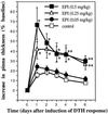
Acute administration of epinephrine enhances skin DTH. A time course of changes in the thickness of right pinnae of previously sensitized animals challenged with DNFB is shown. Epinephrine (EPI, 0.05, 0.25, or 0.5 mg/kg) was administered acutely to ADX animals. Control animals were treated with vehicle (deionized-distilled H2O). Epinephrine-treated animals showed a dose-dependent increase in skin DTH. (∗, P < 0.05; ∗∗, P < 0.005, independent t test.)
Figure 5
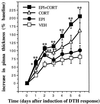
Epinephrine and corticosterone additively enhance skin DTH. A time course of changes in the thickness of right pinnae of previously sensitized animals challenged with OXA is shown. Epinephrine (EPI, 0.5 mg/kg), corticosterone (CORT, 5 mg/kg) or epinephrine plus corticosterone (EPI+CORT, 0.5 mg/kg plus 5 mg/kg, respectively) were administered acutely to ADX animals. Control animals were treated with vehicle (VEH, 30% HBC). Epinephrine- or corticosterone-treated animals showed an enhanced DTH response. Moreover, simultaneous administration of the two hormones resulted in an additive enhancement of skin DTH. (∗, P < 0.05; ∗∗, P < 0.005, independent t test.)
Figure 6
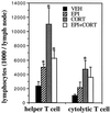
Acute administration of stress hormones increases the cellularity of cervical lymph nodes that drain the site of the skin DTH reaction. Epinephrine (EPI, 0.5 mg/kg), corticosterone (CORT, 5 mg/kg), or epinephrine plus corticosterone (EPI+CORT, 0.5 mg/kg plus 5 mg/kg, respectively) were administered to ADX animals. Control animals were treated with vehicle (VEH, 30% HBC). Lymph nodes were collected, and lymphocytes were isolated 48 h after the induction of DTH. Compared with vehicle-treated animals, hormone-treated animals showed higher T lymphocyte numbers in cervical lymph nodes that drain the site of the DTH reaction. (∗, P < 0.05, independent t test.)
Similar articles
- Acute stress enhances while chronic stress suppresses skin immunity. The role of stress hormones and leukocyte trafficking.
Dhabhar FS. Dhabhar FS. Ann N Y Acad Sci. 2000;917:876-93. doi: 10.1111/j.1749-6632.2000.tb05454.x. Ann N Y Acad Sci. 2000. PMID: 11268419 Review. - Stress, leukocyte trafficking, and the augmentation of skin immune function.
Dhabhar FS. Dhabhar FS. Ann N Y Acad Sci. 2003 May;992:205-17. doi: 10.1111/j.1749-6632.2003.tb03151.x. Ann N Y Acad Sci. 2003. PMID: 12794060 Review. - Acute stress enhances while chronic stress suppresses cell-mediated immunity in vivo: a potential role for leukocyte trafficking.
Dhabhar FS, McEwen BS. Dhabhar FS, et al. Brain Behav Immun. 1997 Dec;11(4):286-306. doi: 10.1006/brbi.1997.0508. Brain Behav Immun. 1997. PMID: 9512816 - Stress-induced augmentation of immune function--the role of stress hormones, leukocyte trafficking, and cytokines.
Dhabhar FS. Dhabhar FS. Brain Behav Immun. 2002 Dec;16(6):785-98. doi: 10.1016/s0889-1591(02)00036-3. Brain Behav Immun. 2002. PMID: 12480507 Review. - Stress-induced enhancement of skin immune function: A role for gamma interferon.
Dhabhar FS, Satoskar AR, Bluethmann H, David JR, McEwen BS. Dhabhar FS, et al. Proc Natl Acad Sci U S A. 2000 Mar 14;97(6):2846-51. doi: 10.1073/pnas.050569397. Proc Natl Acad Sci U S A. 2000. PMID: 10706626 Free PMC article.
Cited by
- Remote-controlled dexamethasone-duration on eye-surface with a micelle-magnetic nanoparticulate co-delivery system for dry eye disease.
Zheng Q, Ge C, Li K, Wang L, Xia X, Liu X, Mehmood R, Shen J, Nan K, Chen W, Lin S. Zheng Q, et al. Acta Pharm Sin B. 2024 Aug;14(8):3730-3745. doi: 10.1016/j.apsb.2024.05.004. Epub 2024 May 10. Acta Pharm Sin B. 2024. PMID: 39220865 Free PMC article. - Prevalence and associated factors of suspected occupational skin diseases among restaurant workers in peninsular Malaysia: secondary data analysis of Registry for Occupational Disease Screening (RODS).
Ahmad Fuad MH, Samsudin EZ, Yasin SM, Ismail N, Mohamad M, Muzaini K, Anuar MR, Govindasamy K, Ismail I, Elias A, Taib KM, Mohamed AS, Azhari Noor AF, Abdullah Hair AF. Ahmad Fuad MH, et al. BMJ Open. 2024 Aug 13;14(8):e079877. doi: 10.1136/bmjopen-2023-079877. BMJ Open. 2024. PMID: 39142678 Free PMC article. - Mechanisms of stress-attributed breast cancer incidence and progression.
Reznik E, Torjani A. Reznik E, et al. Cancer Causes Control. 2024 Nov;35(11):1413-1432. doi: 10.1007/s10552-024-01884-2. Epub 2024 Jul 16. Cancer Causes Control. 2024. PMID: 39012513 Review. - Stress Biomarkers Transferred Into the Female Reproductive Tract by Seminal Plasma Are Associated with ICSI Outcomes.
Nikolaeva M, Arefieva A, Babayan A, Aksenov V, Zhukova A, Kalinina E, Krechetova L, Sukhikh G. Nikolaeva M, et al. Reprod Sci. 2024 Jun;31(6):1732-1746. doi: 10.1007/s43032-024-01486-y. Epub 2024 Feb 23. Reprod Sci. 2024. PMID: 38393625 - The cancer-immune dialogue in the context of stress.
Ma Y, Kroemer G. Ma Y, et al. Nat Rev Immunol. 2024 Apr;24(4):264-281. doi: 10.1038/s41577-023-00949-8. Epub 2023 Oct 13. Nat Rev Immunol. 2024. PMID: 37833492 Review.
References
- Dhabhar F S, McEwen B S. Brain Behav Immun. 1997;11:286–306. - PubMed
- Munck A, Guyre P M, Holbrook N J. Endocr Rev. 1984;5:25–44. - PubMed
- Borysenko M, Borysenko J. Gen Hosp Psychiatry. 1982;4:59–67. - PubMed
- Khansari D N, Murgo A J, Faith R E. Immunol Today. 1990;11:170–175. - PubMed
- Kort W J. Adv Neuroimmunol. 1994;4:1–11. - PubMed
Publication types
MeSH terms
Substances
LinkOut - more resources
Full Text Sources
Other Literature Sources
Medical


