Opposing actions of intact and N-terminal fragments of the human prolactin/growth hormone family members on angiogenesis: an efficient mechanism for the regulation of angiogenesis - PubMed (original) (raw)
Opposing actions of intact and N-terminal fragments of the human prolactin/growth hormone family members on angiogenesis: an efficient mechanism for the regulation of angiogenesis
I Struman et al. Proc Natl Acad Sci U S A. 1999.
Abstract
Angiogenesis, the process of development of a new microvasculature, is regulated by a balance of positive and negative factors. We show both in vivo and in vitro that the members of the human prolactin/growth hormone family, i.e., human prolactin, human growth hormone, human placental lactogen, and human growth hormone variant are angiogenic whereas their respective 16-kDa N-terminal fragments are antiangiogenic. The opposite actions are regulated in part via activation or inhibition of mitogen-activated protein kinase signaling pathway. In addition, the N-terminal fragments stimulate expression of type 1 plasminogen activator inhibitor whereas the intact molecules have no effect, an observation consistent with the fragments acting via separate receptors. The concept that a single molecule encodes both angiogenic and antiangiogenic peptides represents an efficient model for regulating the balance of positive and negative factors controlling angiogenesis. This hypothesis has potential physiological importance for the control of the vascular connection between the fetal and maternal circulations in the placenta, where human prolactin, human placental lactogen, and human growth hormone variant are expressed.
Figures
Figure 1
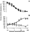
BBCE cell proliferation. Cells were treated with bFGF (1 ng/ml) and the recombinant proteins. The data are expressed as percentages of the stimulation obtained with bFGF alone, 0% being the basal growth level. (A) Inhibition of BBCE cell proliferation. ■, 16K hPRL; ♦, 16K hPL; ●, 16K hGH; ▴, 16K hGH-V. (B) Stimulation of BBCE cell proliferation in presence of bFGF. □, hPRL; ⋄, hPL; ○, hGH; ▵, hGH-V. Each point represents the mean of triplicate wells. The experiments were repeated at least three times, with similar results.
Figure 2
Early-stage CAM assay showing inhibition of angiogenesis in the CAM: Representative examples of CAM are taken from a typical experiment. The disks are visible by light reflection, and the black arrow shows the border of the disk or the border of the avascular area, if present. The lower left panel shows the quantification of the assay performed by measuring the area devoid of capillaries in the region surrounding the disk. Values are means ± SEM. ∗, P < 0.01 vs. BSA.
Figure 3
Late-stage CAM assay showing stimulation of blood vessel formation in the CAM: Representative examples of CAM are taken from a typical experiment. The black arrow shows the border of the disk. The lower left panel shows the quantification of the assay performed by counting the number of blood vessels emerging from the disk. Vessels were scored in function of size from 1 (small) to 3 (large). Values are means ± SEM. ∗, P < 0.05 vs. BSA.
Figure 4
Fragments (16 kDa) inhibit bFGF-dependent MAPK tyrosine phosphorylation and activity whereas full length hormones stimulate these processes in the absence of bFGF. BBCE cells were treated for 5 min with the purified 16-kDa fragments (10 nM) and bFGF (250 pM) or for 10 min with the intact hormones (10 nM) but without bFGF or were left untreated for control. (A) P-Tyr, Western blots with the antityrosine antibody at the level of the MAPK p42 and p44; MAPK, the Western blot shown in the P-Tyr panel, stripped and reprobed with an anti-MAPK antibody. (B)In-gel MAPK activity. Numbers at the bottom of the histograms represent the number of experiments performed.
Figure 5
Stimulation of PAI-1 expression in BBCE cells by the 16-kDa fragments. Mouse antibovine PAI-1 Western blotting were performed on extracts of BBCE cells prestimulated by 20 nM of the recombinant proteins or were left untreated for control. (A) (Upper) Fragments (16 kDa) stimulate PAI-1 expression; full length hormones do not. (Lower) Quantification. (B) Competition between hGH (22K hGH) and 16K hGH. BBCE cells were treated with 20 nM 16K hGH (16K hGH lanes), with 20 nM hGH (22K hGH lanes), or with 20 nM both 16K hGH and hGH (16K hGH plus 22K hGH lanes) or were untreated (control lanes).
Figure 6
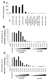
Immunodepletion experiments. PAI-1 levels were stimulated by the addition of 16-kDa fragments. The stimulation was inhibited after immunoneutralization of the 16-kDa fragments with their respective antisera. PAI-1 accumulation in the BBCE cell medium was measured by a mouse antibovine PAI-1 ELISA. (A) BBCE cells were treated with 50 nM of the indicated 16-kDa fragment in the presence or absence of its respective antiserum. (B) Stimulation of PAI-1 levels with 20 nM 16K hPRL was inhibited in a dose-dependent manner by the addition of increasing amounts of anti-hPRL antiserum. (C) Similarly, stimulation of PAI-1 levels by 20 nM 16K hGH-V was inhibited in a graded fashion by the anti-hGH antiserum. The data (in A) are expressed as optical density (O.D.) per microgram of cell protein, 0 being the level obtained with DMEM. The PAI-1 levels (in B and_C_) are expressed as the percentage of the stimulation obtained with the 16-kDa fragment alone, 0% being the level obtained with DMEM. Numbers at the bottom of the histogram represent the antiserum dilution in the well. Data represent the mean of duplicate measures (in A_–_C).
Similar articles
- Roles of prolactin and related members of the prolactin/growth hormone/placental lactogen family in angiogenesis.
Corbacho AM, Martínez De La Escalera G, Clapp C. Corbacho AM, et al. J Endocrinol. 2002 May;173(2):219-38. doi: 10.1677/joe.0.1730219. J Endocrinol. 2002. PMID: 12010630 Review. - The 16-kilodalton N-terminal fragment of human prolactin is a potent inhibitor of angiogenesis.
Clapp C, Martial JA, Guzman RC, Rentier-Delure F, Weiner RI. Clapp C, et al. Endocrinology. 1993 Sep;133(3):1292-9. doi: 10.1210/endo.133.3.7689950. Endocrinology. 1993. PMID: 7689950 - Inhibition of angiogenesis and angiogenesis-dependent tumor growth by the cryptic kringle fragments of human apolipoprotein(a).
Kim JS, Chang JH, Yu HK, Ahn JH, Yum JS, Lee SK, Jung KH, Park DH, Yoon Y, Byun SM, Chung SI. Kim JS, et al. J Biol Chem. 2003 Aug 1;278(31):29000-8. doi: 10.1074/jbc.M301042200. Epub 2003 May 13. J Biol Chem. 2003. PMID: 12746434 - Vasoinhibins: endogenous regulators of angiogenesis and vascular function.
Clapp C, Aranda J, González C, Jeziorski MC, Martínez de la Escalera G. Clapp C, et al. Trends Endocrinol Metab. 2006 Oct;17(8):301-7. doi: 10.1016/j.tem.2006.08.002. Epub 2006 Aug 23. Trends Endocrinol Metab. 2006. PMID: 16934485 Review. - Growth/differentiation factor-5 induces angiogenesis in vivo.
Yamashita H, Shimizu A, Kato M, Nishitoh H, Ichijo H, Hanyu A, Morita I, Kimura M, Makishima F, Miyazono K. Yamashita H, et al. Exp Cell Res. 1997 Aug 25;235(1):218-26. doi: 10.1006/excr.1997.3664. Exp Cell Res. 1997. PMID: 9281371
Cited by
- Therapeutic strategies for enhancing angiogenesis in wound healing.
Veith AP, Henderson K, Spencer A, Sligar AD, Baker AB. Veith AP, et al. Adv Drug Deliv Rev. 2019 Jun;146:97-125. doi: 10.1016/j.addr.2018.09.010. Epub 2018 Sep 26. Adv Drug Deliv Rev. 2019. PMID: 30267742 Free PMC article. Review. - Large uremic toxins: an unsolved problem in end-stage kidney disease.
Wolley MJ, Hutchison CA. Wolley MJ, et al. Nephrol Dial Transplant. 2018 Oct 1;33(suppl_3):iii6-iii11. doi: 10.1093/ndt/gfy179. Nephrol Dial Transplant. 2018. PMID: 30281131 Free PMC article. Review. - Paradigm-shifters: phosphorylated prolactin and short prolactin receptors.
Huang K, Ueda E, Chen Y, Walker AM. Huang K, et al. J Mammary Gland Biol Neoplasia. 2008 Mar;13(1):69-79. doi: 10.1007/s10911-008-9072-x. Epub 2008 Jan 25. J Mammary Gland Biol Neoplasia. 2008. PMID: 18219563 Review. - Non-pituitary GH regulation of the tissue microenvironment.
Chesnokova V, Melmed S. Chesnokova V, et al. Endocr Relat Cancer. 2023 Jun 1;30(7):e230028. doi: 10.1530/ERC-23-0028. Print 2023 Jul 1. Endocr Relat Cancer. 2023. PMID: 37066857 Free PMC article. - Prolactin and vasoinhibin are endogenous players in diabetic retinopathy revisited.
Triebel J, Bertsch T, Clapp C. Triebel J, et al. Front Endocrinol (Lausanne). 2022 Sep 9;13:994898. doi: 10.3389/fendo.2022.994898. eCollection 2022. Front Endocrinol (Lausanne). 2022. PMID: 36157442 Free PMC article. Review.
References
- Nicoll C S, Mayer G L, Russel S M. Endocr Rev. 1986;7:169–203. - PubMed
- Clarke W C, Bern H A. In: Hormonal Proteins and Peptides. Li C H, editor. New York: Academic; 1980. pp. 105–107.
- Murphy W J, Rui H, Longo D L. Life Sci. 1995;57:1–14. - PubMed
- Lewis U J. Trends Endocrinol Metab. 1992;3:117–121. - PubMed
- Goffin V, Shiverick K T, Kelly P A, Martial J A. Endocr Rev. 1996;17:385–410. - PubMed
Publication types
MeSH terms
Substances
LinkOut - more resources
Full Text Sources
Other Literature Sources


