Functional MRI reveals spatially specific attentional modulation in human primary visual cortex - PubMed (original) (raw)
Functional MRI reveals spatially specific attentional modulation in human primary visual cortex
D C Somers et al. Proc Natl Acad Sci U S A. 1999.
Abstract
Selective visual attention can strongly influence perceptual processing, even for apparently low-level visual stimuli. Although it is largely accepted that attention modulates neural activity in extrastriate visual cortex, the extent to which attention operates in the first cortical stage, striate visual cortex (area V1), remains controversial. Here, functional MRI was used at high field strength (3 T) to study humans during attentionally demanding visual discriminations. Similar, robust attentional modulations were observed in both striate and extrastriate cortical areas. Functional mapping of cortical retinotopy demonstrates that attentional modulations were spatially specific, enhancing responses to attended stimuli and suppressing responses when attention was directed elsewhere. The spatial pattern of modulation reveals a complex attentional window that is consistent with object-based attention but is inconsistent with a simple attentional spotlight. These data suggest that neural processing in V1 is not governed simply by sensory stimulation, but, like extrastriate regions, V1 can be strongly and specifically influenced by attention.
Figures
Figure 1
Cortical reconstruction and flattening. (a) Lateral and (b) medial views of a mathematically inflated cortical surface reveal buried sulci (gyri, light gray; sulci, dark gray). The posterior portion is cut off (green, yellow, and blue lines) and cut along the fundus of the calcarine sulcus (red line in b). (c) The resulting cortical patch is unfurled and laid flat for data visualization.
Figure 2
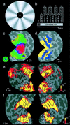
Attentional modulation in mid-eccentricity regions of primary and higher visual cortex. (a) Visual stimuli were composed of an annulus and a central target. Radial wedge patterns were rotated in the annulus. Single letters or a fixation point appeared as the central target (see Methods). (b) Scans consisted of nine 28-sec epochs. A fixation target alone was passively viewed in the first epoch. Attention was alternately directed to foveal and extrafoveal regions of the stimulus (a) in subsequent epochs. (c) Functional mapping of visual eccentricity reveals a foveal representation (shown in red; see color key) in the center of the patch, with more peripheral eccentricities (up to 15o–20o) represented inferiorly (upper visual field) and superiorly (lower visual field). (d) Functional labeling of retinotopic visual cortical areas identifies areas V1−, V2−, V3−, and V3A, superior to the calcarine sulcus, and areas V1+, V2+, VP+, and V4v+, inferior to the calcarine. Upper and lower visual field representations are indicated by + and −, respectively. (e and_f_) Patterns of statistically significant increased activation for attend extrafoveal motion vs. attend foveal letters for both hemispheres of two example subjects (color map shows_P_ values). Mid-eccentricity regions (green in_c_) of all four visual field quadrants of V1 and higher visual cortical areas exhibit highly significant increases in activation. Dashed lines mark iso-eccentricity contours. Solid lines mark boundaries between neighboring cortical areas.
Figure 3
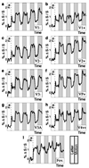
Time course data by cortical area, averaged across subjects (n = 12 hemispheres) for attend foveal letters vs. attend extrafoveal motion. (a_–_h) Mid-eccentricity ROIs in V1−, V1+, V2−, V2+, V3−, VP+, V3A, and V4v+ exhibited greater activation during attend-extrafoveal-motion epochs. (i) Confluent foveal representation (e.g., red region of Fig. 2_b_) exhibited greater activation during attend-foveal-letters epochs.
Figure 4
Average attentional modulation amplitudes. Attend extrafoveal motion − passive viewing shown in black. Passive viewing − attend foveal letters shown in white stacked on top of black bars. Attend extrafoveal motion − attend foveal letters shown in gray.
Figure 5
Eye tracking control data. X and Y eye position traces are shown for two subjects monitored in the MR scanner (see_Methods_). Thick horizontal lines indicate location of the inner radius of the motion annulus (three degrees eccentric).
Similar articles
- Attentional inhibition of visual processing in human striate and extrastriate cortex.
Slotnick SD, Schwarzbach J, Yantis S. Slotnick SD, et al. Neuroimage. 2003 Aug;19(4):1602-11. doi: 10.1016/s1053-8119(03)00187-3. Neuroimage. 2003. PMID: 12948715 - Directing attention to a location in space results in retinotopic activation in primary visual cortex.
Munneke J, Heslenfeld DJ, Theeuwes J. Munneke J, et al. Brain Res. 2008 Jul 30;1222:184-91. doi: 10.1016/j.brainres.2008.05.039. Epub 2008 May 27. Brain Res. 2008. PMID: 18589405 - Multiple spotlights of attentional selection in human visual cortex.
McMains SA, Somers DC. McMains SA, et al. Neuron. 2004 May 27;42(4):677-86. doi: 10.1016/s0896-6273(04)00263-6. Neuron. 2004. PMID: 15157427 - Human cortical areas underlying the perception of optic flow: brain imaging studies.
Greenlee MW. Greenlee MW. Int Rev Neurobiol. 2000;44:269-92. doi: 10.1016/s0074-7742(08)60746-1. Int Rev Neurobiol. 2000. PMID: 10605650 Review. - Functional analysis of primary visual cortex (V1) in humans.
Tootell RB, Hadjikhani NK, Vanduffel W, Liu AK, Mendola JD, Sereno MI, Dale AM. Tootell RB, et al. Proc Natl Acad Sci U S A. 1998 Feb 3;95(3):811-7. doi: 10.1073/pnas.95.3.811. Proc Natl Acad Sci U S A. 1998. PMID: 9448245 Free PMC article. Review.
Cited by
- Attention rhythmically samples multi-feature objects in working memory.
Chota S, Leto C, van Zantwijk L, Van der Stigchel S. Chota S, et al. Sci Rep. 2022 Aug 29;12(1):14703. doi: 10.1038/s41598-022-18819-z. Sci Rep. 2022. PMID: 36038570 Free PMC article. - Neuroimaging weighs in: humans meet macaques in "primate" visual cortex.
Tootell RB, Tsao D, Vanduffel W. Tootell RB, et al. J Neurosci. 2003 May 15;23(10):3981-9. doi: 10.1523/JNEUROSCI.23-10-03981.2003. J Neurosci. 2003. PMID: 12764082 Free PMC article. Review. No abstract available. - Rapid prefrontal-hippocampal habituation to novel events.
Yamaguchi S, Hale LA, D'Esposito M, Knight RT. Yamaguchi S, et al. J Neurosci. 2004 Jun 9;24(23):5356-63. doi: 10.1523/JNEUROSCI.4587-03.2004. J Neurosci. 2004. PMID: 15190108 Free PMC article. - Long-term neurobiological consequences of early postnatal hCMV-infection in former preterms: a functional MRI study.
Dorn M, Lidzba K, Bevot A, Goelz R, Hauser TK, Wilke M. Dorn M, et al. Hum Brain Mapp. 2014 Jun;35(6):2594-606. doi: 10.1002/hbm.22352. Epub 2013 Sep 11. Hum Brain Mapp. 2014. PMID: 24027137 Free PMC article. - Dynamic cognitive remediation for a Traumatic Brain Injury (TBI) significantly improves attention, working memory, processing speed, and reading fluency.
Lawton T, Huang MX. Lawton T, et al. Restor Neurol Neurosci. 2019;37(1):71-86. doi: 10.3233/RNN-180856. Restor Neurol Neurosci. 2019. PMID: 30741708 Free PMC article.
References
- James W. The Principles of Psychology. New York: Dover; 1890.
- Posner M I, Snyder C R R, Davidson B J. J Exp Psychol. 1980;109:160–174. - PubMed
- Hawkins H L, Hillyard S A, Luck S J, Mouloua M, Downing C, Woodward D P. J Exp Psychol Hum Percept Perform. 1990;16:802–811. - PubMed
- Cavanagh P. Science. 1992;257:1563–1565. - PubMed
Publication types
MeSH terms
LinkOut - more resources
Full Text Sources


