Validation of diagnostic accuracy using digital slides in routine histopathology (original) (raw)
Abstract
Background: Robust hardware and software tools have been developed in digital microscopy during the past years for pathologists. Reports have been advocated the reliability of digital slides in routine diagnostics. We have designed a retrospective, comparative study to evaluate the scanning properties and digital slide based diagnostic accuracy.
Figures (9)
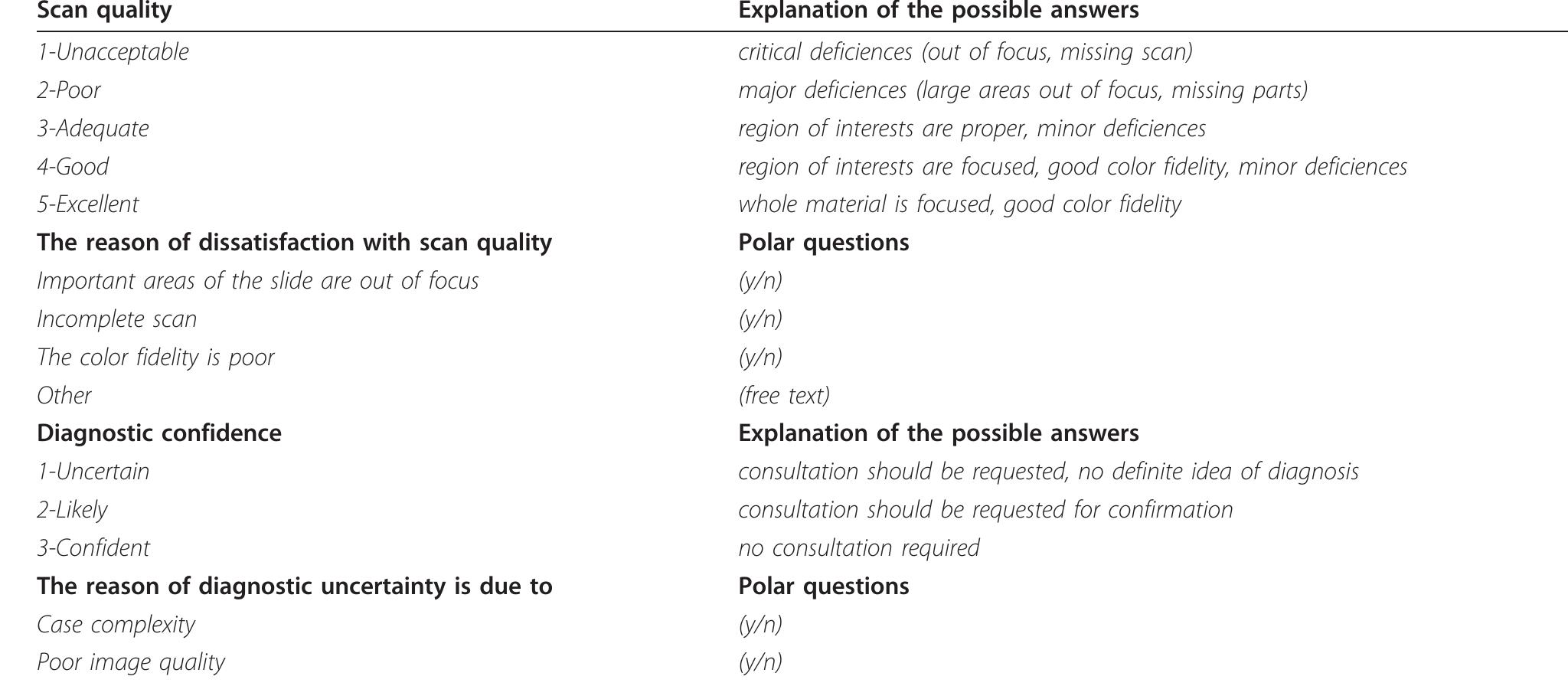
Table 2 Clinical research form
Figure 1 Results of the slide quality query. Column |. all slides, Cll: all slides of the incoherent cases, Cllll:all H&E slides, C.IV.; H&E slides of the incoherent cases, C.V.: all IHC slides, CVI. IHC slides of the incoherent cases, C.VIl.: all Giemsa slides, C.VIll.: Giemsa slides of the incoherent cases, CX. all slides with other stains, C.X.: all slides with other stains of the incoherent cases.
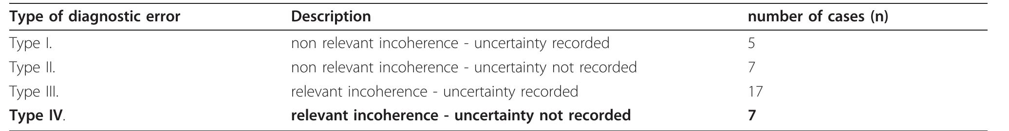
Table 3 Four types of incoherency
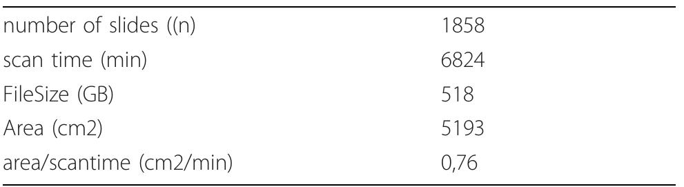
Table 4 Detailed parameters of the scanning properties of 1858 slides
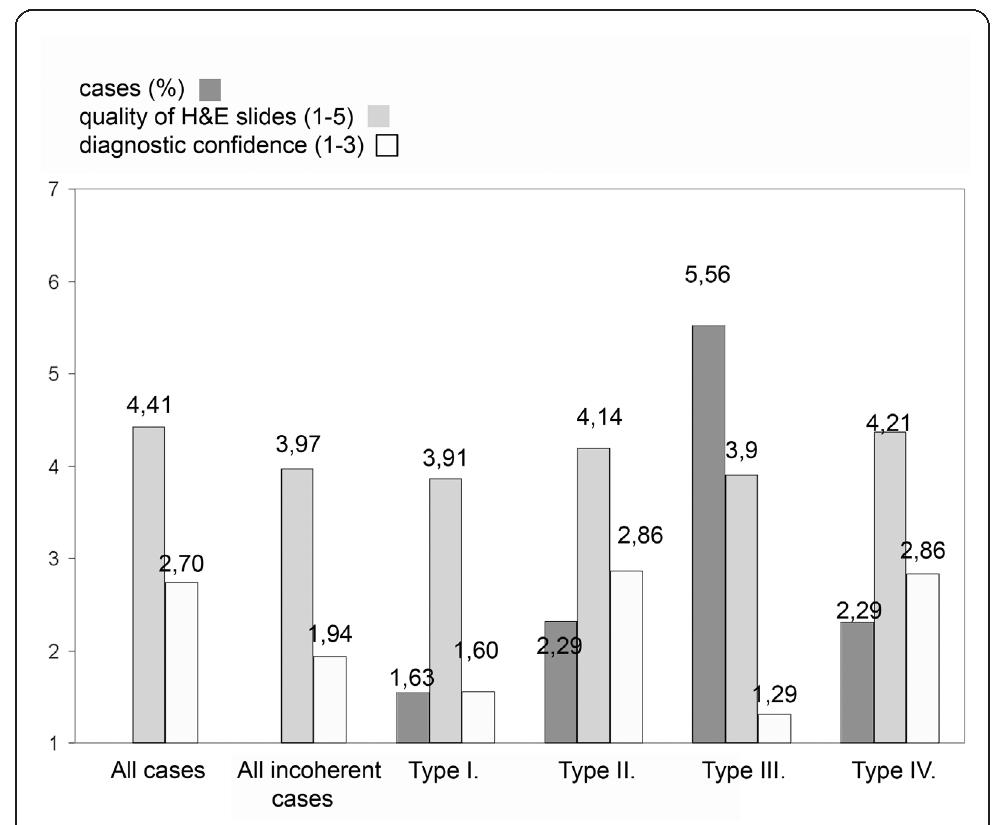
Figure 2 Quality of H&E slides and diagnostic confidence according to the type of incoherency.
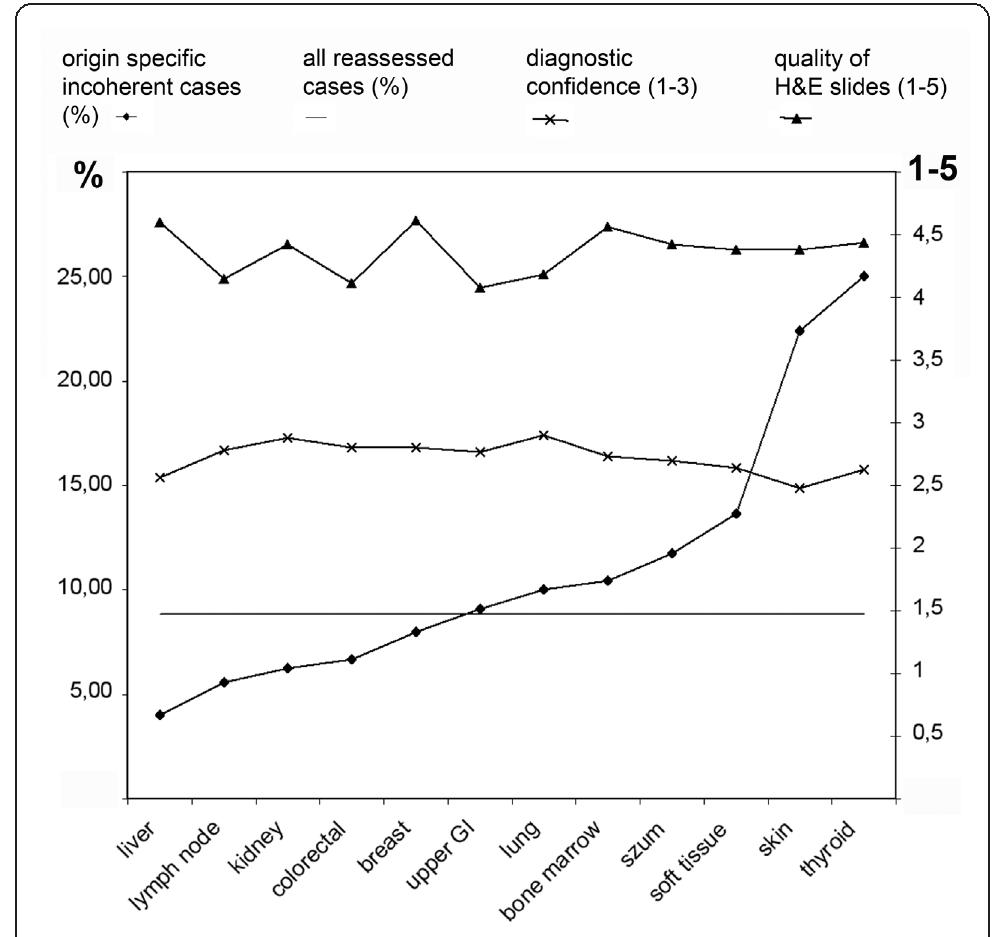
Figure 3 Incoherency, quality of H&E slides and diagnosti confidence according to the origin of the sample. |n 8,82% of the cases the consensus diagnoses were coherent with the digital diagnoses and overwrote the original OM-based diagnoses (reassessed case - straight line)
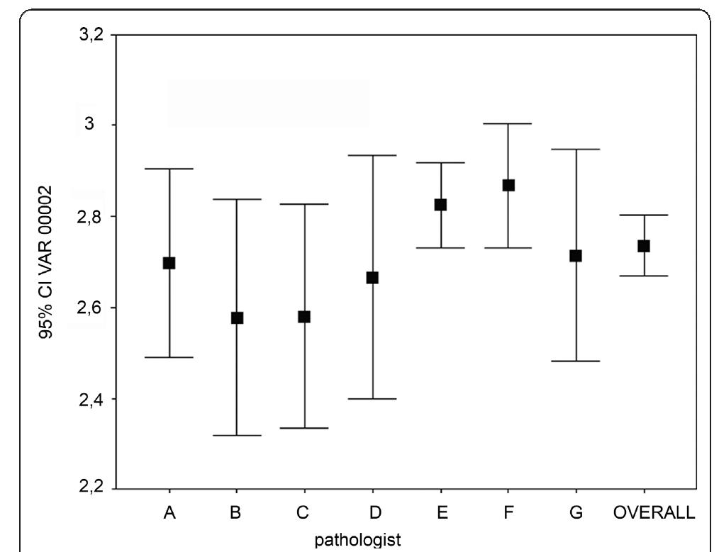
Figure 5 Correlation between the experience (in years) and the diagnostic confidence. overall = sum of all estimates. (Spearman rank R: R = -0.140, t(N-2) = -2.346, p = 0.019). Experience in years: PathA-20, PathB-25, PathC-22, PathD-24, PathE-13, PathF-15, PathG-28 incoherency-ratio of the specific samples to the overall ratio of the reassessed cases. The incoherency-ratio was below 8.82% - therefore we state that DM could be used equivalently to OM in our Institute in this set of cir- cumstance - in cases with samples from liver, lymph node, kidney, colon, and breast. In this series the results nicely correlate with the incidence of the type-IV errors and confirm our statement. Interestingly hematology cases of lymph nodes fell into this category. As the qual- ity of IHC digital slides were evaluated very good, the explanation of this observation could be the explicit importance of the IHC-profiles in haematopathology. This series of samples are specific for our institute in
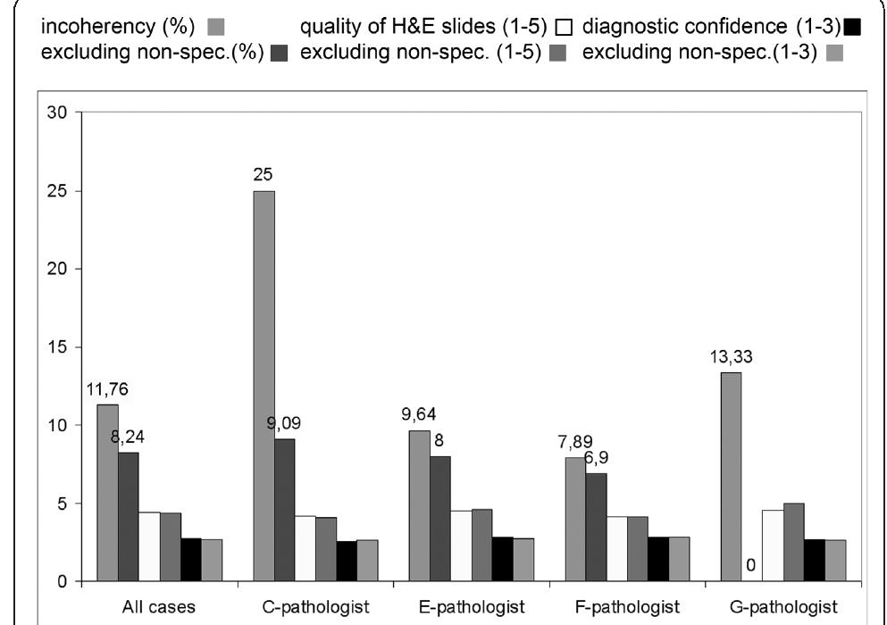
Figure 4 Importance of pathologists’ competence. Excluding the non-field specific cases from each pathologists’ record resulted in better coherency. No significant differences were found in the diagnostic confidence and how the pathologists rated the quality of the slides.

Loading Preview
Sorry, preview is currently unavailable. You can download the paper by clicking the button above.
References (24)
- Weinstein RS: Prospects for telepathology. Hum Pathol 1986, 17(5):433-434.
- Weinberg , Allaert , Dussere : Telepathology diagnosis by means of digital still images: An international validation study (vol 27, pg 111, 1996). Hum Pathol 1996, 27(9):1001-1001.
- Kaplan KJ, Burgess JR, Sandberg GD, Myers CP, Bigott TR, Greenspan RB: Use of robotic Telepathology for frozen-section diagnosis: A retrospective trial of a telepathology system for intraoperative consultation. Mod Pathol 2002, 15(11):1197-1204.
- Kayser K, Molnar B, Weinstein R: Virtual Microscopy: Fundamentals, Applications, Perspectives of Electronic Tissue-based Diagnosis Berlin: VSV Interdisciplinary Medical Publishing; 2006.
- Leong F, McGee J: Automated complete slide digitization: a medium for simultaneous viewing by multiple pathologists. J Pathol 2001, 195(4):508-514.
- Molnar B, Berczi L, Diczhazy C, Tagscherer A, Varga SV, Szende B, Tulassay Z: Digital slide and virtual microscopy based routine and telepathology evaluation of routine gastrointestinal biopsy specimens. J Clin Pathol 2003, 56(6):433-438.
- Wells CA, Sowter C: Telepathology: a diagnostic tool for the millennium? J Pathol 2000, 191(1):1-7.
- Kayser K, Gortler J, Goldmann T, Vollmer E, Hufnagl P, Kayser G: Image standards in Tissue-Based Diagnosis (Diagnostic Surgical Pathology). Diagn Pathol 2008, 3:9.
- Gabril MY, Yousef GM: Informatics for practicing anatomical pathologists: marking a new era in pathology practice. Mod Pathol 2010, 23(3):349-358.
- Papay J, Krenacs T, Moldvay J, Stelkovics E, Furak J, Molnar B, Kopper L: Immunophenotypic profiling of nonsmall cell lung cancer progression using the tissue microarray approach. Appl Immunohistochem Mol Morphol 2007, 15(1):19-30.
- Fonyad L, Gerely L, Cserneky M, Molnar B, Matolcsy A: Shifting gears higher -digital slides in graduate education -4 years experience at Semmelweis University. Diagn Pathol 2010, 5:73.
- Della Mea V, Demichelis F, Viel F, Palma PD, Beltrami CA: User attitudes in analyzing digital slides in a quality control test bed: A preliminary study. Comput Methods Programs Biomed 2006, 82(2):177-186.
- Ho J, Parwani AV, Jukic DM, Yagi Y, Anthony L, Gilbertson JR: Use of whole slide imaging in surgical pathology quality assurance: design and pilot validation studies. Hum Pathol 2006, 37(3):322-331.
- Mireskandari M, Kayser G, Hufnagl P, Schrader T, Kayser K: Teleconsultation in diagnostic pathology: experience from Iran and Germany with the use of two European telepathology servers. J Telemed Telecare 2004, 10(2):99-103.
- Graham AR, Bhattacharyya AK, Scott KM, Lian F, Grasso LL, Richter LC, Carpenter JB, Chiang S, Henderson JT, Lopez AM, et al: Virtual slide telepathology for an academic teaching hospital surgical pathology quality assurance program. Hum Pathol 2009, 40(8):1129-1136.
- Lopez AM, Graham AR, Barker GP, Richter LC, Krupinski EA, Lian F, Grasso LL, Miller A, Kreykes LN, Henderson JT, et al: Virtual slide telepathology enables an innovative telehealth rapid breast care clinic. Hum Pathol 2009, 40(8):1082-1091.
- Horbinski C, Fine JL, Medina-Flores R, Yagi Y, Wiley CA: Telepathology for intraoperative neuropathologic consultations at an Academic Medical Center: A 5-year report. J Neuropathol Exp Neurol 2007, 66(8):750-759.
- Renshaw AA, Gould EW: Measuring errors in surgical pathology in real- life practice -Defining what does and does not matter. Am J Clin Pathol 2007, 127(1):144-152.
- Yagi Y, Gilbertson JR: The importance of optical optimization in whole slide imaging (WSI) and digital pathology imaging. Diagn Pathol 2008, 3:S1.
- Mori I, Ozaki T, Taniguchi E, Kakudo K: Study of parameters in focus simulation functions of virtual slide. Diagn Pathol 2011, 6(Suppl 1):S24.
- Tawfik OW, Kimler BF, Davis M, Donahue JK, Persons DL, Fan F, Hagemeister S, Thomas P, Connor C, Jewell W, Fabian CJ: Comparison of immunohistochemistry by automated cellular imaging system (ACIS) versus fluorescence in-situ hybridization in the evaluation of HER-2/neu expression in primary breast carcinoma. Histopathology 2006, 48(3):258-267.
- Turbin DA, Leung S, Cheang MCU, Kennecke HA, Montgomery KD, McKinney S, Treaba DO, Boyd N, Goldstein LC, Badve S, et al: Automated quantitative analysis of estrogen receptor expression in breast carcinoma does not differ from expert pathologist scoring: a tissue microarray study of 3,484 cases. Breast Cancer Res Treat 2008, 110(3):417-426.
- Fine JL, Grzybicki DM, Silowash R, Ho J, Gilbertson JR, Anthony L, Wilson R, Parwani AV, Bastacky SI, Epstein JI, Jukic DM: Evaluation of whole slide image immunohistochemistry interpretation in challenging prostate needle biopsies. Hum Pathol 2008, 39(4):564-572. doi:10.1186/1746-1596-7-35
- Cite this article as: Fónyad et al.: Validation of diagnostic accuracy using digital slides in routine histopathology. Diagnostic Pathology 2012 7:35.