Quantum cryptography: Overview, security issues and future challenges (original) (raw)
Abstract
In recent years Quantum Cryptography gets more attention as well as becomes most promising cryptographic field for faster, effective and more secure communications. Quantum Cryptography is an art of science which overlap quantum properties of light under quantum mechanics on cryptographic task instead of current state of algorithms based on mathematical computing technology. Major algorithms for public key encryption and some digital signature scheme such as RSA, El Gamal cryptosystem, hash function are vulnerable at quantum adversaries. Most of the factoring problem can be broken by Shore's algorithm and quantum computer threatens other hand discrete logarithm problem. Our paper describes why modern cryptography contributes quantum cryptography, security issues and future goals of modern cryptography.
FAQs
![]()
AI
What relationship exists between scintillation anisotropy and crystal structure in organic scintillators?add
The measurements indicate that materials with herringbone structures show in-sync behaviors of prompt and delayed light anisotropies, while π-stacked materials like anthracene and p-terphenyl exhibit out-of-sync behaviors, highlighting structural dependencies on anisotropy effects.
How does proton recoil energy influence scintillation anisotropy behavior?add
Anisotropy measurements in anthracene and stilbene reveal that the total light anisotropy decreases with higher energies, while the pulse shape anisotropy shows variable correlations, indicating energy-dependent influences on excitation dynamics.
What experimental methodology was used to analyze scintillation anisotropy across materials?add
The research employed a rotational measurement system aligning detectors with a neutron generator, enabling comprehensive characterization of scintillation responses over 70 proton recoil directions for each crystal.
Can scintillation anisotropy present opportunities in radiation detection applications?add
While anisotropy complicates light output resolution, it may enable organic scintillators to function as directional detectors, offering potential applications in dark matter detection efforts.
What unique insights were gained by separately analyzing prompt and delayed light components?add
This approach provided a clearer understanding of excitation dynamics, revealing that prompt and delayed light emissions behaved independently in certain materials, allowing for nuanced comparisons across crystals.
Figures (22)
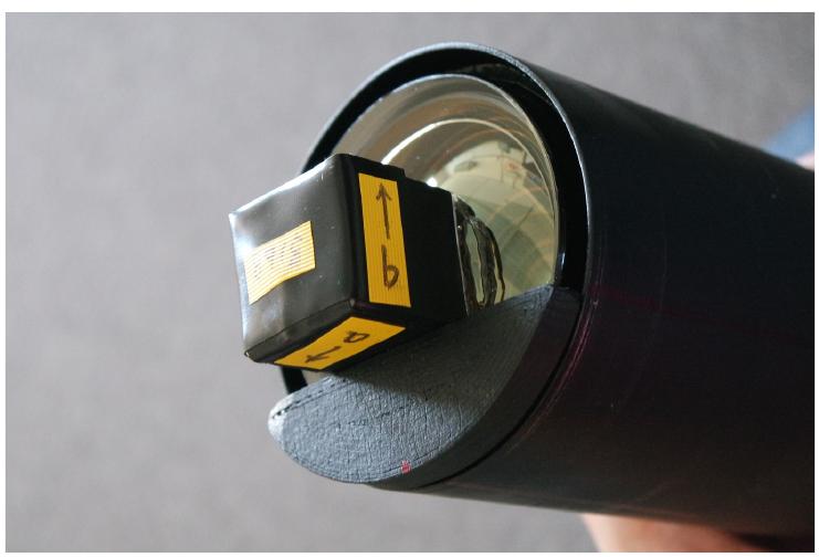
Fig. 1. Wrapped stilbene crystal positioned against a plastic guide and mounted to the PMT face with optical grease.
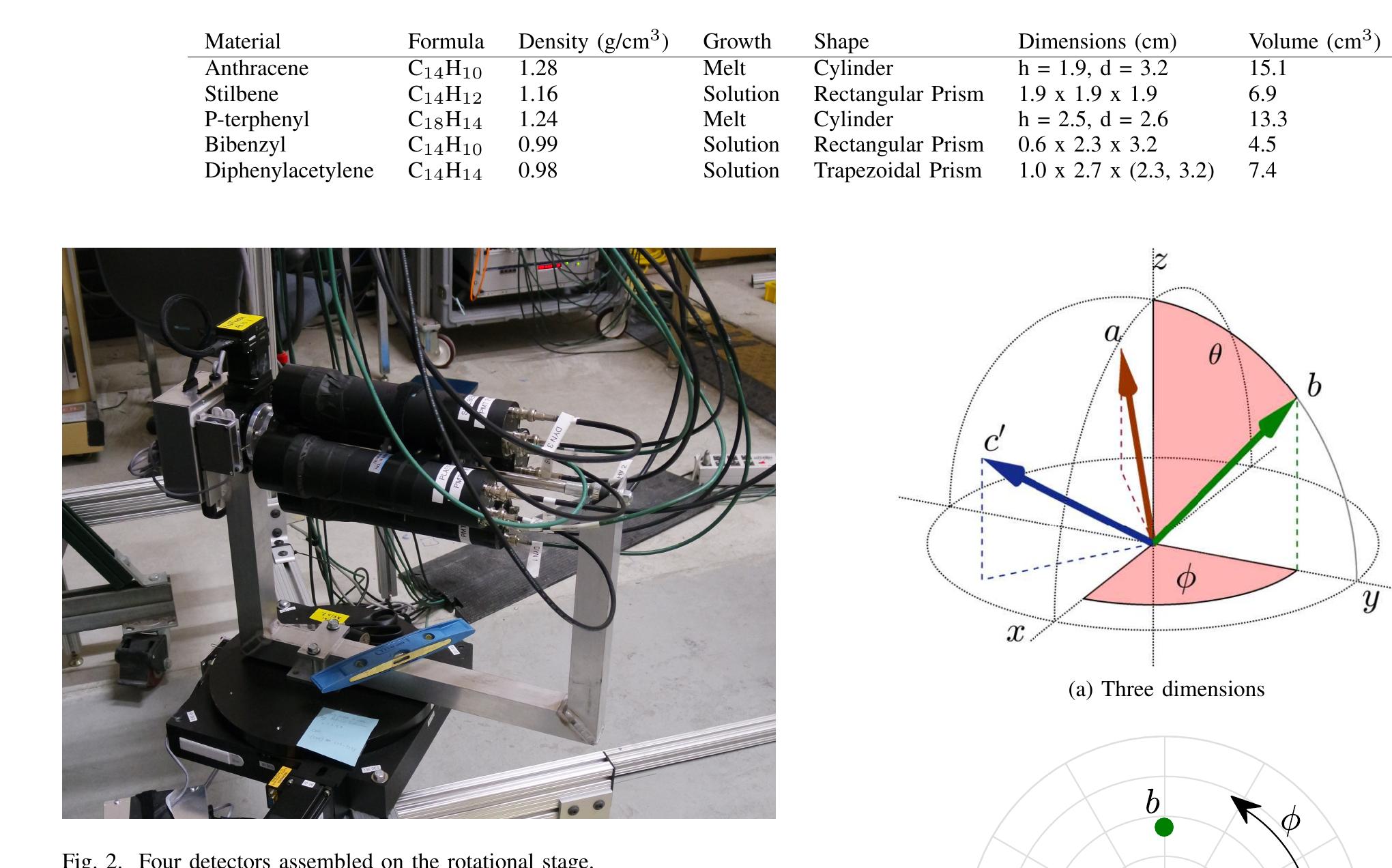
TABLE I B. Rotational measurement system For the measurements presented here, a Thermo Scien- tific MP320 neutron generator was used to produce roughly monoenergetic neutrons with £, = 14.1 MeV (via the DT reaction) and E, = 2.5 MeV (via the DD reaction). In order to measure the light emission in response to proton recoil events at known energies and directions in the crystal axes, only full-energy interactions are used. In these interactions, a neutron deposits its full energy onto the proton and the proton recoil travels in the forward direction. In each measurement, the orientation between the detector and neutron generator was fixed so that one proton recoil direction could be characterized. For each crystal, measurements were taken at many angles in order to characterize the response to many proton recoil directions in the crystal axes.
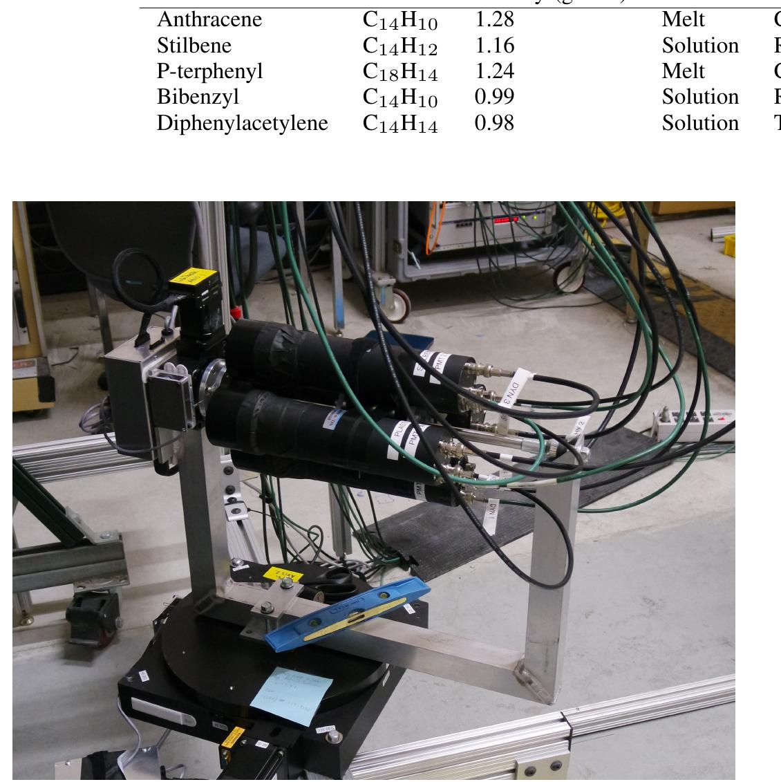
Fig. 2. Four detectors assembled on the rotational stage. SUMMARY OF PHYSICAL CHARACTERISTICS OF ORGANIC CRYSTAL SCINTILLATOR MATERIALS MEASURED IN THIS PAPER.
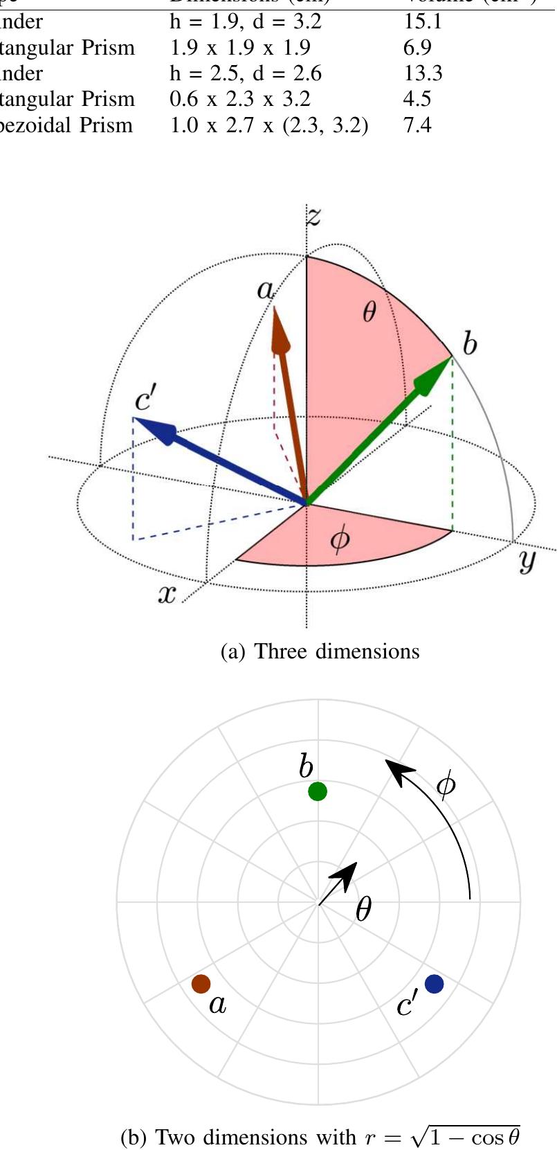
Fig. 3. Visualization of proton recoil directions within a hemisphere of space in (a) three dimensions and (b) two dimensions. Vectors a, b, and c’ correspond to the known axes directions in the measured stilbene sample. The angles @ and @ describe the vector directions in spherical coordinates, where 0° < @ < 360° and 0° < 6 < 90°.
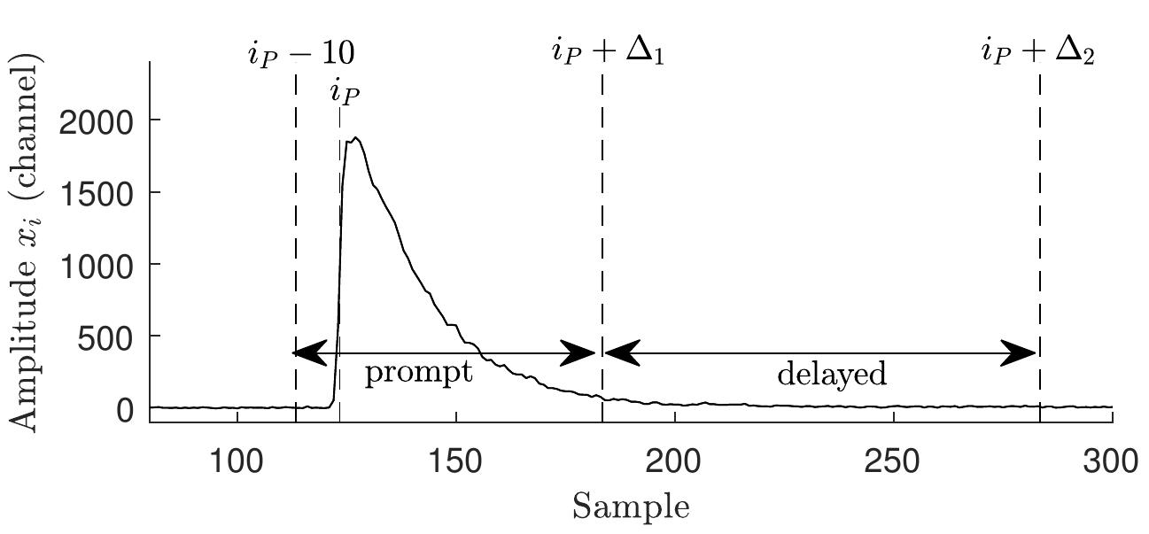
Fig. 4. Illustration of a baseline-subtracted raw pulse in anthracene with timing windows for prompt and delayed light components indicated
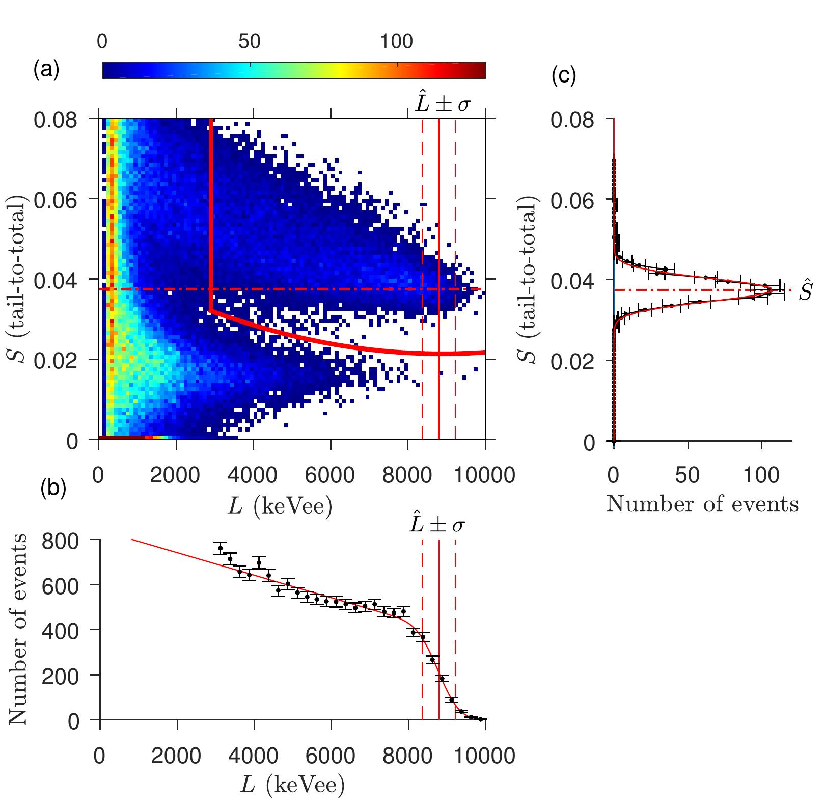
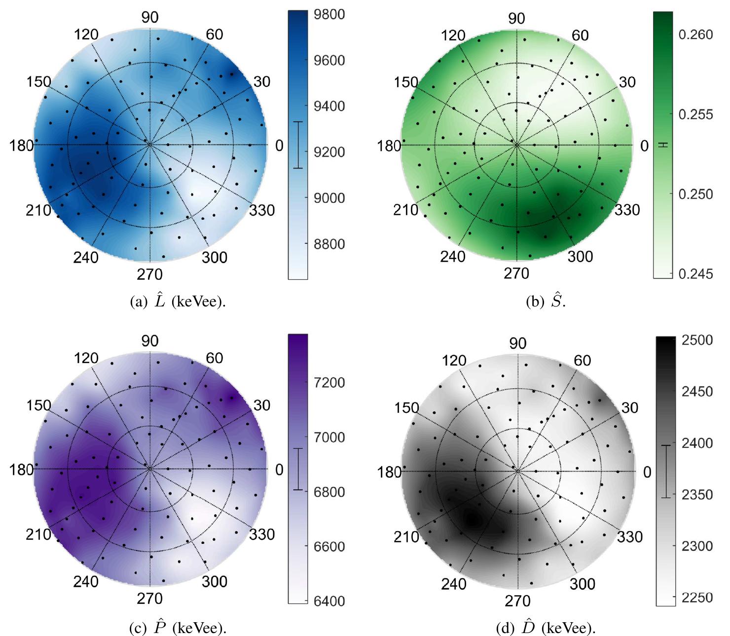
Fig. 5. Distributions produced for calculating E and S for 14.1 MeV proton recoil events in anthracene. (a) Two-dimensional histogram of L vs. S values for all events. Events above and to the right of the bold red curve are classified as neutron events. (b) Light output spectrum of neutron events. The vertical solid and dashed lines in (a) and (b) indicate the range L + o that includes events classified as full-energy neutron interactions. (c) Distribution of pulse shape values for full-energy neutron interactions, displayed with inverted axes to correspond with (a). The horizontal dot-dashed lines in (a) and (c) indicate the expected pulse shape value S for full-energy neutron interactions.
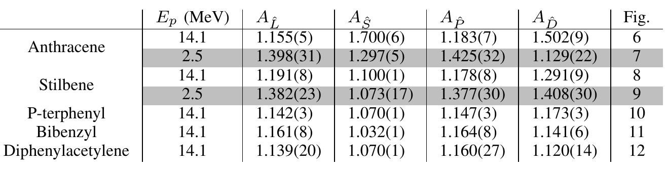
MAGNITUDE OF CHANGE IN L, 8, P, AND D VALUES MEASURED FOR 14.1 MEV (UNSHADED) AND 2.5 MEV (SHADED) PROTON RECOIL EVENTS IN VARIOUS PURE CRYSTAL DETECTORS, AND THE CORRESPONDING FIGURE NUMBER. UNCERTAINTIES ARE STATISTICAL ONLY. stilbene and positively correlated with energy in anthracene. This result adds to questions surrounding the delayed light emission in anthracene. ing statistics. Other systematic errors exist in the measure- ments that are not quantified. These include the following:
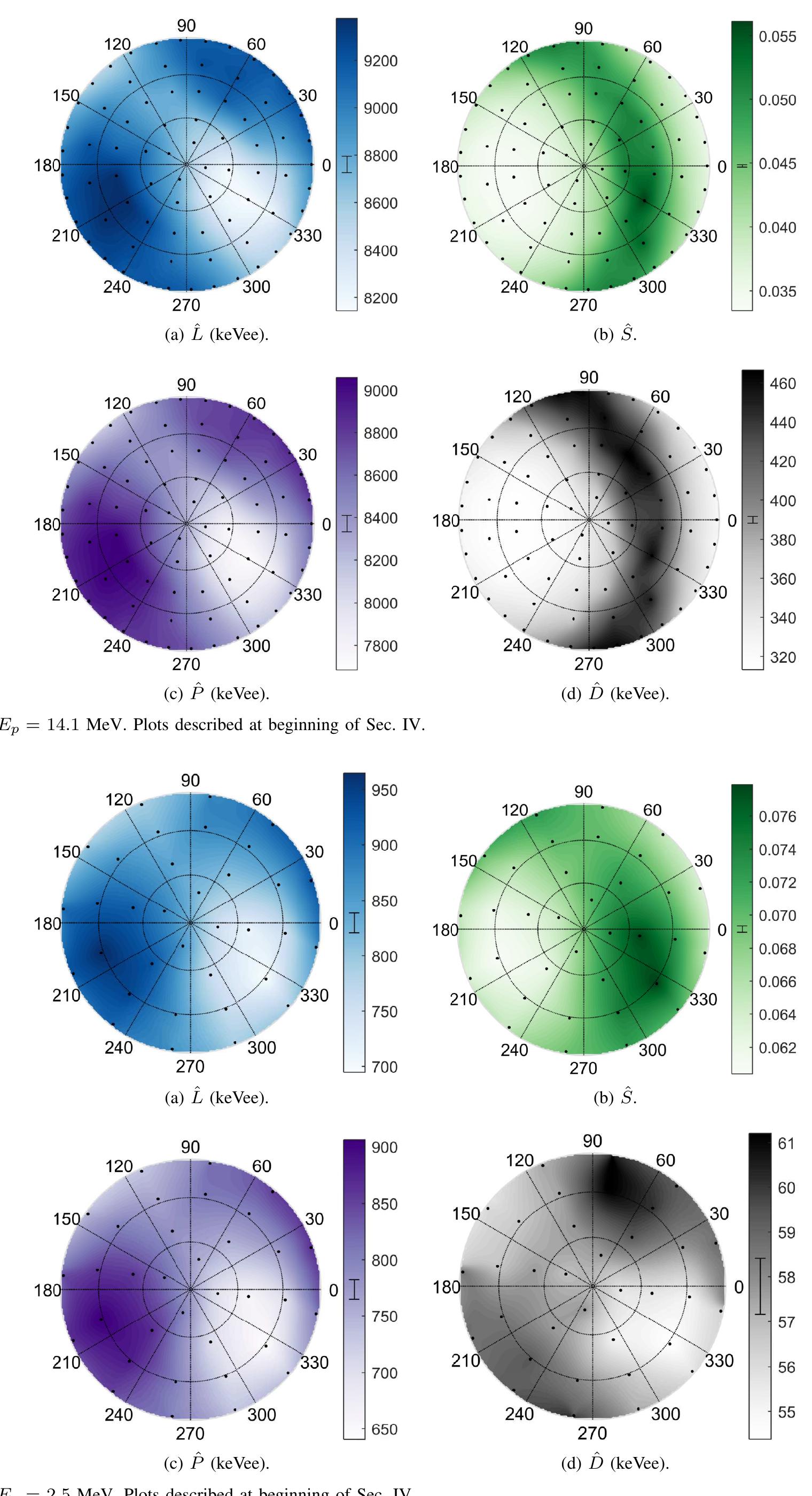
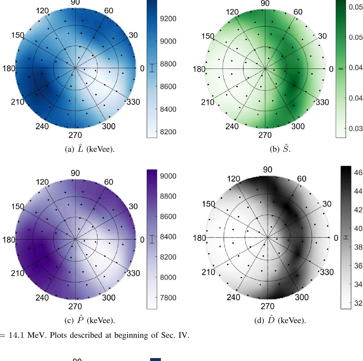
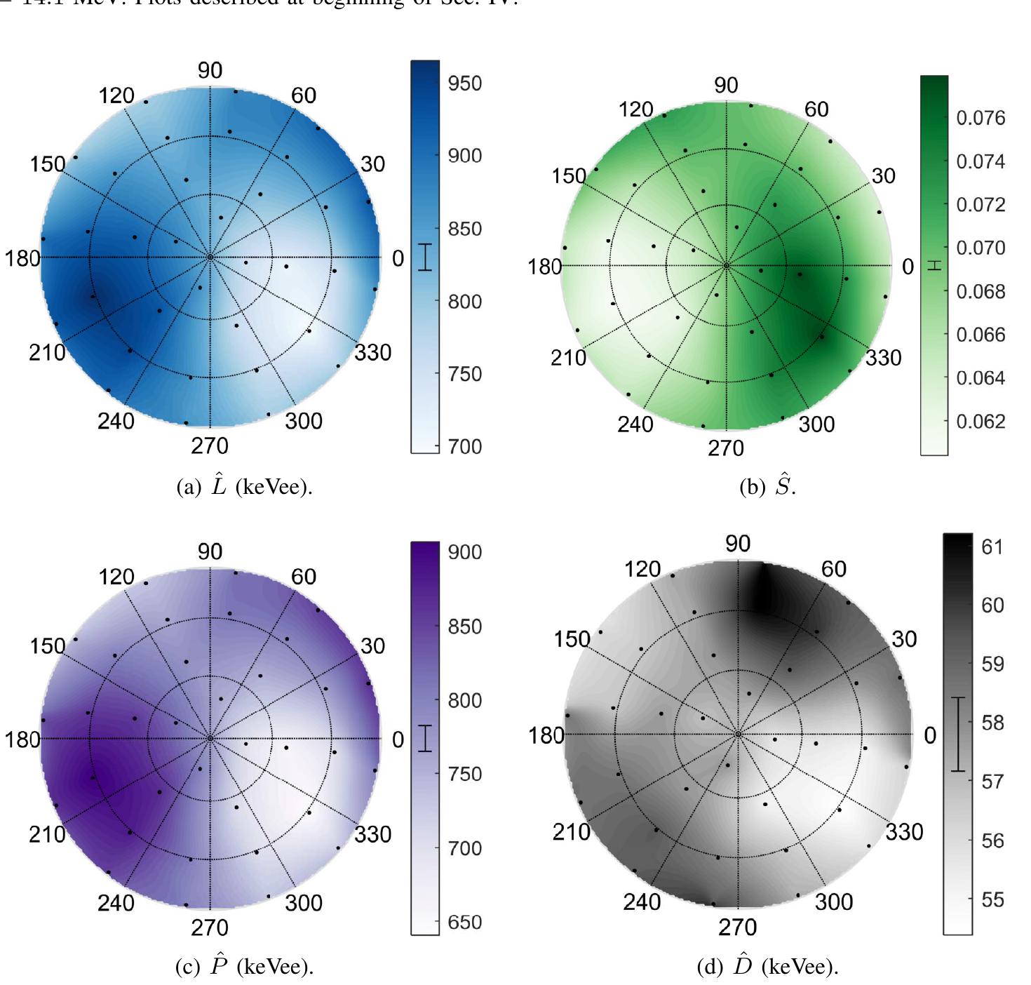
Fig. 7. Anthracene; Ep = 2.5 MeV. Plots described at beginning of Sec. IV.
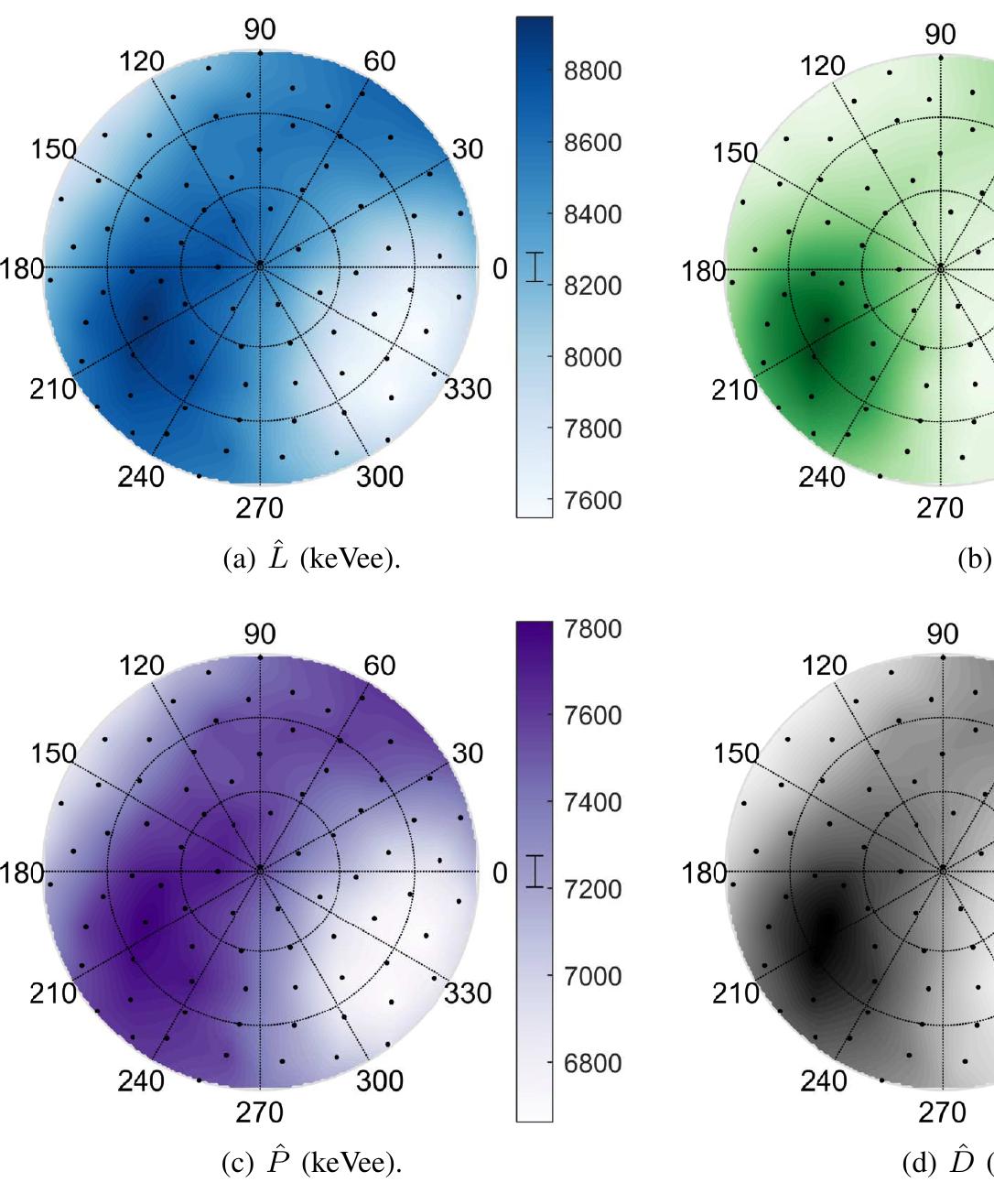
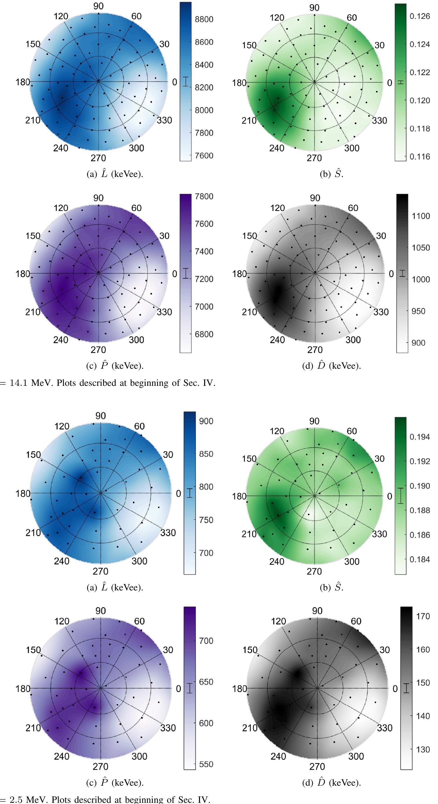
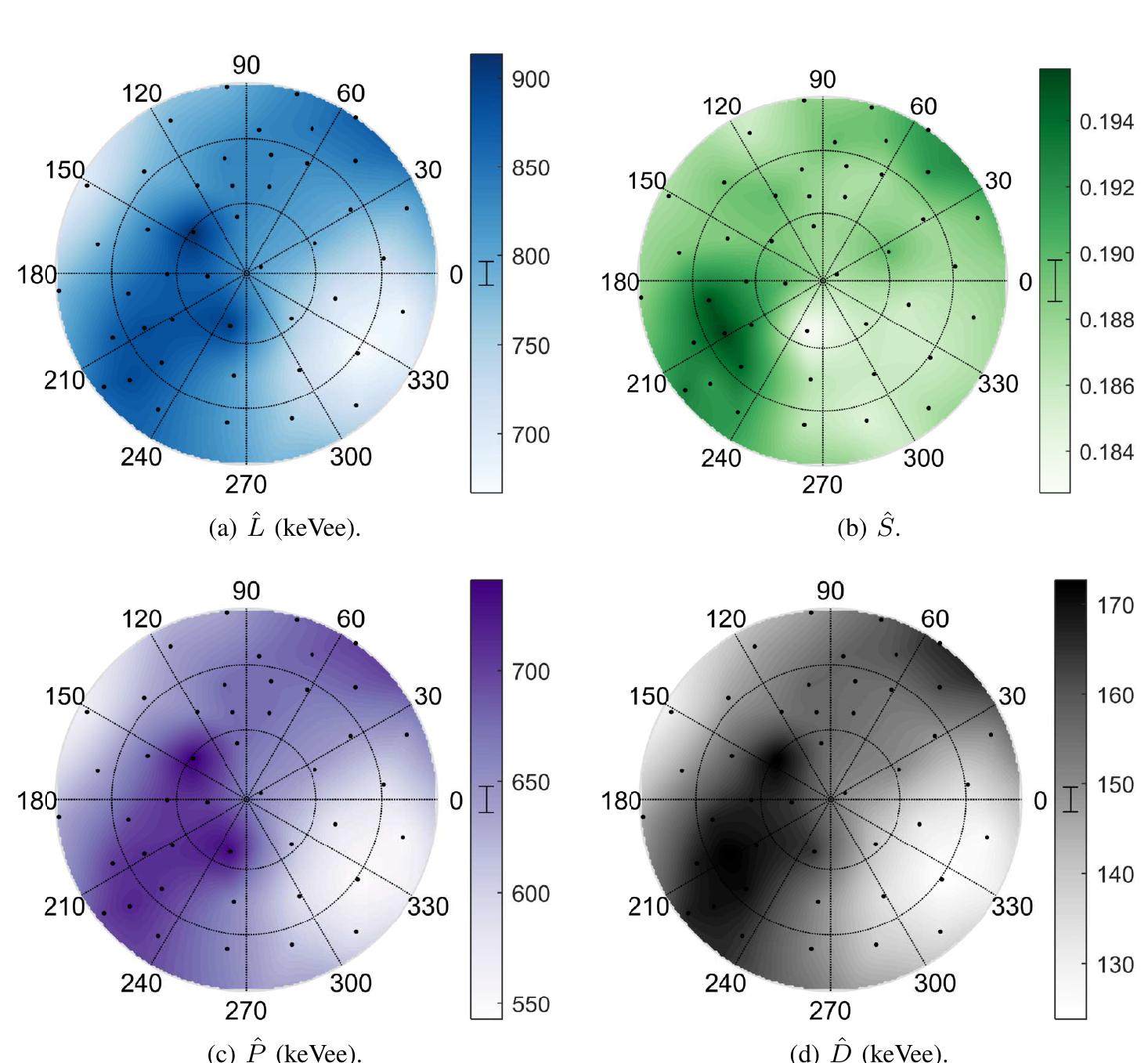
Fig. 8. Stilbene; Ey = 14.1 MeV. Plots described at beginning of Sec. IV. Fig. 9. Stilbene; Ey = 2.5 MeV. Plots described at beginning of Sec. IV.

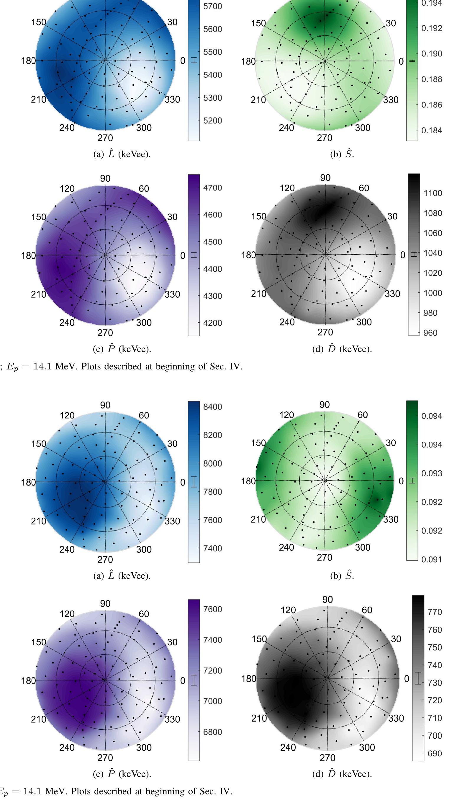
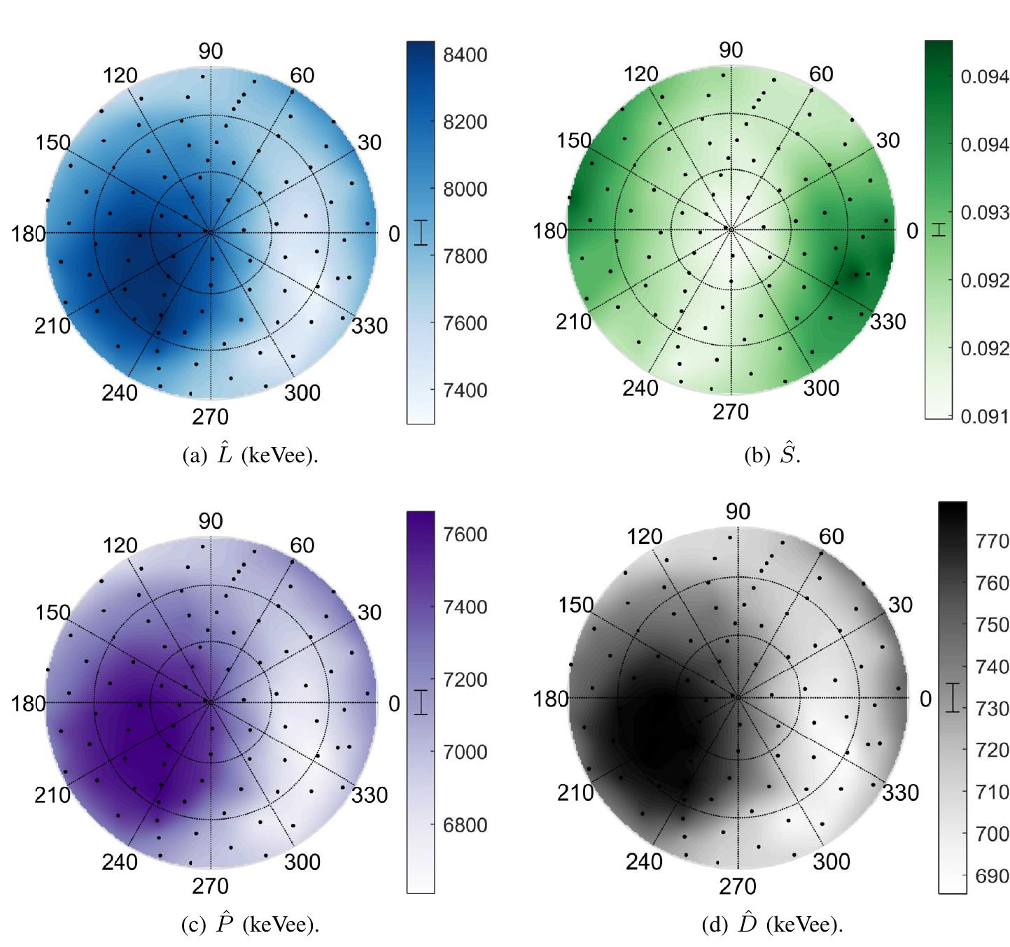
Fig. 11. Bibenzyl; Ep = 14.1 MeV. Plots described at beginning of Sec. IV. Fig. 10. P-terphenyl; E, = 14.1 MeV. Plots described at beginning of Sec. IV.
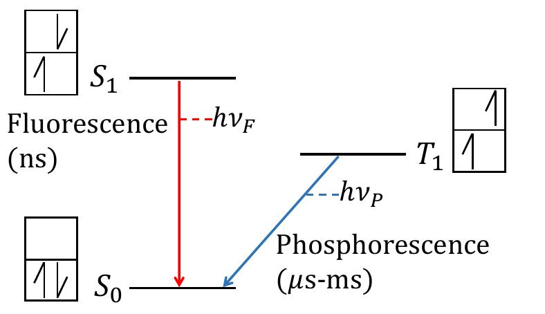
Fig. 13. Simplified Jablonski diagram showing the first electronic excited singlet and triplet states and the ground singlet state. Fig. 12. Diphenylacetylene; Ey = 14.1 MeV. Plots described at beginning of Sec. IV.
but one may proceed as if it does not occur due to its small contribution compared to other processes.
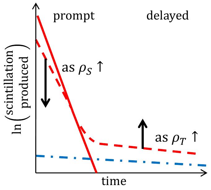
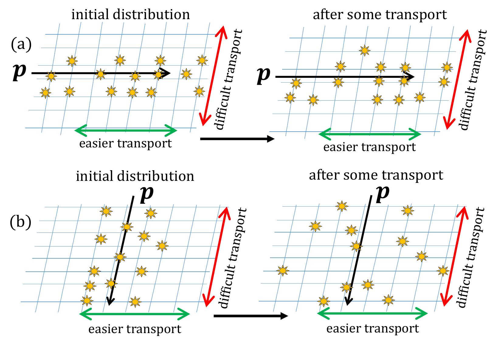
Fig. 15. Illustration of excitation transport over time when excitations are populated by a proton recoil p along crystal axes with (a) easy transport or (b) difficult transport. In each, the left cartoon illustrates the initial excitation distribution and the right cartoon illustrates the distribution after some time has passed and excitation transport has occurred. In (a), the density of excitations remains high over time as the excitations move within their initial spatial distribution, while in (b), the density of excitations drops off over time as excitations move away from their initial spatial distribution. In (a), one would observe higher rates of SIQ and TTA over time, leading to a lower Panda higher D.
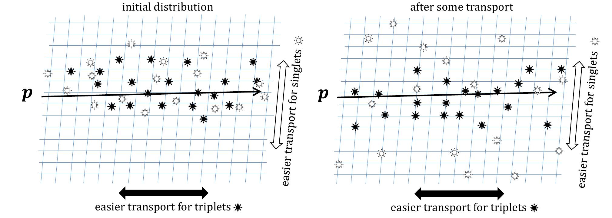
Fig. 16. Illustration of excitation transport over time when excitations are populated by a proton recoil p along a crystal axes with easy triplet (filled) transpor' and difficult singlet (open) transport. After some transport has occurred, the triplet excitation density remains high while the singlet density has dropped leading to higher rates of TTA and lower rates of SIQ. This may lead to P and D being in sync.
![Fig. 17. Crystal structures as visualized by Mercury software [27]. (a) Diphenylacetylene, stilbene, and bibenzyl can be described as herringbone crysti structure and exhibit prompt and delayed light anisotropies that are in sync. (b) Anthracene and p-terphenyl both have z-stacked crystal structure and promy and delayed light anisotropies that are out of sync.](https://figures.academia-assets.com/84119141/figure_021.jpg) ](
](Fig. 17. Crystal structures as visualized by Mercury software [27]. (a) Diphenylacetylene, stilbene, and bibenzyl can be described as herringbone crysti structure and exhibit prompt and delayed light anisotropies that are in sync. (b) Anthracene and p-terphenyl both have z-stacked crystal structure and promy and delayed light anisotropies that are out of sync.

Loading Preview
Sorry, preview is currently unavailable. You can download the paper by clicking the button above.
References (28)
- Beatrice Fraboni, Alessandro Fraleoni-Morgera, and Natalia Zaitseva. "Ionizing radiation detectors based on solution-grown organic single crystals". In: Advanced Functional Materials 26.14 (2016), pp. 2276-2291. ISSN: 16163028. DOI: 10.1002/adfm.201502669.
- Natalia Zaitseva et al. "Scintillation of solution-grown trans-stilbene single crystals". In: Nu- clear Instruments and Methods in Physics Research, Section A: Accelerators, Spectrometers, Detectors and Associated Equipment 789 (2015), pp. 8-15. ISSN: 01689002. DOI: 10.1016/j.nima.2015.03.090.
- G. T. Wright. "Scintillation Response of Organic Phos- phors". In: Physical Review 91.5 (1953), pp. 1282- 1283. ISSN: 0031-899X. DOI: 10 . 1103 / PhysRev. 91 . 1282.2.
- F. D. Brooks. "Scintillation Counters with Pulse Shape Selection to Distinguish Neutrons from Gamma-Rays". In: Liquid Scintillation Counting (1957), pp. 268-269.
- Takayuki Yanagida, Kenichi Watanabe, and Yutaka Fu- jimoto. "Comparative study of neutron and gamma-ray pulse shape discrimination of anthracene, stilbene, and p-terphenyl". In: Nuclear Instruments and Methods in Physics Research, Section A: Accelerators, Spectrome- ters, Detectors and Associated Equipment 784 (2015), pp. 111-114. ISSN: 01689002. DOI: 10 . 1016 / j . nima . 2014.12.031.
- Joseph S. Carlson and Patrick L. Feng. "Melt-cast organic glasses as high-efficiency fast neutron scintilla- tors". In: Nuclear Instruments and Methods in Physics Research Section A: Accelerators, Spectrometers, De- tectors and Associated Equipment 832 (2016), pp. 152- 157. ISSN: 01689002. DOI: 10.1016/j.nima.2016.06. 116.
- Paul Henrich Heckmann. "Richtungsabhaengigkeit der Szintillations-Lichtausbeute von Anthracenkristallen beim Beschuss mit alpha-Strahlen". In: Zeitschrift fuer Physik 157.2 (1959), pp. 139-148. ISSN: 0044-3328. DOI: 10.1007/BF02055746.
- P. H. Heckmann, H. Hansen, and A. Flammers- feld. "Die Richtungsabhaengigkeit der Szintillations- Lichtausbeute von duennen Anthrazen-und Stilben- Kristallen beim Beschuss mit alpha-Strahlen". In: Zeitschrift fuer Physik 162.1 (1961), pp. 84-92. ISSN: 1434-6001. DOI: 10.1007/BF01342470.
- P. H. Heckmann, W. Sander, and A. Flammers- feld. "Die Szintillations-Anisotropie von Anthrazen bei tiefen Temperaturen". In: Zeitschrift fuer Physik 165.1 (1961), pp. 12-16. ISSN: 1434-6001. DOI: 10 . 1007 / BF01378081.
- W. F. Kienzle and A. Flammersfeld. "Die Anisotropie der Szintillationslichtausbeute organischer Moleküleinkristalle für α-Strahlen". In: Zeitschrift fuer Physik 165.1 (1961), pp. 1-11. DOI: 10 . 1007 / BF01378080.
- Kineo Tsukada and Shiroh Kikuchi. "Directional anisotropy in the characteristics of the organic-crystal scintillators". In: Nuclear Instruments and Methods 17.3 (1962), pp. 286-288. ISSN: 0029554X. DOI: 10.1016/ 0029-554X(62)90007-1.
- K. Tsukada, S. Kikuchi, and Y. Miyagawa. "Directional anisotropy in the characteristics of the organic-crystal scintillators, II". In: Nuclear Instruments and Methods 37 (1965), pp. 69-76. ISSN: 0029554X. DOI: 10.1016/ 0029-554X(65)90339-3.
- F.D. Brooks and D.T.L. Jones. "Directional anisotropy in organic scintillation crystals". In: Nuclear Instru- ments and Methods 121.1 (1974), pp. 69-76. ISSN: 0029554X. DOI: 10.1016/0029-554X(74)90141-4.
- D. B. Oliver and G. F. Knoll. "Anisotropy of Scintil- lation Response of Anthracene to Neutron Generated Recoil Protons and Carbon Ions". In: IEEE Transactions on Nuclear Science 15.3 (1968), pp. 122-126. ISSN: 0018-9499. DOI: 10.1109/TNS.1968.4324926.
- Hiroyuki Sekiya et al. "Measurements of anisotropic scintillation efficiency for carbon recoils in a stilbene crystal for dark matter detection". In: Physics Letters, Section B: Nuclear, Elementary Particle and High- Energy Physics 571.3-4 (2003), pp. 132-138. ISSN: 03702693. DOI: 10.1016/j.physletb.2003.07.077. arXiv: 0307384 [astro-ph].
- Patricia Frances Schuster. "Investigating the Anisotropic Scintillation Response in Organic Crystal Scintillator Detectors." PhD thesis. University of California, Berke- ley, 2016.
- Patricia Schuster and Erik Brubaker. "Investigating the Anisotropic Scintillation Response in Anthracene through Neutron, Gamma-Ray, and Muon Measure- ments". In: IEEE Transactions on Nuclear Science 63.3 (2016), pp. 1942-1954. ISSN: 0018-9499. DOI: 10.1109/ TNS.2016.2542589.
- Erik Brubaker and John Steele. "Neutron imaging using the anisotropic response of crystalline organic scintilla- tors". In: IEEE Nuclear Science Symposium Conference Record (2010), pp. 1647-1652. ISSN: 10957863. DOI: 10.1109/NSSMIC.2010.5874055.
- P. Schuster and E. Brubaker. "Characterization of the scintillation anisotropy in crystalline stilbene scintillator detectors". In: Nuclear Instruments and Methods in Physics Research Section A: Accelerators, Spectrome- ters, Detectors and Associated Equipment (2016). ISSN: 01689002. DOI: 10.1016/j.nima.2016.11.016.
- P. Belli et al. "Identifying a "dark matter" signal by non- isotropic scintillation detector". In: Il Nuovo Cimento C 15.4 (1992), pp. 473-479. ISSN: 0390-5551. DOI: 10.1007/BF02511747.
- J. B. Birks. The Theory and Practice of Scintillation Counting. Vol. 148. 3667. Elsevier, 1964, p. 662.
- Dwaine O. Cowan and Ronald L. Drisko. Elements of Organic Photochemistry. New York: Plenum Press, 1976. ISBN: 0-306-30821-5.
- F.D. Brooks. "Development of organic scintillators". In: Nuclear Instruments and Methods 162.1-3 (1979), pp. 477-505. ISSN: 0029554X. DOI: 10 . 1016 / 0029 - 554X(79)90729-8.
- Tohru Azumi and Sean P. McGlynn. "Delayed Fluo- rescence of Solid Solutions of Polyacenes. II. Kinetic Considerations". In: The Journal of Chemical Physics 39.5 (1963), p. 1186. ISSN: 00219606. DOI: 10.1063/1. 1734411.
- F.D. Brooks. "A scintillation counter with neutron and gamma-ray discriminators". In: Nuclear Instruments and Methods 4.3 (1959), pp. 151-163. ISSN: 0029554X. DOI: 10.1016/0029-554X(59)90067-9.
- J.M. Adams and G. White. "A versatile pulse shape discriminator for charged particle separation and its application to fast neutron time-of-flight spectroscopy". In: Nuclear Instruments and Methods 156.3 (1978), pp. 459-476. ISSN: 0029554X. DOI: 10 . 1016 / 0029 - 554X(78)90746-2.
- Clare F. Macrae et al. "Mercury CSD 2.0 -New features for the visualization and investigation of crystal structures". In: Journal of Applied Crystallography 41.2 (2008), pp. 466-470. ISSN: 00218898. DOI: 10.1107/ S0021889807067908.
- Gleb M Akselrod et al. "Visualization of exciton trans- port in ordered and disordered molecular solids." In: Nature communications 5 (2014), p. 3646. ISSN: 2041- 1723. DOI: 10.1038/ncomms4646.