Anatomy and Physiology of Human Eye (original) (raw)
Last Updated : 25 May, 2026
The eye is a highly specialised sensory organ responsible for vision. It allows us to detect light, perceive images, and interpret the surrounding environment. The study of the eye involves both its structure (anatomy) and its function (physiology), which work together to produce the sense of sight. The human eye is nearly spherical in shape and is located within the bony socket called the orbit. It is protected by eyelids, eyelashes, and tear glands.
Layers of the Eye
The wall of the eye consists of three main layers:
1. Outer Layer (Fibrous Layer)
This layer provides protection and shape to the eye.
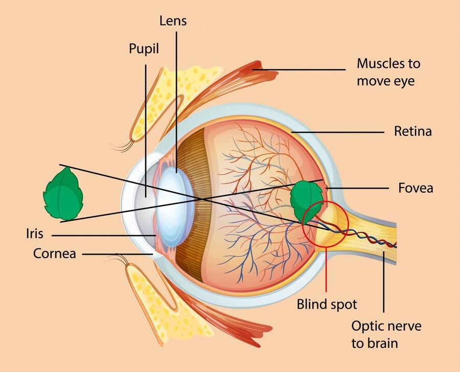
- **Sclera: The white, tough outer covering that protects the eye and maintains its shape.
- **Cornea: The transparent front part of the eye that allows light to enter and helps in focusing.
2. Middle Layer (Vascular Layer / Uvea)
This layer is rich in blood vessels and supplies nutrients.
- **Choroid: Contains blood vessels that nourish the retina and absorb excess light to prevent reflection.
- **Ciliary Body: Contains ciliary muscles that control the shape of the lens during focusing.
- **Iris: The coloured part of the eye that regulates the size of the pupil.
3. Inner Layer (Retina)
The retina is the innermost layer responsible for vision. Contains light-sensitive cells called photoreceptors:
- **Rods: Help in vision in dim light (night vision).
- **Cones: Responsible for colour vision and sharpness.
- **Fovea: A small region with a high concentration of cones for sharp vision.
- **Optic Nerve: Carries visual information from the retina to the brain.
Physiology of Vision
The physiology of the eye explains how vision occurs.
**Step 1: Entry of Light
Light rays enter the eye through the cornea and pass through the aqueous humour, pupil, and lens.
**Step 2: Refraction and Focusing
The cornea and lens bend (refract) the light rays. The lens adjusts its shape (accommodation) to focus light onto the retina.
**Step 3: Image Formation
A real, inverted image is formed on the retina.
**Step 4: Conversion into Nerve Signals
Photoreceptors (rods and cones) convert light into electrical signals.
**Step 5: Transmission to the Brain
These signals are transmitted through the optic nerve to the brain.
**Step 6: Interpretation of Vision
The brain interprets the signals, allowing us to perceive a clear and upright image.
Important Structures
- **Lens: A transparent, flexible structure that focuses light onto the retina.
- **Aqueous Humour: Fluid present between the cornea and lens; maintains pressure and nourishes the eye.
- **Vitreous Humour: A gel-like substance fills the space behind the lens and maintains the shape of the eye.
- **Pupil: Opening in the centre of the iris through which light enters.