Hemibrain (original) (raw)
janelia7_blocks-janelia7_fake_breadcrumb | block
node_title | node_title
node_body | node_body
The hemibrain connectome includes many of the brain areas that scientists are most interested in studying, such as circuits that control learning, memory, and key fly behaviors.
The hemibrain connectome is the largest synaptic-level connectome ever reconstructed. It covers a large portion of the central fly brain, including the mushroom body and central complex circuits critical for associative learning and fly navigation. Clock neuron circuity included in the volume could also provide insights on mechanisms underlying sleep. Furthermore, given available light microscopy image data and previous partial connectome reconstruction in the optic lobe, we also identified most optic lobe neurons entering the cental brain regions enabling analysis of downstream visual processing.
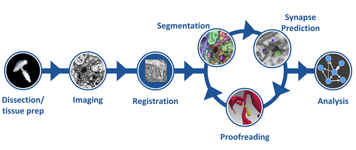
This connectome reconstruction contains around 25,000 neurons, which can be grouped into thousands of distinct cell types spanning several brain regions. This connectome required advances in imaging, segmentation (by the Connectomics Group at Google), and proofreading and analysis software.

Getting started
- Read the overview paper describing the hemibrain and underlying technology.
- Hemibrain is licensed under CC-BY.
- Explore the dataset using neuPrint: an analysis ecosystem for exploring connectomes.
- See the tutorial video below as well as more detailed documentation.
- View data in neuroglancer
- See relevant resources for intensive programmatic access of connectome data below.
- If you have questions, problems, and comments
- Post on relevant software github pages or in the neuPrint user group.
- Connectome reconstruction feedback is possible through neuPrint's neuroglancer interface. We aim to address important issues with our team of proofreaders. See our instructional video.
The FlyEM team has developed free tools to sort through the connectome data, including NeuPrint. Using this tool, scientists can search for particular cell types and identify connection pathways between specific areas or neurons.
Resources and relevant technologies
| Biological exploration & analysis | Compact connection matrix summary v1.2 release (v1.1 release) (v1.0 release) CSV files containing over 20,000 neurons, connections between them, decomposition of connections by brain region, and cell type information How are fly connectomes different from vertebrates? See section 1.2 of our paper View tutorials for using neuPrint or read our technical user guide Detailed notes of dataset revisions and errata |
|---|---|
| Programming and data access | Python access to neuPrint Background on the neuPrint query language in the user guide Documentation of the different types of data available Download neuPrint data for local use Use neutu to proofread data locally Download all reconstruction data and serve locally through DVID neuPrint HTTP API |
| Technologies | Sample preparation Fib-sem imaging Flood-filling segmentation (Google) Synapse prediction Data management with DVID Semi-automated proofreading tools and workflows: neu3 and neutu neuPrint connectome analysis |
Videos of hemibrain circuitry
Neuroscientists are already using connectome data to understand the mechanistic underpinnings of fly behavior. The circuitry shown here is from the fly’s central complex, a region that controls navigation.
To image the Drosophila hemibrain, researchers precisely cut the fly brain into relatively thick slabs using a hot knife. Each slab is then imaged using focused ion beam and scanning electron microscopy (FIB-SEM) to generate data with nanometer isotropic resolution. Afterwards, images from different sections were carefully stitched together. The goal: To create an image volume with as little aberration along the boundaries as possible, to make tracing the paths of neurons through the brain possible.
Researchers have identified more than 4,000 distinct types of neurons in the Drosophila hemibrain. Here are a few of them.
Completing the hemibrain connectome required optimizing every part of the process, from the microscopes that imaged the brain to the algorithms that automatically traced neurons through the dataset. This video gives an overview of the endeavor.
janelia7_blocks-janelia7_block_right_hand_rail | block

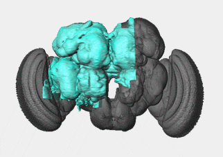
The hemibrain dataset encompasses the part of the fly brain highlighted here in blue. This region includes neurons involved in learning, navigation, smell, vision, and many other functions.
- 2020-12-23 | hemibrain v1.2 released
- neuprint => neuprint+ and mitochondria prediction
- Update: Read our mitochondria analysis here.
- 2020-06 | hemibrain v1.1 released
- 2020-01-22 | hemibrain v1.0 now available for exploring and downloading.
- 2019-12-19 | Reconstruction to make version 1.0 of the hemibrain is complete and being prepared for the upcoming release.
- 2019-10-15 | Hemibrain public data release scheduled for January 22nd