Decades of field data reveal that turtles senesce in the wild (original) (raw)
Significance
Turtles are icons of longevity, popularly characterized as lacking aging and remaining robust as they get older. Indeterminate growth and a positive relationship between body size and fecundity suggest that a greater proportion of reproductive output could come from older, rather than younger, individuals. However, studies of turtle populations are typically too short to empirically test these assertions. We tracked >1,000 painted turtles for >20 y in a population in northwest Illinois, United States. Contrary to traditional thought, successful reproduction and survival declined as the turtles aged. Consistent with life-history theory, the observed senescence in reproduction and survival in this population may be attributable to relatively high extrinsic mortality as a result of human disruption.
Keywords: aging, lifespan, painted turtle, reproduction, senescence
Abstract
Lifespan and aging rates vary considerably across taxa; thus, understanding the factors that lead to this variation is a primary goal in biology and has ramifications for understanding constraints and flexibility in human aging. Theory predicts that senescence—declining reproduction and increasing mortality with advancing age—evolves when selection against harmful mutations is weaker at old ages relative to young ages or when selection favors pleiotropic alleles with beneficial effects early in life despite late-life costs. However, in many long-lived ectotherms, selection is expected to remain strong at old ages because reproductive output typically increases with age, which may lead to the evolution of slow or even negligible senescence. We show that, contrary to current thinking, both reproduction and survival decline with adult age in the painted turtle, Chrysemys picta, based on data spanning >20 y from a wild population. Older females, despite relatively high reproductive output, produced eggs with reduced hatching success. Additionally, age-specific mark–recapture analyses revealed increasing mortality with advancing adult age. These findings of reproductive and mortality senescence challenge the contention that chelonians do not age and more generally provide evidence of reduced fitness at old ages in nonmammalian species that exhibit long chronological lifespans.
Why do some organisms show little to no signs of aging as they get older, whereas others exhibit substantial physiological deterioration and reproductive senescence with advancing age (1, 2)? This question has motivated many studies of aging in the wild, ranging from reviews of demographic aging (3, 4) to compilations of mechanistic studies (e.g., ref. 5 and references therein). Senescence should evolve when selection is weaker on deleterious traits expressed at an old age relative to those expressed at a young age (6). Indeed, mutations with senescent effects can persist because of a tradeoff between beneficial effects early in life and pleiotropic detrimental effects late in life (7, 8). Importantly, the persistence of age-specific deleterious mutations will therefore be influenced by levels and sources of extrinsic mortality (e.g., predation, resource scarcity, infectious disease) and the particular ages at which extrinsic sources cause death (9, 10). For example, the onset of physiological or reproductive deterioration is expected to occur relatively early in populations exposed to high levels of extrinsic mortality of adults. Accordingly, such organisms are expected to evolve rapid development/growth, high reproductive effort at young ages, and a shortened lifespan. For species or populations that experience low extrinsic mortality rates, physiological and reproductive function are expected to decline more slowly with advancing age or with a delay in the onset of senescent decline (7). Moreover, such age-specific selection dynamics can result in negligible (11) and even negative (12) senescence.
To clarify the role of natural selection in shaping both reproductive and general senescence and lifespans, an understanding of not only mortality but also reproductive parameters and their fitness consequences across adult ages is required. For instance, although declining fertility in old age indicates reproductive senescence (13), how the resultant age-related changes in reproductive output translate to lifetime fitness is largely unknown. Indeed, even constant reproductive output with age may not necessarily translate into increasing Darwinian fitness with advancing age; reproduction would actually need to increase if selection against deleterious mutations is to remain strong across the lifespan (14). Intuitively, one would expect that relatively low reproductive output (attributable to declining fertility) would result in reduced fitness, but such an effect may not hold if the quality of offspring (and hence the likelihood that offspring contribute to future generations) differs as individuals age. That is, the relative number of progeny produced by old versus young females may not necessarily reflect age differences in reproductive fitness if there is age-related variation in offspring quality and survival. Finally, whether a postreproductive life stage exists in any given species and how such a postreproductive stage affects an individual’s lifetime reproductive fitness are pressing questions in evolutionary biology (15, 16).
Here, we analyze 24 y of individual-based birth, death, and reproductive data gathered for more than 1,000 marked individuals since 1988 from a population of painted turtles (Chrysemys picta) inhabiting the backwaters of the Mississippi River in the state of Illinois, United States (17). We quantify age-related changes in mortality and reproduction from capture–mark–recapture and reproductive/nesting behavior data of known-age individuals. We assessed hatching success of eggs each September (18, 19), which we use as a measure of fitness. With these unique long-term data we ask the following. (i) How does reproductive output and reproductive behavior change with advancing age? (ii) How does fitness change with advancing age? (iii) How does mortality change with advancing age? We then interpret our findings in the context of evolutionary theories for rates of senescence.
Results
Age-Specific Reproduction.
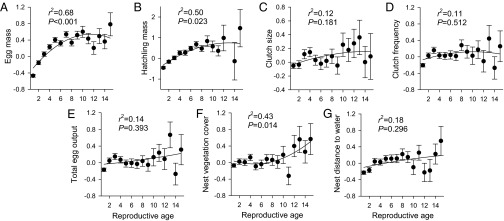
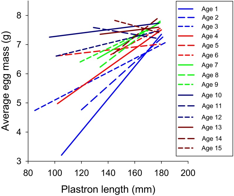
Survival and physical data were collected on 2,234 nests constructed by 600 painted turtles with known identities and reproductive ages (Table S1). Although reproductive and nest microhabitat variables generally increased with age, only egg mass and nest vegetation cover exhibited a statistically significant increase (Fig. 1). Importantly, however, our analyses detected a significant interactive effect of age × plastron length on egg mass (_F_14,891 = 6.3; P < 0.001) such that the generally positive relationship between female body size and egg size waned with older ages: the slopes declined and the y intercepts increased with advancing age (Fig. 2). These findings imply that investment into egg size is similar between relatively small and large late-age females, but large late-age females invest more than smaller females of all other age classes [_sensu_ (20)]. Thus, the relatively large eggs produced by old females resulted in larger hatchlings than those produced by young females, irrespective of maternal body size (Fig. 1_B_).
Table S1.
Age-specific sample sizes of females and nests used in our statistical analyses and percentage of depredated nests
| Reproductive age (y) | Excluding depredated nests | Including depredated nests | Nests predated (%) | ||
|---|---|---|---|---|---|
| Females | Nests | Females | Nests | ||
| 1 | 226 | 276 | 590 | 718 | 61.6 |
| 2 | 105 | 151 | 231 | 319 | 52.7 |
| 3 | 59 | 87 | 168 | 245 | 64.5 |
| 4 | 55 | 75 | 166 | 233 | 67.8 |
| 5 | 35 | 51 | 133 | 184 | 72.3 |
| 6 | 42 | 68 | 100 | 145 | 53.1 |
| 7 | 40 | 51 | 85 | 119 | 57.1 |
| 8 | 17 | 35 | 58 | 89 | 60.7 |
| 9 | 6 | 11 | 39 | 59 | 81.4 |
| 10 | 5 | 10 | 21 | 31 | 67.7 |
| 11 | 8 | 15 | 20 | 32 | 53.1 |
| 12 | 7 | 7 | 13 | 17 | 58.8 |
| 13 | 1 | 1 | 9 | 15 | 93.3 |
| 14 | 1 | 2 | 13 | 17 | 88.2 |
| 15 | 4 | 5 | 7 | 11 | 54.6 |
| Total unique females or nests | 355 | 845 | 600 | 2,234 | 62.2 |
Fig. 1.
Age-specific variation in reproductive and nesting parameters in adult painted turtles (C. picta). Graphs show age differences in egg mass (A), hatchling mass (B), clutch size (C), clutch frequency (D), total egg output (E), vegetation cover over nest (F), and distance of nest to water (G). Dependent variables are standardized to a mean of 0 and unit variance. Statistical values are results from quadratic regressions. Data represent least-square means (and 1 SE) calculated from linear mixed models, with plastron length as a covariate and maternal identity as a random effect. Reproductive ages 1–14 reflect actual ages of 5–7 to 18–20 y old, and reproductive age 15 includes individuals that are ≥20 y old.
Fig. 2.
Age-specific relationships between plastron length and egg mass. Graphs show the age-specific slopes (A) and y intercepts (B) from regression analyses of plastron length versus egg mass (C). The negative relationship (_r_2 = 0.53; P = 0.002) between reproductive age and slope coupled with the positive relationship (_r_2 = 0.75; P < 0.001) between reproductive age and y intercept illustrate that late-age females invest less per egg than do young females at relatively large body sizes. At small body sizes, old females invest more per egg than do young females. C illustrates the relationships between plastron length and egg mass for young (reproductive ages 1–3) and old (reproductive ages 13–15) mothers. The range of body sizes for old mothers was relatively narrow, but young mothers exhibited a large range of sizes that encompassed the size range of old mothers. Regression lines for all ages are in Fig. S1.
Fig. S1.
Relationship between plastron length and egg mass for females of all reproductive ages. As reproductive age increases, the range of body sizes for mothers narrows, but young mothers exhibited a large range of sizes that encompassed the size range of old mothers.
Age-Specific Fitness.
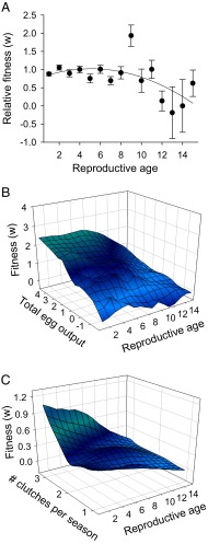
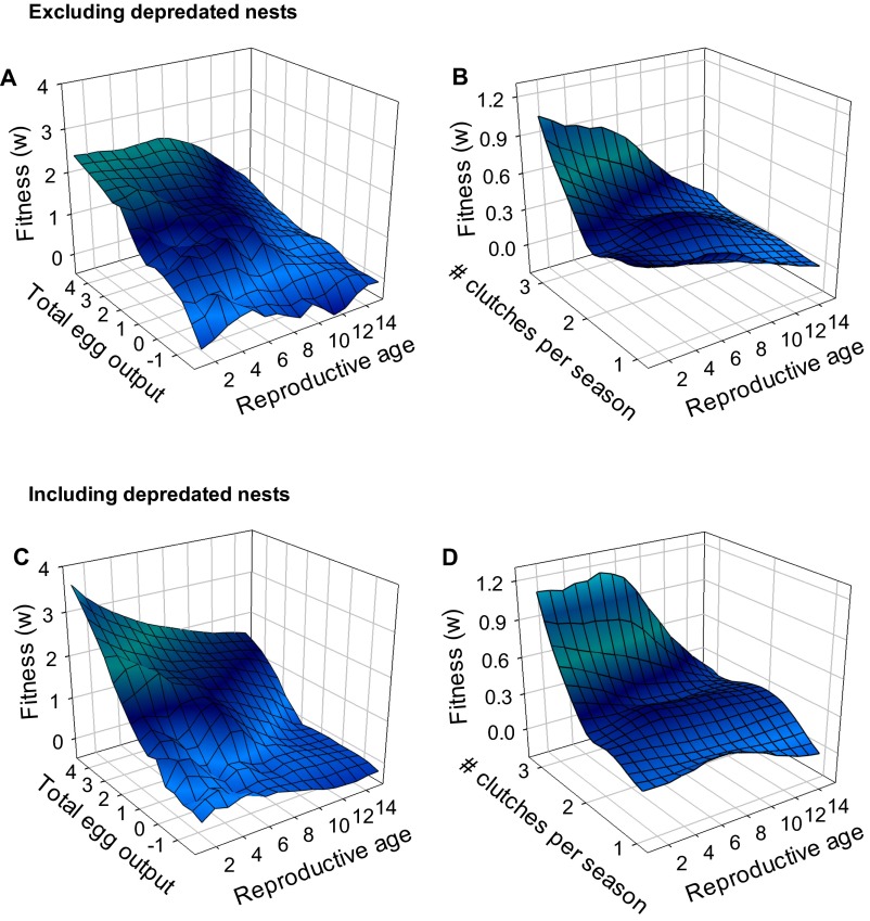
Females that produced large eggs and frequent clutches yielded more successfully hatched offspring than individuals that produced small eggs and infrequent clutches (P values, <0.05; Table 1). Fitness (number of eggs hatched per year) declined in relatively old turtles despite a moderate age-related increase in total egg output (number of eggs produced each year; Fig. 3). Moreover, fitness was influenced by the interactions of total egg output and the number of clutches produced at each reproductive age (Fig. 3). Specifically, fitness declined with age, particularly for individuals with relatively high seasonal egg output (i.e., defined as either total egg output or clutch frequency per season); in animals with overall low egg output, an age-related decline in fitness was not evident. Because depredated nests were excluded from these analyses, extrinsic mortality of eggs attributable to nest predation did not drive this age-related variation in hatching success (patterns were generally similar when depredated nests were included in the analyses; Figs. S2–S4).
Table 1.
Results from two final models of selection on age-specific reproductive traits in painted turtles (C. picta)
| Effect | Parameter estimate, SE | ndf, ddf | F | P |
|---|---|---|---|---|
| Excluding depredated nests | ||||
| Reproductive age | −0.0520 (0.01) | 1, 166 | 18.5 | <0.001 |
| Plastron length | 0.0751 (0.04) | 1, 166 | 3.6 | 0.059 |
| No. of clutches produced | −0.2828 (0.13) | 1, 166 | 5.1 | 0.025 |
| Egg mass | 0.0913 (0.04) | 1, 166 | 5.3 | 0.023 |
| Clutch size | −0.0183 (0.06) | 1, 166 | 0.1 | 0.763 |
| Total egg output per season | 0.5483 (0.13) | 1, 166 | 16.7 | <0.001 |
| Nest vegetation cover | 0.0611 (0.03) | 1, 166 | 3.5 | 0.063 |
| Nest distance to water | −0.0312 (0.03) | 1, 166 | 0.9 | 0.439 |
| Age × number of clutches produced | 0.0453 (0.02) | 1, 166 | 6.9 | 0.009 |
| Age × total egg output per season | −0.0400 (0.02) | 1, 166 | 6.8 | 0.010 |
| Including depredated nests | ||||
| Reproductive age | −0.0432 (0.02) | 1, 763 | 6.0 | 0.014 |
| Plastron length | 0.0694 (0.07) | 1, 763 | 1.1 | 0.293 |
| No. of clutches produced | 0.0500 (0.13) | 1, 763 | 0.2 | 0.691 |
| Egg mass | 0.0323 (0.06) | 1, 763 | 0.3 | 0.616 |
| Clutch size | −0.1237 (0.08) | 1, 763 | 2.2 | 0.140 |
| Total egg output per season | 0.4019 (0.14) | 1, 763 | 8.1 | 0.005 |
| Nest vegetation cover | 0.1580 (0.05) | 1, 763 | 10.0 | 0.002 |
| Nest distance to water | −0.0097 (0.05) | 1, 763 | 0.0 | 0.852 |
Fig. 3.
Age-specific variation in relative fitness for adult painted turtles (C. picta). Reproductive ages 1–14 reflect actual ages of 5–7 to 18–20 y old, and reproductive age 15 includes individuals that are ≥20 y old. (A) Change in relative fitness (egg-hatching success) with reproductive age (least-square means ± 1 SE). (B) Interaction between total egg output and reproductive age. Reproductive output is represented as the total number of eggs produced (per female) within a season. (C) Interaction between total number of clutches produced per season and reproductive age. Statistical results are in Table 1. These graphs are based on analyses that excluded depredated nests.
Fig. S2.
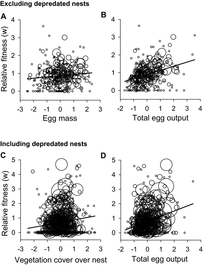
(A and B) Bubble plots for relative fitness versus egg mass (_r_2 = 0.02; P = 0.019) (A) and total egg output (_r_2 = 0.16; P < 0.001) (_B_) for analyses that excluded depredated nests. (_C_ and _D_) Bubble plots for nest vegetation cover (_r_2 = 0.01; _P_ = 0.010) (_C_) and total egg output (_r_2 = 0.05; _P_ < 0.001) (_D_) for analyses that included depredated nests. For graphs _C_ and _D_, six outliers with fitness >7 were removed for visualization but were retained in the statistical analyses. Regression analyses were weighted by the sample size for each female, which reflect the relative size of the bubble (e.g., large bubbles contain multiple data points from single females). Additional statistical results for these relationships are in Table 1.
Fig. S4.
Selection surfaces for reproductive age and reproductive output for adult female painted turtles (C. picta). The top graphs (A and B) illustrate results from analyses that excluded depredated nests, and the bottom graphs (C and D) illustrate results from analyses that included depredated nests. Reproductive output is represented as the total number of eggs produced (per female) within a season (A and C) and number of clutches produced per season (B and D). Statistical results are in Table 1.
Fig. S3.
Age-specific variation in relative fitness for analyses that excluded (A) and included (B) depredated nests. Data represent least-square means (and 1 SE) calculated from linear mixed models and maternal identity as a random effect. Reproductive ages 1–14 reflect actual ages of 5–7 to 18–20 y old, and reproductive age 15 includes individuals that are ≥20 y old.
Age-Specific Mortality.
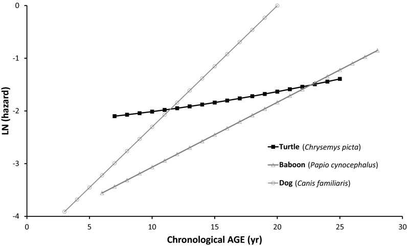
Mortality analysis that included encounter histories (capture–mark–recapture) for 1,031 females from 1993 to 2012 revealed measurable mortality senescence [i.e., accelerating mortality probability with advancing age (Fig. 4)]. The Gompertz function [u x = _A_e_b_x, where u x is the age-specific mortality hazard, A is the initial mortality rate at first nesting (i.e., the first year of the adult stage), and b is the rate of increasing mortality] had the lowest Deviance Information Criterion (DIC) value among those models considered (Table 2). Estimated parameters were A (initial mortality rate/year) = 0.102 (95% credible interval: 0.090–0.116) and b = 0.050 (95% credible interval: 0.034–0.067). This result corresponds to a doubling of mortality every 13.8 y (10.3–20.2 y). Two additional mortality functions are included in Fig. 4 to provide comparison with mammalian-type curves for baboons [_u_ _x_ = 0.028e0.123x (21)] and domestic dogs [_u_ _x_ = 0.02e0.23x (22)].
Fig. 4.
Fitted Gompertz models (natural logarithm transformed) of age-specific mortality for painted turtles and comparison species [data for baboons (21, 52) and dogs (Gompertz parameters from ref. 22, data from ref. 53); see Results, Age-Specific Mortality for parameter estimates]. Age is chronological age for all species. For turtles, although initial adult mortality was relatively high, mortality rates increased at a much slower rate than seen in many mammals.
Table 2.
Hierarchical capture–mark–recapture models used to describe mortality patterns for turtles
| Model | DIC | Δ_DIC_ | pD | k | ||
|---|---|---|---|---|---|---|
| Function | Makeham? | Temporal? | ||||
| Exponential | No | no | 8,198.8 | 833.7 | 292.8 | 2 |
| Exponential | No | yes | 8,004.9 | 639.8 | 328.1 | 21 |
| Gompertz | No | no | 7,466.0 | 100.9 | 242.2 | 3 |
| Gompertz | No | yes | 7,365.1 | 0.0 | 300.3 | 22 |
| Gompertz | Yes | no | 7,440.9 | 75.8 | 220.4 | 4 |
| Gompertz | Yes | yes | 7,435.6 | 70.5 | 381.6 | 23 |
| Logistic | No | no | 7,473.6 | 108.5 | 246.6 | 4 |
| Logistic | No | yes | 7,427.5 | 62.4 | 368.8 | 23 |
| Logistic | Yes | no | * | * | * | 5 |
| Logistic | Yes | yes | * | * | * | 24 |
| Weibull | No | no | 8,170.8 | 805.6 | 395.6 | 3 |
| Weibull | No | yes | 7,880.2 | 515.1 | 294.7 | 22 |
| Weibull | Yes | no | * | * | * | 4 |
| Weibull | Yes | yes | * | * | * | 23 |
Discussion
Survival senescence has generally been thought to be negligible or absent in various reptiles (refs. 23–27) but see ref. 3) because traits that either protect against predators (such as external ribcages and venom; reviewed in ref. 28) or that increase reproductive success with advancing age (such as indeterminate growth and fecundity) may overcome the declining power of natural selection with advancing age that characterizes natural populations (10, 14). In the specific case of C. picta, which has both adult-protective morphology and indeterminate growth and fecundity, lifetime fitness would increase if individuals maintained successful and increasing reproduction through adulthood. Such steady or even increasing reproduction with advancing age is particularly likely for species that exhibit low rates of extrinsic mortality at old ages, indeterminate growth, or a positive relationship between body size and fecundity (e.g., ref. 26) [i.e., many reptiles, including turtles (27)]. Although our aim was to understand whether and how indeterminate growth and fecundity may offset declining late-life selection, our 24-y dataset on C. picta revealed instead a significant decline in both reproductive success and adult survival for females. Moreover, when we tested among competing models for mortality acceleration, some of which would allow deceleration later in life, the Gompertz model had the most support (Table 2), suggesting increasing mortality acceleration over the entire adult lifespan (Fig. 4).
Our results differ from those for another C. picta population, which exhibits both overall low mortality and no differences in survivorship of old- versus middle-aged females (23). Although the survivorship curves of ref. 23 are consistent with gently increasing mortality with advancing age in that population, for direct tests of senescence, it is important to estimate and analyze age-specific mortality hazards as we have done here (29). Thus, estimating the rate and form of mortality senescence by computing instantaneous mortality rates revealed that the present study population of C. picta is characterized by mortality senescence (i.e., accelerating mortality with advancing age). Our study population differs from that of ref. 23 in that ours has experienced a relatively long history of human-driven mortality. The extent to which this human-induced mortality is in any way age-dependent is unknown. Specific human-induced mortality derives primarily from injuries to nesting females (e.g., when crossing roads) and injuries to adults of both sexes in the water column (e.g., by boating activity). Were these injuries to manifest in an age-dependent manner across the lifespan, the age-specific selection gradients could be altered. In contrast, if the increased mortality affected all adult ages equally, we would expect acceleration of the entire life history, because the population age structure would become skewed toward young ages. A comparison of the high mortality population (this study) and a lower mortality population (23) supports this contention. Specifically, here we detected rapid young-adult growth rates (Fig. S5), increased reproductive effort at young adult ages, and reduced lifespan of turtles in our population. Moreover, extensive demographic analyses of prematuration growth in our population indicated faster individual growth and earlier maturation than in less human-impacted populations (30). Thus, these two painted turtle populations fit strikingly well with predictions from life-history theory based on the force and sensitive ages of mortality (14, 31) (Fig. S4). Overall then, these studies support the notion that the life-history strategy is a set of correlated traits that respond predictably to natural selection.
Fig. S5.
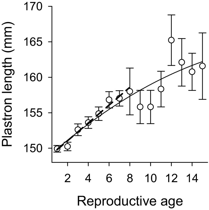
Relationship between reproductive age and plastron length (quadratic regression: _r_2 = 0.82; P < 0.001). For meaningful comparison with other studies, we also quantified this relationship up to reproductive age 8 using linear regression (dotted line). Linear regression equation: plastron length = 1.24*(age) + 149; _r_2 = 0.97; P < 0.001. The slope of the relationship between reproductive age and plastron length is substantially higher in our study population than that reported in Michigan populations [slope = 1.2 at our site versus 0.5, 0.55, and 0.75 at other sites (54–56)], suggesting relatively fast growth rates in our population. The same pattern is evident when plastron length is standardized to a mean of 0 and unit variance within years.
Interestingly, in contrast to reproduction and survival, measures related to body size and behavior did not exhibit senescence (Fig. 1 and Fig. S5). This result accords with evolutionary senescence theory in that such traits (i.e., growth and learning, respectively) could buffer selective forces that accelerate death and infertility rates (9, 32). At the same time, hatching success and adult mortality—traits that are substantially influenced by physiological processes (27)—exhibited senescence. Long-term research that not only assesses shifts in survival and reproductive parameters with age but also quantifies age-related changes in physiology and offspring fitness may reveal that senescent patterns in long-lived reptiles are common (see also refs. 33, 34). In addition, long-term studies that explore tradeoffs between early- and late-life performance will provide crucial information for better understanding these declines in reproduction or survival as individuals age. For example, greater resource allocation to early-life growth or reproduction is expected to come at a cost to late-life performance that can lead to age-related declines in reproduction and survival [i.e., disposable soma theory of aging (35)]. These patterns have been observed in many vertebrates (36), including reptiles (37, 38), and could contribute to the reproductive and survival senescence observed here in C. picta.
Determining the universality of reproductive aging has received great recent interest both in terms of documenting its existence and calculating whether reproductive aging occurs at similar (vs. faster) rates than general senescence (13). Analyses have taken the form of either measuring the rate of declining fertility and reproductive success with advancing age (39, 40) or measuring the distribution of ages of last successful reproduction and comparing that to the distribution of deaths (16). Our analysis is similar to the former of these approaches, and we indeed document reproductive decline with advancing age, although not in measures traditionally used (such as rates of conception) but rather by calculating egg-hatching success. Similarly, Congdon et al. (24) reported greater embryo mortality attributable to arrested development in eggs produced by the oldest female age class in Blanding’s turtles. Other long-lived species tend to exhibit reproductive senescence at rates that are faster than somatic aging and that ends at an earlier age than death, which yields a postreproductive lifespan [primates including humans (16), killer whales (41), but see elephants (42, 43)]. These mammalian species may exhibit reproductive senescence attributable to a shelf-life of their primary oocytes, but this possibility is unlikely the case in C. picta because reptiles are not oocyte-limited (44). One firm conclusion is that reproductive senescence in turtles and other ectothermic amniotes cannot be attributable to adaptive explanations invoked in the mammalian literature [e.g., the “grandmother hypothesis,” which largely rests on the role of parental care to increase one’s own fitness or the fitness of relatives (45)].
A critical question is whether senescent changes in mortality (as in primates) or in reproduction drive declines in fitness. In reptiles, which are often characterized as indeterminate growers, few studies document either mortality senescence or the decline in some physiological trait with advancing adult age (3). Because many species with indeterminate growth increase fecundity with increasing age (attributable in part to increasing body size), the question becomes, at what level of increased fecundity will selection cease declining with advancing age? Although we demonstrate mortality senescence in our population of C. picta, the age-specific increase in mortality is considerably less rapid than that reported for mammalian taxa (e.g., Fig. 4), which is consistent with the view that rates of senescence in these long-lived reptiles are relatively slow. These divergent patterns may be explained by very different rates of extrinsic mortality in turtles (or their ancestors) compared with mammals. Indeed, because of their protective morphology (shell) and lack of senescence in immune function (46), turtles likely experience considerably less late-age mortality attributable to extrinsic factors than do mammals. This comparison between turtles and mammals fits with theoretical expectations for patterns of survival senescence in these taxa, but our results challenge the traditional view that long-lived ectotherms, such as turtles, do not exhibit declining reproductive function with advancing age. Indeed, declines in fitness with age will be exacerbated by a combination of increased mortality rate and decreased reproductive success, as we demonstrate in these long-lived ectotherms.
Methods
Since 1988, nesting patterns of painted turtles (C. picta) have been continually monitored at the Thomson Causeway Recreation Area in northwestern Illinois (Carroll County; 41°57′N, 90°7′W) (10–12). Unmarked females exhibiting 5–7 annuli on the pectoral scutes were classified as primiparous (i.e., a reproductive age of one, chronological age 5–7 y old). Thus, all future recaptures of these animals were at a known age (20). Individuals that could not be assigned ages confidently were excluded from analyses of reproduction unless the turtles were recaptured across a 15+ y time span, in which case, data collected from captures ≥15 y after initial capture were included in the oldest age class (i.e., chronological age ≥20 y old). During each nesting season (mid-May to early July), the study area was monitored intensively for nesting turtles. All females at the nesting grounds were individually marked, and their eggs were weighed and counted (typically within 4 h of oviposition). All eggs were returned to their nests and allowed to incubate naturally, and nests were checked for predation almost daily until the end of June of each year. Microhabitat data (shade cover and distance from the water) were also collected for each nest to assess age-related changes in nesting behavior. Nesting behaviors are important because maternal choice of nest microhabitat differs among age classes (47) and can influence egg survival (18). These data are available in the Dryad Digital Repository (dx.doi.org/10.5061/dryad.g2r87).
This research has been approved by the Iowa State University Institutional Animal Care and Use Committee and the Illinois Department of Natural Resources.
Age-Specific Reproduction.
Analyses of age-specific fitness included 600 individuals with known ages captured from 1997 to 2010. Of these individuals, 65% were observed nesting more than once, and most were first-time nesters (Table S1). Thus, our comparisons across ages were largely based on longitudinally sampled individuals. Linear mixed models were used to evaluate age-specific variation in reproductive variables (i.e., egg mass, clutch size, clutch frequency, total egg output per female per season, hatchling mass) and nest microhabitat variables (i.e., nest shade cover, distance from nest site to nearest water). In cases where females nested two or three times per season, within-season mean female values were used for egg mass, clutch size, nest shade cover, and nest distance to water. Each dependent variable was standardized to a mean of 0 and unit variance within each year before analysis. Reproductive age (1–15 y) was defined as a fixed main effect, and plastron length was a covariate. The interaction term of plastron length × reproductive age was removed from models when it was not significant, which occurred in all cases except in the analysis of egg mass. Maternal identity was included as a random effect to account for repeated measurements on females across ages. Linear and second-order polynomial regressions were used to detect age-specific trends in reproductive and nest microhabitat variables.
Age-specific Fitness.
We used generalized linear mixed models to test for linear selection on each reproductive variable and interactions with reproductive age using egg-hatching success per female as a measure of fitness, standardized within season [i.e., ωrelative = ω/ωmean (14)]. This measure of fitness (number of successfully hatched eggs per clutch) strongly correlates with the likelihood of offspring recruiting into future adult age classes (17) and is therefore a meaningful proxy for maternal reproductive success (i.e., fitness). Independent variables (standardized to a mean of 0 with unit variance) included plastron length, egg mass, clutch size, clutch frequency, nest shade cover, distance from the nest site to nearest water, and total egg output per female per season, as well as all two-way interactions with reproductive age. Three-way and higher-order interactions were not included in the model because they were not relevant to our specific questions, and models with higher-level interactions often would not converge, because degrees of freedom were reduced by including these factors. Nonsignificant interaction terms were sequentially removed from the initial model, and final models were selected based on the lowest Akaike Information Criterion (AIC) score. Again, maternal identity was included as a random effect to account for repeated measurements on females across ages. Parameter estimates for fixed effects were calculated using restricted maximum likelihood and statistical significance was determined with F tests.
The selection analyses described above were performed twice. First, to assess non–predation-related variation in fitness, all depredated nests were excluded from our analyses. Second, to assess how nest predation impacts the relationship between the reproductive parameters and fitness (i.e., selection), all depredated nests (depredated nests had a fitness of 0) were included in the second set of analyses. Standardizations of all traits (including fitness) were recalculated accordingly for the subsets of nests used in each analysis. Results from analyses that included versus excluded depredated nests were compared qualitatively. Because the potential fate of eggs in depredated nests (had they been left intact) is unknown, this qualitative comparison assumes that nest predation is random with respect to the variables measured. A generalized linear mixed model using reproductive traits (egg mass, clutch size, clutch number) and nest microhabitat (vegetation cover, nest distance to water) as independent variables and maternal identity as a random effect showed that this assumption was met (Table S2).
Table S2.
Effect of reproductive and nest variables on nest predation
| Trait | ndf | ddf | F | P |
|---|---|---|---|---|
| Egg mass | 1 | 1,876 | 0.01 | 0.925 |
| Clutch size | 1 | 1,876 | 0.14 | 0.707 |
| Clutch no. | 1 | 1,876 | 2.54 | 0.112 |
| Nest vegetation cover | 1 | 1,876 | 0.37 | 0.545 |
| Nest distance to water | 1 | 1,876 | 3.02 | 0.083 |
Additional analyses were performed to confirm and visualize significant effects. Regression analyses (weighted by sample size because of repeated measures per female) were used to assess the relationship between fitness and reproductive and nest variables. Second-order polynomial regressions were used to assess age-specific trends in fitness. Lastly, to visualize two-way interactions between age and reproductive output on fitness, selection surfaces were calculated with a first-degree polynomial locally weighted scatterplot smoothing technique (for total egg output) and a negative exponential smoothing technique (for number of clutches produced) in Sigma Plot.
Age-Specific Mortality.
Analyses of age-specific mortality included 1,031 individuals. These individuals included the 600 used in the analyses above but also included turtles with unknown ages that were captured from 1993 to 2012. To test for a signature of demographic senescence in mortality, accelerating mortality models were fit to mark–recapture data for both known age and unknown age females using a Bayesian hierarchical analysis (48, 49). DIC was used to choose among alternative models for the survival function fit using the BaSTA (Bayesian Survival Trajectory Analysis) package (48) (version 1.3) in R (version 2.14; R development Core Team 2011) (Table 2). Four survival functions were considered: exponential, Gompertz, logistic, and Weibull functions (49, 50). In the exponential model, survival is constant across ages, whereas the other three models all allow for decreasing survival as individuals become older. Thus, the alternative models permitted assessment of whether there was evidence for demographic senescence and, if so, what form the senescence took. In addition, models that included an age-independent constant to the survival function (Makeham coefficient) were considered for the Gompertz, logistic, and Weibull models. The approach does not allow for temporal or cohort effects to be incorporated. However, previous work detected no evidence for an overall temporal trend in survival across the study period (51) or for cohort effects on survival related to developmental conditions in the population (38). Finally, alternatives that differed in whether detection was constant or variable among all years were examined.
Acknowledgments
We thank the many volunteers, students, and postdoctoral researchers who have been involved with fieldwork at Turtle Camp over the past decades. We thank members of F.J.J.’s laboratory and T. Schwartz for helpful comments on earlier drafts of this paper and J. Sherwood for assistance with ArcGIS software. We acknowledge ongoing support from the US Fish and Wildlife Service and the US Army Corp of Engineers. Funding for this research was provided by National Science Foundation Grants DEB-9629529, DEB-0089680, DEB-0640932, and DEB-1242510 (to F.J.J.). Additional support was provided by a National Institutes of Health Grant R01AG049416.
Footnotes
The authors declare no conflict of interest.
This article is a PNAS Direct Submission.
Data deposition: The reproductive data and encounter histories reported in this paper have been deposited in the Dryad Digital Repository (dx.doi.org/10.5061/dryad.g2r87).
See Commentary on page 6328.
References
- 1.Austad SN. Methusaleh’s Zoo: How nature provides us with clues for extending human health span. J Comp Pathol. 2010;142(Suppl 1):S10–S21. doi: 10.1016/j.jcpa.2009.10.024. [DOI] [PMC free article] [PubMed] [Google Scholar]
- 2.Holmes DJ, Ottinger MA, Ricklefs RE, Finch CE. Sosa-2: Introduction to the proceedings of the second symposium on organisms with slow aging. Exp Gerontol. 2003;38(7):721–722. [Google Scholar]
- 3.Nussey DH, Froy H, Lemaitre J-F, Gaillard J-M, Austad SN. Senescence in natural populations of animals: Widespread evidence and its implications for bio-gerontology. Ageing Res Rev. 2013;12(1):214–225. doi: 10.1016/j.arr.2012.07.004. [DOI] [PMC free article] [PubMed] [Google Scholar]
- 4.Roach DA, Carey JR. Population biology of aging in the wild. Annu Rev Ecol Evol Syst. 2014;45:421–443. [Google Scholar]
- 5.Fletcher QE, Selman C. Aging in the wild: Insights from free-living and non-model organisms. Exp Gerontol. 2015;71:1–3. doi: 10.1016/j.exger.2015.09.015. [DOI] [PubMed] [Google Scholar]
- 6.Charlesworth B. Fisher, Medawar, Hamilton and the evolution of aging. Genetics. 2000;156(3):927–931. doi: 10.1093/genetics/156.3.927. [DOI] [PMC free article] [PubMed] [Google Scholar]
- 7.Hamilton WD. The moulding of senescence by natural selection. J Theor Biol. 1966;12(1):12–45. doi: 10.1016/0022-5193(66)90184-6. [DOI] [PubMed] [Google Scholar]
- 8.Williams GC. Pleiotropy, natural selection, and the evolution of senescence. Evolution. 1957;11(4):398–411. [Google Scholar]
- 9.Williams PD, Day T. Antagonistic pleiotropy, mortality source interactions, and the evolutionary theory of senescence. Evolution. 2003;57(7):1478–1488. doi: 10.1111/j.0014-3820.2003.tb00356.x. [DOI] [PubMed] [Google Scholar]
- 10.Baudisch A. Hamilton’s indicators of the force of selection. Proc Natl Acad Sci USA. 2005;102(23):8263–8268. doi: 10.1073/pnas.0502155102. [DOI] [PMC free article] [PubMed] [Google Scholar]
- 11.Finch CE. Update on slow aging and neglibible senescence - a mini-review. Gerontology. 2009;55(3):307–313. doi: 10.1159/000215589. [DOI] [PubMed] [Google Scholar]
- 12.Vaupel JW, Baudisch A, Dölling M, Roach DA, Gampe J. The case for negative senescence. Theor Popul Biol. 2004;65(4):339–351. doi: 10.1016/j.tpb.2003.12.003. [DOI] [PubMed] [Google Scholar]
- 13.Kirkwood TBL, Shanley DP. The connections between general and reproductive senescence and the evolutionary basis of menopause. Ann N Y Acad Sci. 2010;1204:21–29. doi: 10.1111/j.1749-6632.2010.05520.x. [DOI] [PubMed] [Google Scholar]
- 14.Charlesworth BC. Evolution in Age-Structured Populations. 2nd Ed Cambridge Univ Press; Cambridge, UK: 1994. [Google Scholar]
- 15.Cohen AA. Female post-reproductive lifespan: A general mammalian trait. Biol Rev Camb Philos Soc. 2004;79(4):733–750. doi: 10.1017/s1464793103006432. [DOI] [PubMed] [Google Scholar]
- 16.Alberts SC, et al. Reproductive aging patterns in primates reveal that humans are distinct. Proc Natl Acad Sci USA. 2013;110(33):13440–13445. doi: 10.1073/pnas.1311857110. [DOI] [PMC free article] [PubMed] [Google Scholar]
- 17.Schwanz LE, Spencer RJ, Bowden RM, Janzen FJ. Climate and predation dominate juvenile and adult recruitment in a turtle with temperature-dependent sex determination. Ecology. 2010;91(10):3016–3026. doi: 10.1890/09-1149.1. [DOI] [PubMed] [Google Scholar]
- 18.Warner DA, Jorgensen CF, Janzen FJ. Maternal and abiotic effects on egg mortality and hatchling size of turtles: Temporal variation in selection over seven years. Funct Ecol. 2010;24(4):857–866. [Google Scholar]
- 19.Janzen FJ, Warner DA. Parent-offspring conflict and selection on egg size in turtles. J Evol Biol. 2009;22(11):2222–2230. doi: 10.1111/j.1420-9101.2009.01838.x. [DOI] [PubMed] [Google Scholar]
- 20.Bowden RM, Harms HK, Paitz RT, Janzen FJ. Does optimal egg size vary with demographic stage because of a physiological constraint? Funct Ecol. 2004;18(4):522–529. [Google Scholar]
- 21.Bronikowski AM, et al. Aging in the natural world: Comparative data reveal similar mortality patterns across primates. Science. 2011;331(6022):1325–1328. doi: 10.1126/science.1201571. [DOI] [PMC free article] [PubMed] [Google Scholar]
- 22.Finch CE, Pike MC, Witten M. Slow mortality rate accelerations during aging in some animals approximate that of humans. Science. 1990;249(4971):902–905. doi: 10.1126/science.2392680. [DOI] [PubMed] [Google Scholar]
- 23.Congdon JD, et al. Testing hypotheses of aging in long-lived painted turtles (Chrysemys picta) Exp Gerontol. 2003;38(7):765–772. doi: 10.1016/s0531-5565(03)00106-2. [DOI] [PubMed] [Google Scholar]
- 24.Congdon JD, Nagle RD, Kinney OM, van Loben Sels RC. Hypotheses of aging in a long-lived vertebrate, Blanding’s turtle (Emydoidea blandingii) Exp Gerontol. 2001;36(4-6):813–827. doi: 10.1016/s0531-5565(00)00242-4. [DOI] [PubMed] [Google Scholar]
- 25.Jones OR, et al. Diversity of ageing across the tree of life. Nature. 2014;505(7482):169–173. doi: 10.1038/nature12789. [DOI] [PMC free article] [PubMed] [Google Scholar]
- 26.Olsson M, Shine R. Does reproductive success increase with age or with size in species with indeterminate growth? A case study using sand lizards (Lacerta agilis) Oecologia. 1996;105(2):175–178. doi: 10.1007/BF00328543. [DOI] [PubMed] [Google Scholar]
- 27.Schwartz TS, Bronikowski AM. Molecular stress pathways and the evolution of life histories in reptiles. In: Flatt T, Heyland A, editors. Molecular Mechanisms of Life History Evolution: The Genetics and Physiology of Life History Traits and Trade-Offs. Oxford Univ Press; Oxford: 2011. pp. 193–209. [Google Scholar]
- 28.Blanco MA, Sherman PW. Maximum longevities of chemically protected and non-protected fishes, reptiles, and amphibians support evolutionary hypotheses of aging. Mech Ageing Dev. 2005;126(6-7):794–803. doi: 10.1016/j.mad.2005.02.006. [DOI] [PubMed] [Google Scholar]
- 29.Bronikowski AM, Flatt T. Aging and its demographic measurement. Nat Educ Knowl. 2010;3(10):3. [Google Scholar]
- 30.Spencer RJ, Janzen FJ. Demographic consequences of adaptive growth and the ramifications for conservation of long-lived organisms. Biol Conserv. 2010;143(9):1951–1959. [Google Scholar]
- 31.Stearns SC, Ackermann M, Doebeli M, Kaiser M. Experimental evolution of aging, growth, and reproduction in fruitflies. Proc Natl Acad Sci USA. 2000;97(7):3309–3313. doi: 10.1073/pnas.060289597. [DOI] [PMC free article] [PubMed] [Google Scholar]
- 32.Bronikowski AM, Promislow DEL. Testing evolutionary theories of aging in wild populations. Trends Ecol Evol. 2005;20(6):271–273. doi: 10.1016/j.tree.2005.03.011. [DOI] [PubMed] [Google Scholar]
- 33.Massot M, et al. An integrative study of ageing in a wild population of common lizards. Funct Ecol. 2011;25(4):848–858. [Google Scholar]
- 34.Nussey DH, Coulson T, Festa-Bianchet M, Gaillard JM. Measuring senescence in wild animal populations: Towards a longitudinal approach. Funct Ecol. 2008;22(3):393–406. [Google Scholar]
- 35.Kirkwood TBL, Rose MR. Evolution of senescence: Late survival sacrificed for reproduction. Philos Trans R Soc Lond B Biol Sci. 1991;332(1262):15–24. doi: 10.1098/rstb.1991.0028. [DOI] [PubMed] [Google Scholar]
- 36.Lemaitre JF, et al. Early-late life trade-offs and the evolution of ageing in the wild. Proc R Soc Lond B. 2015;282(1806):20150209. doi: 10.1098/rspb.2015.0209. [DOI] [PMC free article] [PubMed] [Google Scholar]
- 37.Massot M, Aragón P. Phenotypic resonance from a single meal in an insectivorous lizard. Curr Biol. 2013;23(14):1320–1323. doi: 10.1016/j.cub.2013.05.047. [DOI] [PubMed] [Google Scholar]
- 38.Miller DAW, Janzen FJ, Fellers GM, Kleeman PM, Bronikowski A. Biodemography of ectothermic tetrapods provides insights into the evolution and plasticity of mortality trajectories. In: Weinstein M, Lane MA, editors. Sociality, Hierarchy, Health: Comparative Demography Advances in Biodemography: Cross-Species Comparisons of Social Environments and Social Behaviors, and Their Effects on Health and Longevity. Natl Acad Press; Washington, DC: 2014. pp. 295–314. [Google Scholar]
- 39.Packer C, Tatar M, Collins A. Reproductive cessation in female mammals. Nature. 1998;392(6678):807–811. doi: 10.1038/33910. [DOI] [PubMed] [Google Scholar]
- 40.Sparkman AM, Arnold SJ, Bronikowski AM. An empirical test of evolutionary theories for reproductive senescence and reproductive effort in the garter snake Thamnophis elegans. Proc Biol Sci. 2007;274(1612):943–950. doi: 10.1098/rspb.2006.0072. [DOI] [PMC free article] [PubMed] [Google Scholar]
- 41.Ward EJ, Parsons K, Holmes EE, Balcomb KC, 3rd, Ford JKB. The role of menopause and reproductive senescence in a long-lived social mammal. Front Zool. 2009;6:4. doi: 10.1186/1742-9994-6-4. [DOI] [PMC free article] [PubMed] [Google Scholar]
- 42.Moss CJ. The demography of an African elephant (Loxodonta africana) population in Amboseli, Kenya. J Zool (Lond) 2001;255(2):145–156. [Google Scholar]
- 43.Robinson MR, Mar KU, Lummaa V. Senescence and age-specific trade-offs between reproduction and survival in female Asian elephants. Ecol Lett. 2012;15(3):260–266. doi: 10.1111/j.1461-0248.2011.01735.x. [DOI] [PubMed] [Google Scholar]
- 44.Jones SM. Hormonal regulation of ovarian function in reptiles. In: Norris DO, Lopez KH, editors. Hormones and Reproduction of Vertebrates. Vol 3. Academic; Amsterdam: 2011. pp. 89–115. [Google Scholar]
- 45.Hawkes K, O’Connell JF, Jones NGB, Alvarez H, Charnov EL. Grandmothering, menopause, and the evolution of human life histories. Proc Natl Acad Sci USA. 1998;95(3):1336–1339. doi: 10.1073/pnas.95.3.1336. [DOI] [PMC free article] [PubMed] [Google Scholar]
- 46.Schwanz L, Warner DA, McGaugh S, Di Terlizzi R, Bronikowski A. State-dependent physiological maintenance in a long-lived ectotherm, the painted turtle (Chrysemys picta) J Exp Biol. 2011;214(Pt 1):88–97. doi: 10.1242/jeb.046813. [DOI] [PubMed] [Google Scholar]
- 47.Harms HK, Paitz RT, Bowden RM, Janzen FJ. Age and season impact resource allocation to eggs and nesting behavior in the painted turtle. Physiol Biochem Zool. 2005;78(6):996–1004. doi: 10.1086/432920. [DOI] [PubMed] [Google Scholar]
- 48.Colchero F, Jones OR, Rebke M. BaSTA: An R package for Bayesian estimation of age-specific survival from incomplete mark-recapture/recovery data with covariates. Methods Ecol Evol. 2012;3(3):466–470. [Google Scholar]
- 49.Colchero F, Clark JS. Bayesian inference on age-specific survival for censored and truncated data. J Anim Ecol. 2012;81(1):139–149. doi: 10.1111/j.1365-2656.2011.01898.x. [DOI] [PubMed] [Google Scholar]
- 50.Pletcher SD. Model fitting and hypothesis testing for age-specific mortality data. J Evol Biol. 1999;12(3):430–439. [Google Scholar]
- 51.Jergenson A, Miller DAW, Neuman-Lee LA, Warner DA, Janzen FJ. Swimming against the tide: Resilience of a riverine turtle to extreme environment events. Biol Lett. 2014;10(3):20130782. doi: 10.1098/rsbl.2013.0782. [DOI] [PMC free article] [PubMed] [Google Scholar]
- 52.Bronikowski AM, et al. 2011 Data from: Aging in the natural world: Comparative data reveal similar mortality patterns across primates. Dryad Digital Repository. Available at dx.doi.org/10.5061/dryad.8682.
- 53.Comfort A. 1979. The Biology of Senescence (Elsevier, New York), 3rd Ed.
- 54.Gibbons JW. Population structure and survivorship in painted turtle Chrysemys picta. Copeia. 1968;1968:260–268. [Google Scholar]
- 55.Gibbons JW. Variation in growth rates in three populations of the painted turtle, Chrysemys picta. Herpetologica. 1967;23:296–303. [Google Scholar]
- 56.Wilbur HM. A growth model for the painted turtle Chrysemys picta. Copeia. 1975;1975:337–343. [Google Scholar]
