Signal Sequences Activate the Catalytic Switch of SRP RNA (original) (raw)
. Author manuscript; available in PMC: 2009 Oct 26.
Published in final edited form as: Science. 2009 Jan 2;323(5910):127–130. doi: 10.1126/science.1165971
Abstract
The signal recognition particle (SRP) recognizes polypeptide chains bearing a signal sequence as they emerge from the ribosome, and then binds its membrane-associated receptor (SR), thereby delivering the ribosome–nascent chain complex to the endoplasmic reticulum in eukaryotic cells and the plasma membrane in prokaryotic cells. SRP RNA catalytically accelerates the interaction of SRP and SR, which stimulates their guanosine triphosphatase (GTPase) activities, leading to dissociation of the complex. We found that although the catalytic activity of SRP RNA appeared to be constitutive, SRP RNA accelerated complex formation only when SRP was bound to a signal sequence. This crucial control step was obscured because a detergent commonly included in the reaction buffer acted as a signal peptide mimic. Thus, SRP RNA is a molecular switch that renders the SRP-SR GTPase engine responsive to signal peptide recruitment, coupling GTP hydrolysis to productive protein targeting.
Secretory and transmembrane proteins are delivered to the membrane cotranslationally by the signal recognition particle (SRP) and its membrane-associated receptor (SR) (1). SRP recognizes signal sequences as they emerge from the ribosome (2) and then associates with SR at the membrane where the ribosome is transferred to the translocon. The guanosine triphosphatase (GTPase) domains of SRP and SR mediate this interaction cycle (3). Interaction of SRP with SR leads to the reciprocal stimulation of their GTPase activities, and GTP hydrolysis dissociates the complex (4, 5). In Escherichia coli, SR is a single protein, FtsY, and SRP consists of 4.5_S_ RNA and a single protein, Ffh (6). 4.5_S_ RNA catalyzes the interaction of Ffh and FtsY, accelerating both on and off rates by a factor of more than 100 (7).
If the energy of GTP hydrolysis is to be harnessed for protein targeting, recruitment of targeting substrates by SRP should be coupled to the SRP-SR interaction cycle. Both signal sequences and 4.5_S_ RNA bind to the M domain of Ffh, which suggests that the catalytic activity of 4.5_S_ RNA could be responsive to signal sequence binding (8). However, under typical assay conditions, 4.5_S_ RNA is constitutively active, negating this role for the RNA (4, 7, 9, 10). A small amount of the nonionic detergent octaethyleneglycol dodecylether (C12E8) has been used in assays for SRP function, including kinetic characterization of the Ffh-FtsY interaction (4, 7, 9–11). We found that C12E8 was required for the stimulation of Ffh-FtsY binding rate caused by 4.5_S_ RNA (Fig. 1A and table S1).
Fig. 1.
Detergent activates 4.5_S_ RNA to catalyze the Ffh-FtsY interaction. (A) C12E8 stimulates the binding of Ffh and FtsY only in the presence of 4.5_S_ RNA. Observed binding rates for formation of Ffh-FtsY complexes are plotted as a function of Ffh concentration, [Ffh], in the presence and absence of 4.5_S_ RNA and 185 μM C12E8. Lines represent fits to the equation _k_obs = k_off + k_on[Ffh]. Inset shows the slow reactions on an expanded scale. (B) C12E8 activates 4.5_S RNA stimulation of Ffh-FtsY complex dissociation. Dissociation rate constants are plotted in the absence and presence of C12E8. (C) Chemical structures of C12E8, E8, CTABr, and SDS. (D) Association rate constants for Ffh–4.5_S RNA–FtsY complex formation with no detergent, 185 μM C12E8, 100 μM E8, 70 μM CTABr, or 100 μM SDS. Error bars in (B) and (D) are SEs of the fits.
Assembly of the Ffh-FtsY complex can be measured by tryptophan fluorescence (7, 9). In the presence of 4.5_S_ RNA, C12E8 stimulated the rate of Ffh-FtsY association by a factor of 70 (Fig. 1A). Likewise, the stimulation of Ffh-FtsY disassembly caused by 4.5_S_ RNA required C12E8 (faster with C12E8 than without by a factor of 23; Fig. 1B and table S1). C12E8 had no effect on the assembly or disassembly reactions in the absence of 4.5_S_ RNA (Fig. 1, A and B). Thus, C12E8 is not a passive stabilizing additive but “activates” 4.5_S_ RNA to accelerate Ffh-FtsY complex formation. Moreover, as most previous studies characterizing 4.5_S_ RNA catalysis of the Ffh-FtsY interaction were carried out with detergent, they monitored this activated state.
The molecular properties of C12E8 that are important for activating 4.5_S_ RNA suggested that it acts as a signal peptide mimic. We tested E8, the nonionic head group of C12E8, and the detergents cetyltrimethylammonium bromide (CTABr) and sodium dodecyl sulfate (SDS), which share a long carbon chain with C12E8 but are positively and negatively charged, respectively (Fig. 1, C and D). CTABr stimulated binding similarly to C12E8, whereas SDS and E8 did not (Fig. 1D). Thus, the long carbon chain of C12E8 with a neutral or positively charged head group is sufficient to activate 4.5_S_ RNA. This suggests that C12E8 acts as a signal peptide mimic, because signal peptides generally have a hydrophobic core and positively but not negatively charged amino acids (12). Additionally, Ffh was crystallized with detergents (13), and density in the signal sequence–binding groove may have been attributable to the detergent. Finally, the Hill coefficient (n = 5.8) for C12E8 stimulation of Ffh–4.5_S_ RNA–FtsY complex formation (fig. S1A) suggested that at least six detergent molecules cooperate to activate each Ffh–4.5_S_ RNA and corresponded well with the size of the putative signal sequence–binding pocket in Ffh (fig. S1B).
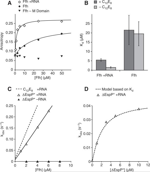
We sought to determine whether signal peptides activate 4.5_S_ RNA in the absence of C12E8. Because most signal peptides are insoluble (14), we chose the ΔEspP signal peptide (15), which is less hydrophobic than most signal peptides. We measured binding of ΔEspP peptide labeled with carboxyfluorescein (ΔEspP-FAM) to Ffh by fluorescence anisotropy. ΔEspP-FAM bound Ffh–4.5_S_ RNA with an equilibrium dissociation constant (K_d) of 1.5 ± 0.4 μM (Fig. 2A). The K_d for Ffh alone was 19.6 ± 6.4 μM (Fig. 2A), which confirms that 4.5_S RNA contributes to the binding of signal peptides as predicted (8). The addition of C12E8 weakened ΔEspP-FAM binding to Ffh–4.5_S RNA (K_d = 5.5 ± 1.5 μM) but not to Ffh alone (21.6 ± 7.9 μM) (Fig. 2B), suggesting that ΔEspP and detergent compete for binding to SRP. ΔEspP-FAM did not bind Ffh lacking its signal sequence–binding M domain (Fig. 2A), binding was reversible (fig. S2A), and ΔEspP-FAM did not impair the solubility of Ffh–4.5_S RNA (fig. S2, B and C).
Fig. 2.
ΔEspP binds SRP with micromolar affinity and stimulates 4.5_S_ RNA catalysis of Ffh-FtsY interaction. (A) Fluorescence anisotropy of ΔEspP-FAM is plotted as a function of [Ffh]. Lines represent fits to the equation Anisotropy = Anisotropyfree + Anisotropybound([Ffh]/(_K_d + [Ffh])). (B) C12E8 increased the K_d of ΔEspP for Ffh–4.5_S RNA. K_d values for ΔEspP binding to Ffh from fluorescence anisotropy in the presence and absence of 4.5_S RNA are plotted. Dark bars represent K_d in the presence of 185 μM C12E8. Error bars are SEs of the fits. (C) In the presence of 4.5_S RNA, ΔEspP stimulates the association rate for Ffh-FtsY complex formation. Observed rate constants are plotted as a function of [Ffh]. Lines are fits to the equation _k_obs = k_off + k_on[Ffh]. The dashed line is a reference to the binding rate in the presence of C12E8 from Fig. 1. (D) ΔEspP* activates 4.5_S RNA by binding to SRP. Observed rates for 1 μM Ffh–4.5_S RNA binding to 1 μM FtsY are plotted as a function of ΔEspP* concentration. The dashed line represents the equation _k_obs = [(fraction bound) (maximum stimulated rate)] + [(fraction unbound)(unstimulated rate)], where the fraction bound was calculated from the _K_d measured in (A); χ2 = 5.4 × 10⁻⁶.
To test whether saturating concentrations of ΔEspP stimulate the activity of 4.5_S_ RNA, we used ΔEspP with added lysines at the C terminus to improve its solubility [ΔEspP* (16)]. Like C12E8, ΔEspP* accelerated Ffh–4.5_S_ RNA–FtsY association (by a factor of >40; Fig. 2C and table S1) and dissociation (by a factor of ~10; table S1) but had no effect in the absence of 4.5_S_ RNA (Fig. 2C and table S1). In the presence of both C12E8 and ΔEspP*, the rate of Ffh–4.5_S_ RNA–FtsY complex formation was not substantially changed relative to individual additions (table S1). Thus, the ΔEspP peptide and C12E8 act by the same mechanism.
If ΔEspP* activates 4.5_S_ RNA by associating with SRP, then the rate of Ffh–4.5_S_ RNA–FtsY interaction should correlate with the fraction of ΔEspP*-bound SRP (calculated from the K_d in Fig. 2A). We measured the rate of Ffh–4.5_S RNA and FtsY interaction as a function of ΔEspP* concentration (Fig. 2D). When we compared the observed Ffh-FtsY binding rates to the rate predicted from the fraction of SRP bound to ΔEspP* (Fig. 2A), the data matched this model exceptionally well (Fig. 2D).
In addition to accelerating Ffh-FtsY association, 4.5_S_ RNA increases the rate of GTP hydrolysis by FfhGTP-FtsYGTP complexes (4) (fig. S3). However, neither ΔEspP* nor C12E8 affected this rate (fig. S3). Thus, signal peptides specifically affect the ability of 4.5_S_ RNA to accelerate Ffh-FtsY complex formation.
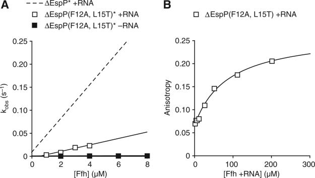
To assess the specificity of 4.5_S_ RNA activation, we used a version of ΔEspP* bearing Phe12 → Ala and Leu15 → Thr mutations [ΔEspP(F12A, L15T)*] that reduce SRP-dependent targeting in vivo (15). In the presence of 10 μM ΔEspP(F12A, L15T)*, the 4.5_S_ RNA–stimulated association and dissociation of Ffh and FtsY was slower than that measured with “wild-type” ΔEspP* by a factor of ~5 (Fig. 3A and table S1). Similar to ΔEspP*, ΔEspP (F12A, L15T)* had no effect in the absence of 4.5_S_ RNA (Fig. 3A). To determine whether this was due to reduced binding of ΔEspP(F12A, L15T)* to SRP, we measured the _K_d by fluorescence anisotropy and found that binding was substantially weaker (_K_d = 87 ± 18 μM, Fig. 3B). Consistent with this result, increasing concentrations of ΔEspP(F12A, L15T)* increased the observed rate for SRP-FtsY association (fig. S4).
Fig. 3.
Mutations in ΔEspP that impair SRP-mediated targeting show decreased binding to SRP and decreased stimulation of 4.5_S_ RNA. (A) ΔEspP(F12A, L15T)* stimulates SRP-FtsY complex formation less than does ΔEspP*. The dashed line represents the ΔEspP* + RNA peptide binding rate from Fig. 2C. (B) Fluorescence anisotropy of FAM-labeled ΔEspP bearing Phe12 → Ala and Leu15 → Thr mutations [ΔEspP(F12A, L15T)] is plotted as a function of [Ffh +RNA]. Line is fit as in Fig. 2A.
Thus, SRP RNA acts as a switchable regulatory module at the center of the SRP protein-targeting machine to link recruitment of cargo (a signal peptide) to the next step in the targeting reaction (binding to SR). If free SRP and SR interacted efficiently with each other, they would undergo futile cycles of binding and GTP hydrolysis. Cargo-dependent activation of SRP RNA prevents this, harnessing the energy of GTP hydrolysis for protein targeting.
High-affinity interaction of SRP with ribosomes can occur before SRP interaction with the signal peptide when a short nascent chain is still inside the ribosome, raising the question of how SRP selectively targets signal sequence–containing substrates (17). Our results show that the interaction of the signal peptide with SRP accelerates SRP-SR complex formation, thereby providing a mechanism for selective delivery of appropriate substrates to the membrane. This is conceptually analogous to the kinetic mechanism by which translation achieves fidelity, where cognate codon-anticodon pairing accelerates GTP hydrolysis by elongation factor Tu (EF-Tu) (18, 19).
Our results provide an intuitive model for how each step of the targeting process activates the next to achieve productive, directional targeting. Signal peptides bind to SRP's conformationally flexible M domain that forms a continuous surface with SRP RNA (8, 13). Binding induces a conformational change that activates SRP RNA (20). Activated SRP RNA facilitates the displacement of the N-terminal helices of SRP and SR that slow their association without SRP RNA (21). This commits the ribosome–nascent chain complex to membrane targeting. The kinetic control described here, where substrate recruitment accelerates downstream interactions, provides a generalizable principle for coordination of multi-step pathways.
Supplementary Material
supporting materials
Footnotes
References and Notes
- 1.Keenan RJ, Freymann DM, Stroud RM, Walter P. Annu. Rev. Biochem. 2001;70:755. doi: 10.1146/annurev.biochem.70.1.755. [DOI] [PubMed] [Google Scholar]
- 2.Zopf D, Bernstein HD, Johnson AE, Walter P. EMBO J. 1990;9:4511. doi: 10.1002/j.1460-2075.1990.tb07902.x. [DOI] [PMC free article] [PubMed] [Google Scholar]
- 3.Miller JD, Wilhelm H, Gierasch L, Gilmore R, Walter P. Nature. 1993;366:351. doi: 10.1038/366351a0. [DOI] [PubMed] [Google Scholar]
- 4.Peluso P, Shan SO, Nock S, Herschlag D, Walter P. Biochemistry. 2001;40:15224. doi: 10.1021/bi011639y. [DOI] [PubMed] [Google Scholar]
- 5.Powers T, Walter P. Science. 1995;269:1422. doi: 10.1126/science.7660124. [DOI] [PubMed] [Google Scholar]
- 6.Poritz MA, et al. Science. 1990;250:1111. doi: 10.1126/science.1701272. [DOI] [PubMed] [Google Scholar]
- 7.Peluso P, et al. Science. 2000;288:1640. doi: 10.1126/science.288.5471.1640. [DOI] [PubMed] [Google Scholar]
- 8.Batey RT, Rambo RP, Lucast L, Rha B, Doudna JA. Science. 2000;287:1232. doi: 10.1126/science.287.5456.1232. [DOI] [PubMed] [Google Scholar]
- 9.Jagath JR, Rodnina MV, Wintermeyer W. J. Mol. Biol. 2000;295:745. doi: 10.1006/jmbi.1999.3427. [DOI] [PubMed] [Google Scholar]
- 10.Zhang X, Kung S, Shan SO. J. Mol. Biol. 2008;381:581. doi: 10.1016/j.jmb.2008.05.049. [DOI] [PMC free article] [PubMed] [Google Scholar]
- 11.Walter P, Blobel G. Proc. Natl. Acad. Sci. U.S.A. 1980;77:7112. doi: 10.1073/pnas.77.12.7112. [DOI] [PMC free article] [PubMed] [Google Scholar]
- 12.von Heijne G. J. Mol. Biol. 1985;184:99. doi: 10.1016/0022-2836(85)90046-4. [DOI] [PubMed] [Google Scholar]
- 13.Keenan RJ, Freymann DM, Walter P, Stroud RM. Cell. 1998;94:181. doi: 10.1016/s0092-8674(00)81418-x. [DOI] [PubMed] [Google Scholar]
- 14.Swain JF, Gierasch LM. J. Biol. Chem. 2001;276:12222. doi: 10.1074/jbc.M011128200. [DOI] [PubMed] [Google Scholar]
- 15.Peterson JH, Woolhead CA, Bernstein HD. J. Biol. Chem. 2003;278:46155. doi: 10.1074/jbc.M309082200. [DOI] [PubMed] [Google Scholar]
- 16.See supporting material on Science Online.
- 17.Bornemann T, Jockel J, Rodnina MV, Wintermeyer W. Nat. Struct. Mol. Biol. 2008;15:494. doi: 10.1038/nsmb.1402. [DOI] [PubMed] [Google Scholar]
- 18.Pape T, Wintermeyer W, Rodnina M. EMBO J. 1999;18:3800. doi: 10.1093/emboj/18.13.3800. [DOI] [PMC free article] [PubMed] [Google Scholar]
- 19.Ogle JM, Ramakrishnan V. Annu. Rev. Biochem. 2005;74:129. doi: 10.1146/annurev.biochem.74.061903.155440. [DOI] [PubMed] [Google Scholar]
- 20.Bradshaw N, Walter P. Mol. Biol. Cell. 2007;18:2728. doi: 10.1091/mbc.E07-02-0117. [DOI] [PMC free article] [PubMed] [Google Scholar]
- 21.Neher SB, Bradshaw N, Floor SN, Gross JD, Walter P. Nat. Struct. Mol. Biol. 2008;15:916. doi: 10.1038/nsmb.1467. [DOI] [PMC free article] [PubMed] [Google Scholar]
- 22.We thank J. Weissman, C. Gross, P. Egea, C. Gallagher, A. Korennykh, A. Acevedo, W. Weare, and J. Kardon for assistance and comments. Supported by NIH grant ROI GM32384 (P.W.), an NSF predoctoral fellowship (N.B.), the Jane Coffin Childs Memorial Fund (S.B.N.), and National Institute of General Medical Sciences fellowship R25 GM56847 (D.S.B.). P.W. is an Investigator of the Howard Hughes Medical Institute.
Associated Data
This section collects any data citations, data availability statements, or supplementary materials included in this article.
Supplementary Materials
supporting materials


