Loss of PTEN promotes resistance to T cell-mediated immunotherapy (original) (raw)
. Author manuscript; available in PMC: 2016 Aug 1.
Abstract
T cell-mediated immunotherapies are promising cancer treatments. However, most patients still fail to respond to these therapies. The molecular determinants of immune resistance are poorly understood. We show that loss of PTEN in tumor cells in preclinical models of melanoma inhibits T cell-mediated tumor killing and decreases T cell trafficking into tumors. In patients, PTEN loss correlates with decreased T cell infiltration at tumor sites, reduced likelihood of successful T cell expansion from resected tumors, and inferior outcomes with PD-1 inhibitor therapy. PTEN loss in tumor cells increased the expression of immunosuppressive cytokines, resulting in decreased T cell infiltration in tumors, and inhibited autophagy, which decreased T cell-mediated cell death. Treatment with a selective PI3Kβ inhibitor improved the efficacy of both anti-PD-1 and anti-CTLA4 antibodies in murine models. Together these findings demonstrate that PTEN loss promotes immune resistance and support the rationale to explore combinations of immunotherapies and PI3K-AKT pathway inhibitors.
Keywords: PTEN loss, Immunotherapy
INTRODUCTION
T cells play an important role in cancer immunosurveillance and tumor destruction, and therapies that enhance anti-tumor T cell responses have achieved encouraging clinical results. PD-1 checkpoint blockade and adoptive T cell therapy (ACT) can induce objective responses in 33–48% of metastatic melanoma patients, many of which are durable (1–3). However, the majority of patients still fail to respond to T cell-mediated immunotherapy and little is known about why such treatment failures occur. Understanding the pathways that cause resistance would improve the clinical application of immunotherapies through improved patient selection. Such understanding may also identify rational, more effective therapeutic combinations. Our group and others have shown that oncogenic signaling by BRAF, which is mutated in ~50% of melanomas, modulates the immune microenvironment to perturb T cell-mediated anti-tumor responses. Mutant BRAF increases the expression of IL-1α and IL-1β by tumor cells, which increases the expression of PD-L1 and PD-L2 in tumor-associated fibroblasts and suppresses the function of tumor-infiltrating T cells (TILs) (4). BRAF inhibition increases the expression of melanocytic antigens (5) and inhibits VEGF production by melanoma cells, thereby enhancing trafficking of tumor-reactive T cells to tumors (6). Clinical trials evaluating the safety and efficacy of BRAF inhibitors in combination with immunotherapies are currently underway. In addition, activation of the β-catenin pathway, another oncogenic pathway, was found to be associated with poor tumor infiltration of T cells in a recent publication (7). Together, these results indicate that the impact of tumor-intrinsic pathways is not always confined to tumor cells and can be extended to anti-tumor immune responses, especially T cell responses.
The phosphatidylinositol 3-kinase (PI3K) pathway plays a critical role in cancer by regulating several critical cellular processes, including proliferation and survival. One of the most common ways that this pathway is activated in cancer is by loss of expression of the tumor suppressor PTEN, which is a lipid phosphatase that dampens the activity of PI3K signaling. Loss of PTEN corresponds with increased activation of the PI3K-AKT pathway in multiple tumor types (8). Loss of PTEN occurs in up to 30% of melanomas, frequently in tumors with a concurrent activating BRAF mutation (9). While expression of mutant BRAF alone fails to transform melanocytes, invasive and spontaneously metastatic lesions develop when this is complemented by loss of PTEN in mouse models (10, 11). Loss of PTEN in melanoma patients with BRAF mutations is associated with worse outcomes in stage III patients, and in stage IV patients treated with FDA-approved BRAF inhibitors (12, 13). Several studies have demonstrated that melanoma cell lines with loss of PTEN can be growth arrested by BRAF and MEK inhibitors but that they are resistant to apoptosis induction (14, 15). These studies support that PTEN loss identifies a distinct, clinically significant subset of melanomas.
In this study, we evaluated the impact of loss of PTEN on T cell-mediated anti-tumor responses. Our studies in preclinical models and clinical specimens demonstrate that loss of PTEN promotes resistance to immunotherapy in melanoma. Our findings provide new insights into the role of PTEN in cancer and identify new strategies to increase the efficacy of immunotherapy in patients.
RESULTS
Silencing PTEN expression in melanoma reduces T cell-mediated tumor killing in vitro and in vivo
Since PTEN loss is most common in melanoma patients with concurrent BRAF mutations, we silenced PTEN expression in established _BRAF_-mutant human melanoma cell lines and evaluated anti-tumor response to T cell-mediated immunotherapy using our previously described melanoma model (Supplementary Fig. S1A)(6). Briefly, the melanoma tumor antigen gp100 and the murine major histocompatibility complex (MHC) class I molecule H-2Db were ectopically and constitutively expressed in the human melanoma cell line A375. Gp100- and H-2Db-expressing A375 cells (A375/GH) can be recognized by T cells derived from pmel-1 mice in a MHC class I-dependent manner. This model enables identification of immune resistance mechanisms that are independent of tumor antigen and/or MHC loss in tumors.
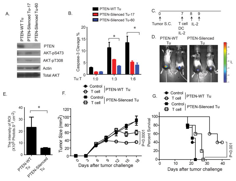
Western blotting confirmed decreased PTEN and increased phosphorylated (activated) AKT (p-AKT) expression in A375 melanoma cells stably transfected with shRNA targeting PTEN (A375/GH/shPTEN) (Fig.1A). Silencing of PTEN significantly reduced the percentage of lysed (cleaved caspase-3+) tumor cells when the melanoma cells were co-cultured with the tumor-reactive pmel-1 T cells in vitro (Fig. 1B). To evaluate the in vivo effects of PTEN loss on T cell-mediated anti-tumor activity, we used an established ACT murine model (6) (Fig.1C). PTEN loss significantly reduced the accumulation of transferred tumor-reactive T cells in A375 melanoma tumors in vivo (Fig.1D–E). The adoptively transferred pmel-1 T cells showed significantly reduced therapeutic activity in mice bearing PTEN-silenced tumors when compared to mice bearing PTEN-expressing tumors (Fig.1F, G). Similarly impaired T cell-mediated anti-tumor activity against PTEN-silenced tumors was also observed in the context of concurrent treatment with a selective BRAF inhibitor (Supplementary Fig. S1B–F). Collectively, our in vitro and in vivo studies indicate that PTEN loss can cause resistance to T cell-mediated anti-tumor immune responses.
Figure 1. Reduced T cell-mediated anti-tumor activity against PTEN-silenced melanoma cells.
(A) PTEN expression and AKT activation in A375/GH cells with and without PTEN silencing. Two PTEN-silenced tumor cell lines (17 and 60) were independently produced by two shRNAs targeting PTEN (shPTEN). Tumor cells expression scrambled shRNA (shNS) were served as control (PTEN-WT Tu). (B) T cell-induced apoptosis rate of melanoma tumor cells with and without PTEN silencing. A375/GH/shPTEN and A375/GH/shNS tumor cells were co-cultured with tumor-reactive pmel-1 T cells, at different ratios of effector and target cells (E:T). The cleavage of caspase-3 in tumor cells was determined by flow cytometry. (C) Experimental setup of the murine ACT protocol to evaluate in vivo T cell-mediated anti-tumor activity. (D) T cell infiltration of melanoma tumors with and without PTEN silencing in vivo. Luciferase-expressing pmel-1 T cells were transferred into B6 nude mice bearing A375/GH tumor with or without PTEN-silencing. To evaluate tumor trafficking of transferred T cells, the luciferase intensity at the tumor site was determined by bioluminescence imaging 6 days after T cell transfer. (E) Summary of quantitative imaging analysis of transferred T cells at the tumor site. Quantification was expressed as the average of photon flux within region of interest (ROI). (F) Tumor growth and (G) Kaplan-Meier survival curves for tumor-bearing B6 nude mice treated with adoptive transfer of pmel-1 T cells. Similar results were obtained in repeated experiments. In D–G, three to five mice per group were used. *P < 0.05.
PTEN loss correlates with decreased numbers, and impaired function of tumor-infiltrating T cells, and inferior outcomes with anti-PD-1 in melanoma patients
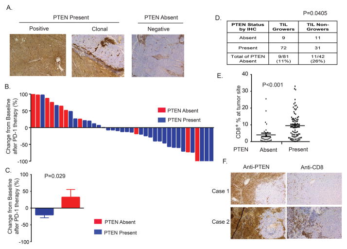
To determine the clinical relevance of these findings, we analyzed PTEN expression in samples from melanoma patients. Tumors with less than 10% of cells with PTEN expression by IHC staining were classified as PTEN absent, as our previous studies demonstrated that this correlates with increased activation of the PI3K-AKT pathway (12); all other tumors were categorized as PTEN present (Fig. 2A). Analysis of a cohort of 39 metastatic melanoma patients treated with FDA-approved anti-PD-1 antibodies (pembrolizumab and nivolumab) demonstrated that patients with PTEN present tumors achieved significantly greater reduction of tumor size than patients with PTEN absent tumors (p=0.029) (Fig. 2B and 2C). No significant differences in gender, age, stage of disease, target tumor size, or serum LDH were detected between patients with PTEN present tumors and PTEN absent tumors (Supplementary Table S1).
Figure 2. Correlation of PTEN loss in melanoma cells with an immune resistance phenotype.
(A) Overview of IHC for PTEN in advanced metastatic melanoma patients. PTEN expression was evaluated by a CLIA-certified PTEN IHC assay. Representative PTEN staining pictures for each category were shown. (B) A waterfall plot of the best objective response in each anti-PD-1 treated patient. 39 cases of melanoma patients treated with anti-PD-1 antibody were stratified based on the expression of PTEN. Tumor burden was measured by the sum of longest diameters of target lesions. The best objective response of anti-PD-1 antibody was evaluated by the maximum change of tumor burden based on baseline. (C) Comparison of tumor reduction after PD-1 therapy in patients with or without PTEN loss. The p value of the comparison was determined by Wilcoxon rank sum test. (D) Increased percentage of PTEN absent in melanoma patients with failed initial expansion of TILs (≤40X106 TIL after initial expansion). A 2X2 contingency table was made based on the frequency distribution of the PTEN expression status and the success of initial TIL growth in the ACT cohort. The p value (p=0.04) was determined by Fisher’s exact t test. (E) Correlation of CD8+ T cell infiltration with PTEN expression status of stage IIIB/C melanoma patient tumors. (F) Examples of CD8 staining in two patients with clonal PTEN expression.
We next attempted to analyze if PTEN status correlated with clinical outcomes with TIL therapy. However, we observed that the overwhelming majority of patients treated with TIL exhibited PTEN expression (44/48) in their harvested tumors, thus precluding the ability to make meaningful conclusions about the impact of PTEN loss on treatment outcomes. Analysis of tumor harvests for TIL expansion, which must be achieved successfully in order for TIL therapy to be feasible(16), showed that 26% of melanomas that did not yield successful TIL growth were PTEN absent tumors, which was more frequent than was observed in tumors that yielded successful TIL growth (11%, p=0.04) (Fig. 2D). Thus, it is likely that the lower success rate for TIL growth from melanomas with PTEN loss leads to the exclusion of PTEN absent patients from the TIL treatment cohort. However, other factors, such as poor prognosis of metastatic melanoma patients with PTEN loss (12), may also contribute to this exclusion.
Consistent with the observed association of unsuccessful TIL expansion with PTEN loss, analysis of a cohort of 135 resected stage IIIB/C melanoma regional metastases (12) found that melanomas with PTEN loss had significantly lower CD8+ T cell tumor infiltration compared to tumors with PTEN expression (p<0.001, Fig. 2E; Supplementary Fig. S2A). CD8+ T cell infiltration did not correlate significantly with BRAF/NRAS mutation status, and both BRAF mutant and BRAF/NRAS wild-type melanomas with loss of PTEN had significantly less CD8+ T cell infiltration than melanomas with PTEN expression (Supplementary Fig. S2B, S2C). Notably, in the small number of tumors (6%) that demonstrated a heterogeneous, ‘clonal-like’ pattern of PTEN expression (Fig. 2A), more CD8+ T cells were observed infiltrating regions with PTEN expression compared to adjacent regions with PTEN loss (Fig. 2F).
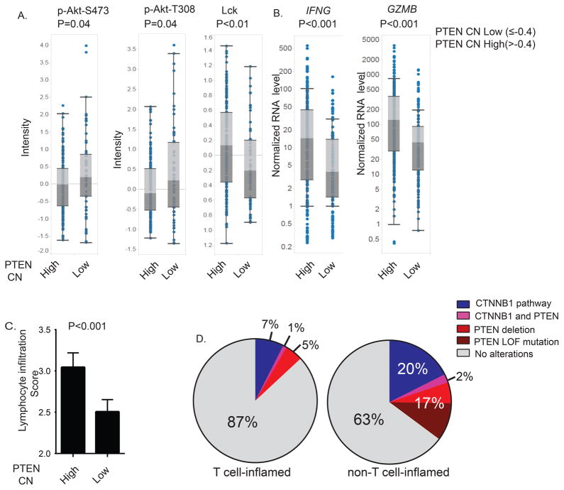
We further evaluated the relationship between PTEN expression and immune infiltrates using the publicly available melanoma Cancer Genome Atlas (TCGA) dataset. As we could not perform IHC, tumors were categorized based on PTEN gene copy number (CN) as gene deletion appears to be one of the most common mechanisms of PTEN loss in melanoma (17). As expected, increased expression of p-AKT was observed in tumors with low (≤ 0.4) PTEN CN, although we cannot exclude the potential contribution of non-tumor cells to this result (Fig. 3A). In contrast the expression of Lck, a protein expressed predominantly by T cells, and the transcripts of the T cell effector molecules IFN-γ and granzyme B were significantly decreased in tumors with low PTEN CN (Fig. 3A–B). The immune cytolytic activity score defined in a recent pan-TCGA study (18) was also significantly reduced in melanomas with low PTEN CN (Supplementary Fig. S2D). Tumors with low PTEN CN also had lower histologically-determined lymphocyte infiltration scores (Lscores) (Fig. 3C) (19). Lscores were also inversely related to expression of p-Akt (Supplementary Fig. S2E).
Figure 3. Reduced number and impaired effector functions of TILs in tumors with PTEN deletion or loss-of-function mutations in PTEN.
Cutaneous melanoma patients whose information was included in TCGA were stratified based on the PTEN copy number (CN) (cutoff, ≤−0.4, which was chosen in order to maximize the difference in the level of activity of the AKT pathway). A box-and-whisker plot was used to demonstrate the differences in expression levels of indicated genes or proteins between these two groups. (A) Comparison of the intensity of phosphorylated Akt and Lck in melanomas obtained from patients with different PTEN CNs according to RPPA. (B) The mRNA expression levels for genes encoding interferon-γ and granzyme B in tumor samples obtained from melanoma patients with different PTEN CNs. (C) Comparison of lymphocytic infiltration score (Lscore), as determined by pathological review, between groups of patients with different PTEN CNs. The p values of the comparisons were determined by unpaired T test. (D) Frequencies of genetic alterations in the β-catenin pathway and PTEN between T cell-inflamed and non-T cell-inflamed tumors. Metastatic melanomas from TCGA were first catalogued based on T cell infiltration and subsequently based on activating mutations in β-catenin itself (CTNNB1) or loss-of-function mutations in negative regulators of the β-catenin pathway (APC, APC2, AXIN1, AXIN2), and PTEN deletion or PTEN mutations. Non-T cell-inflamed tumors have an increased frequency of PTEN alterations compared to the T cell-inflamed tumors (p<0.01 by Fisher’s exact test).
A recent analysis of the melanoma TCGA demonstrated that activating mutations in CTNNB1 and increased activation of the β-catenin pathway are enriched in tumors with a non-T cell-inflamed pattern of gene expression compared to tumors with a T cell-inflamed signature (7). Similarly, we observed that the frequency of deletions and loss-of-function mutations in the PTEN gene were higher in the non-T cell-inflamed tumors than in the T cell-inflamed melanomas (19% vs 6%, p<0.01) (Fig.3D). The alterations in PTEN and the β-catenin pathway were largely non-overlapping, as only 2 tumors (2%) in the non-T cell-inflamed cohort had alterations in both. The association of activation of β-catenin and loss of PTEN was tested by a chi-squared test (χ2=0.81 and p=0.37, indicating that the tested events are not dependent). These data support that tumor-intrinsic activation of β-catenin and loss of PTEN represent two independent events associated with T cell exclusion from the tumor microenvironment. In summary, our analyses of independent cohorts of melanoma clinical samples are consistent with the preclinical observation that loss of PTEN in melanoma reduces the recruitment and function of T cells in tumors and promotes immune resistance.
Effects of PTEN loss on immune modulators expressed on and produced by tumor cells
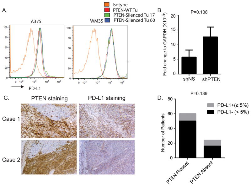
To interrogate the underlying immune suppressive mechanism(s) associated with PTEN loss, we first evaluated PD-L1 expression (20, 21). Silencing PTEN expression in both A375 and WM35 cell lines did not significantly increase the surface expression level of PD-L1 in vitro (Fig. 4A), nor was PD-L1 mRNA expression affected in day 14 PTEN-silenced xenograft tumors in vivo (Fig.4B). We also observed similar patterns of PD-L1 expression between the PTEN-negative and PTEN-positive tumor areas from clinical samples with heterogeneous PTEN expression (Fig. 4C), and PD-L1 expression did not correlate with PTEN status in the cohort of stage IIIB/C melanomas (Fig. 4D). These findings, which are consistent with results in a large panel of human melanoma cell lines (22), suggest that PD-L1 is not the primary mechanism of the suppressed anti-tumor immune response induced by PTEN loss. In addition, PTEN loss did not correlate with changes in the expression of MHC class I molecules in the cohort of stage IIIB/C melanomas (Supplementary Fig. S2F).
Figure 4. PD-L1 expression is not associated with PTEN expression status in melanomas.
(A) The PD-L1 surface expression levels in melanoma cells with and without PTEN silencing. A375 and WM35 tumor cells were transduced with control shRNA or PTEN-specific shRNA. Transduced tumor cells were stained with anti-PD-L1 to evaluate the surface expression of PD-L1. (B) The in vivo PD-L1 expression in melanoma tumor tissues with and without PTEN silencing. B6 nude mice were challenged with A375/GH/shPTEN or A375/GH/shNS tumor cells. Tumor samples were collected from mice bearing 14-day established tumors. The PD-L1 mRNA expression levels in tumor samples were determined using real-time PCR. (C) Examples of IHC for PD-L1 in tumors with clonal PTEN expression. (D) The PD-L1 surface expression levels in tumor samples from patients with stage IIIB/C melanoma. Tumor samples were placed in PTEN absent and PTEN present groups, and the expression of PD-L1 on the tumor surface was determined by IHC.
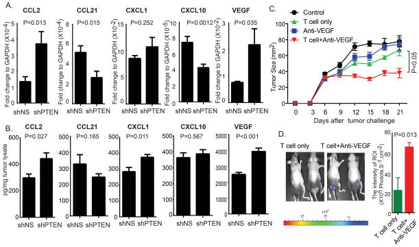
To identify PTEN-regulated immunomodulatory genes, PTEN-silenced or control A375/GH xenografts were harvested in vivo to determine the mRNA and protein expression of chemokines and cytokines involved in the recruitment and/or function of TILs (Supplementary Table S2). Differentially expressed genes are shown in Figure 5A. Both RT-PCR analysis and Luminex assays confirmed that loss of PTEN significantly up-regulates CCL2 and VEGF (Fig. 5A, B). VEGF IHC staining confirmed that melanoma clinical samples with heterogeneous PTEN expression demonstrated increased VEGF in regions with loss of PTEN (Supplementary Fig. S3). Preclinically, functional testing confirmed that anti-VEGF blocking antibody enhanced tumor infiltration and improved anti-tumor activity of transferred tumor-reactive T cells against A375/GH/shPTEN xenografts (Fig. 5C, D). These results and previously published studies (23, 24) suggest that loss of PTEN in melanomas promotes resistance to immune infiltration of tumors through the production of inhibitory cytokines.
Figure 5. Critical role of VEGF in immune resistance associated with loss of PTEN.
(A and B) B6 nude mice were challenged with A375/GH/shPTEN or A375/GH/shNS melanoma cells and tumor samples were collected from 14-day tumor-bearing mice. (A) Transcript expression levels for cytokine and chemokine genes in tumor samples as determined using quantitative RT-PCR. (B) Protein expression of cytokines and chemokines in tumor samples as determined by Luminex analyses. (C and D) B6 nude mice were challenged with A375/GH/shPTEN. Seven days after tumor challenge, luciferase-expressing pmel-1 T cells were transferred into tumor-bearing mice. Bioluminescence imaging was performed to evaluate tumor trafficking of transferred T cells in treated mice. Tumor growth (C) and T cell infiltration (D) of PTEN-silenced tumor in mice treated with ACT and/or anti-VEGF antibody. Representative imaging figures and quantification of the intensity of luciferase at tumor site from all tested mice were shown. Quantification was expressed as the average of photon flux within ROI. In C–D, three to five mice per group were used. *P < 0.05.
We extended our investigation of the factors mediating the immunosuppressive effects of PTEN loss by analyzing the gene expression of 609 inflammation-related genes in tumors from 47 metastatic melanoma patients, which include 10 samples with absent PTEN (Supplementary Fig. S4A). The vast majority of analyzed genes (504/609) exhibited reduced expression in the tumors with PTEN loss, suggesting a lack of inflammatory cells in the tumor microenvironment. To define genes that were consistently altered, we utilized a leave-one-out logistic regression analysis (see supplementary information). This analysis identified TNFRSF10C, PDGFRB, TILR3, MASP2 and CLU as genes significantly correlated with PTEN in at least 8 distinct models, and all 5 genes correlated positively with PTEN expression (Supplementary Fig. S4B). The area under the ROC curve (AUC) obtained by this classifier of the 5 genes was 91.6% (Supplementary Fig. S4C), indicating a strong performance in discriminating between PTEN-replete and PTEN-loss melanomas.
Reduced autophagic activity in PTEN-loss melanoma promotes the resistance to T cell induced tumor apoptosis
Since infiltrating non-transformed cells in both clinical specimens and xenografts could potentially confound our ability to identify tumor-specific gene expression changes mediated by PTEN, we performed a microarray-based gene expression analysis in human melanoma cell lines with PTEN silencing, and compared the results to the analyses described above. The expression levels of the 5 genes identified by the leave-one-out logistic regression analysis in tumor tissue were comparable in tumor cells with and without PTEN silencing, making it likely that these genes were expressed in infiltrating cells in the tumor tissues. However, this approach identified ATG16L as a gene upregulated by PTEN in both melanoma cell lines and clinical specimens (Supplementary Fig. S4A). The ~800-kDa protein complex formed by Atg16L conjugated with Atg12 and Atg5 is required for efficient LC3 lipidation, a critical step in autophagy. Interestingly, several genome-wide association studies suggest that ATG16L mutation is a risk factor for inflammatory bowel disease (25, 26).
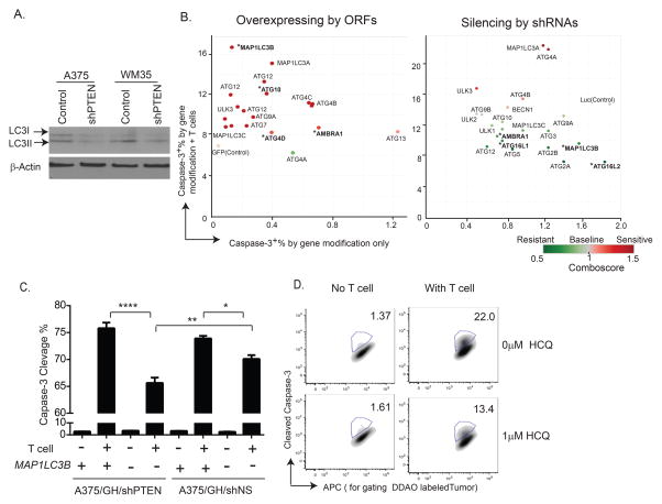
We hypothesized that PTEN loss promotes resistance to T cell-mediated killing by inhibiting autophagy. Western blotting of protein from melanomas with PTEN loss demonstrated reduced LC3 lipidation, as determined by the level of LC3II (Supplementary Fig S5A), and PTEN knockdown in human melanoma cell lines decreased the expression of both LC3I and LC3II (Fig. 6A). We then performed functional testing in three patient-derived melanoma cell lines that undergo apoptosis when exposed to their autologous TILs. We perturbed the expression of genes required for activation of autophagy (Supplementary Table S3) in the tumor cells and exposed them to autologous TILs. A comboscore was calculated based on observed changes in the percentage of T cell-induced apoptosis in tumor cells with or without genetic alterations. Genetic alterations which enhance the sensitivity of tumor cells to T cell-mediated killing have comboscores >1. Enforced expression of almost all autophagy-related genes increased the susceptibility of tumor cells to apoptosis induced by their autologous TIL, whereas silencing their expression caused resistance (Fig. 6B; Supplementary Figure. S5B, S5C).
Figure 6. PTEN expression regulates autophagy in tumor cells, reducing T cell-mediated killing.
(A) Expression of LC3 I and LC3 II in _BRAF-_mutant melanoma cell lines with and without PTEN silencing. The expression levels of LC3 I and LC3 II in protein lysates from A375 and WM35 tumor cells were determined by western blotting analysis. (B) Perturbing the expression of autophagy related genes changes the sensitivity of melanoma to apoptosis induced by tumor-reactive T cells. A patient-derived melanoma cell line, Mel2400, was transduced with lentiviral vectors encoding the ORFs or shRNAs of autophagy-related genes. Virally transduced tumor cells were co-cultured with paired autologous T cells for 3 hours. The percentage of killed (cleaved casp-3+) tumor cells expressing ORF or shRNAs was determined using flow cytometry. A comboscore was calculated as described in the method section and used to evaluate the effect of genetic modifications on sensitivity of tumor cells to T cell-mediated killing. Tumor cells transduced with virus expressing GFP or shRNA targeting luciferase (Luc) were served as controls for overexpression and knockdown experiments, respectively. *genetic modification, which significantly changed the sensitivity of tumor to T cell-mediated killing (p<0.05). (C) PTEN-silenced and control melanoma cells were transduced with a viral vector encoding MAP1LC3B. The percentage of cleaved casp-3+ cells among transduced tumor cells in response to pmel-1 T cells (E:T=10:1) was evaluated. (D) Melanoma cells (Mel 2338) were pretreated with 1μM Hydroxychloroquine (HCQ) for overnight and followed by co-culture with paired TILs for 3 hours. The percentage of apoptotic tumor cells was evaluated by the cleaved caspase-3 assay. Results are representative of data generated in two independent experiments. *P < 0.05; **P < 0.01; ****P < 0.0001.
Overexpressing MAP1LC3B, which is critical in autophagy initiation, improved T cell killing in all three cell lines, while the resistance to T cell killing was observed in _MAP1LC3B_-silenced tumor cells. We further confirmed that the autophagic activity can be modulated by perturbing the expression of MAP1LC3B, as shown in Supplementary Figure. S5D, demonstrating reduced change of autophagic flux (ΔAF) of p62 in _MAP1LC3B_-silenced tumor cells and enhanced ΔAF of p62 in _MAP1LC3B_-overexpressing tumor cells. Therefore, we used MAP1LC3B as a representative gene to test whether increasing autophagic activity can rescue the resistance of PTEN-silenced tumors to T cell killing. Our result indicated that overexpressing MAP1LC3B in PTEN-silenced tumor cells fully restored susceptibility to killing by tumor-reactive T cells (Fig. 6C). Furthermore, pre-treatment of patient-derived melanoma cells with hydroxychloroquine (HCQ), an autophagy inhibitor, reduced the apoptosis induced by autologous TILs (Fig. 6D and Supplementary Fig. S5E, S5F). Together, the results support that PTEN loss can protect tumor cells from T cell killing through an autophagy-dependent mechanism.
PI3Kβ inhibitor improves the activity of checkpoint blockade for PTEN-loss tumors
We hypothesized that inhibiting the PI3K pathway would improve the effectiveness of immunotherapy. Although inhibition of some targets in this pathway are critical to immune cell function and viability, the PI3Kβ isoform can regulate AKT activity in tumors with PTEN loss yet it is dispensable in the activation of the TCR signaling pathway (27, 28). Therefore, we tested whether selective inhibition of PI3Kβ would increase the efficacy of immunotherapy in melanomas with PTEN loss.
In vitro treatment with a selective PI3Kβ small molecule inhibitor, GSK2636771, reduced the activation of the AKT pathway and moderately (<20%) inhibited the growth of three human melanoma cells with loss of PTEN (Supplementary Fig. S6A, S6B). To examine the role of the PI3Kβ on T cell induced apoptosis, the three PTEN null human melanoma cell lines were engineered to express gp100 and murine H2-Db, and then tested for caspase-3 cleavage after co-incubation with the PI3Kβ inhibitor in the presence of pmel-1 T cells. Treatment with the PI3Kβ inhibitor improved the T cell-induced tumor killing of all three PTEN-null melanoma cell lines (Supplementary Fig. S6C).
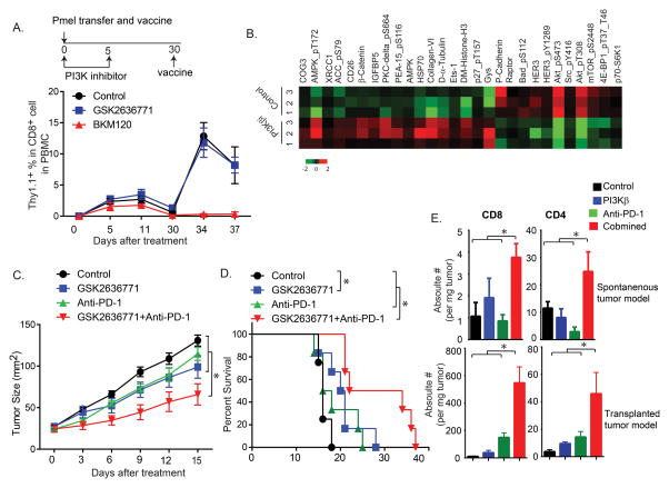
We then compared the effects of the PI3Kβ inhibitor and a pan-PI3K inhibitor (BKM120) on the effector function of antigen-specific T cells using an established murine vaccine model (29). Briefly, C57BL/6 mice were adoptively transferred with splenocytes from pmel-1 mice followed by hgp10025–33 peptide vaccination on day 0 and day 30. Vaccinated mice were treated with PI3Kβ inhibitors daily for 5 days. Treatment with the pan-PI3K BKM120 inhibitor reduced whole blood cell counts and inhibited the proliferation of gp100-specific T cells upon initial immunization, whereas PI3Kβ inhibitor did not affect either parameter (Fig. 7A; Supplementary Fig. S6D). Importantly, the PI3Kβ inhibitor did not affect the percentage of antigen-specific T cells in mice receiving a booster vaccine (Fig. 7A). These results support that the generation of memory T cells remains intact with PI3Kβ inhibitor treatment.
Figure 7. The PI3Kβ inhibitor enhances the anti-tumor activity of T cell-mediated immunotherapy in mice bearing PTEN loss tumor.
(A) C57BL/6 mice were transferred with the splenocytes from Pmel-Thy1.1 mice, followed by gp100 peptide vaccination. Vaccinated mice received either vehicle, GSK2636771 (30mg/kg/d), or BKM120 (60mg/kg/d) for 5 days. After 30 days, mice were boosted with gp100 peptide vaccine. Schematic representation of vaccine and the PI3K inhibitor (PI3Ki) treatment protocol was shown. Thy1.1, a congenic marker for transferred Pmel T cells, was used to determine the number of gp100-specific T cells in peripheral blood from vaccinated mice after the PI3K inhibitor treatment. (B) Analysis of the effect of GSK2636771 on protein signaling networks by RPPA. Melanoma was initiated in a group of Tyr:CreER; PTENlox/lox; BRAF V600E/+ mice. Mice with measureable tumors were randomly treated with either vehicle or GSK2636771 (30mg/kg/d) for 5 days. Protein lysates from treated tumors were harvested. The heatmap demonstrates the changes in proteins differentially expressed in GSK2636771 treated tumors. (C–D) Tyr:CreER; PTENlox/lox; BRAF V600E/+ mice with measureable tumors were randomly treated with either vehicle plus control antibody, GSK2636771 (30mg/kg/d), anti-PD-1(100μg), or the combination of both GSK2636771 and anti-PD-1. (C) Tumor size in each of the treatment groups. Tumor growth was monitored every 3 days. (D) Kaplan-Meier survival curves of mice treated with GSK2636771 and/or anti-PD-1. Log-rank test demonstrates statistical significance (P<0.05): control vs GSK2636771 or GSK2636771+anti-PD-1; GSK2636771 vs GSK2636771+anti-PD-1; anti-PD-1 vs GSK2636771+anti-PD-1 (N=4–8). (E) The numbers of tumor-infiltrating T cells in mice treated with GSK2636771 and/or anti-PD-1. GSK2636771 and/or anti-PD-1 were used to treat mice bearing either a spontaneous tumor or a transplanted tumor as described in Figure 7C and Supplementary Figure S7D respectively. 7 days after treatment, tumor tissues were harvested and weighed. Single cell suspensions from tumor tissues were made for CD8 and CD4 staining. One-Way ANOVA test demonstrates statistical significance (P<0.05): GSK2636771+anti-PD-1 vs control, GSK2636771 or anti-PD-1. *P < 0.05(N=4–5).
The recent establishment of Tyr:CreER; BRAFV600E/+ ; PTENlox/lox mice (BP mice) in C57BL/6 background, which spontaneously develop _BRAF_-mutant, PTEN-null melanomas after the induction of Cre expression, provides a valuable model to evaluate the therapeutic effects of cancer treatments in melanoma with PTEN loss (11). Treatment of BP mice in vivo with the PI3Kβ inhibitor reduced p-AKT expression in the tumors, whereas treatment with the pan-PI3K inhibitor BKM120 at the dose that was suppressive to T cells did not (Supplementary Fig. S6E). Reverse phase protein array (RPPA) analysis confirmed that PI3Kβ inhibitor treatment decreased the phosphorylation of AKT and other PI3K pathway activation markers (e.g., p-mTOR) (Fig. 7B). To test the anti-tumor activity of checkpoint blockade combined with the PI3Kβ inhibitor, BP mice bearing measurable melanoma lesions were randomly treated with solvent/antibody control, PI3Kβ inhibitor, anti-PD-1 antibody, or the combination of PI3Kβ inhibitor and anti-PD-1. Treatment with each single agent had minimal effect, but combined treatment with PI3Kβ inhibitor and anti-PD-1 significantly improved tumor growth inhibition and survival of the mice (Fig. 7C, 7D). PI3Kβ inhibition also demonstrated synergy in combination with anti-CTLA-4 in vivo in the same model (Supplementary Fig. S7A–C). Testing was also performed using a transplanted tumor model in which BP cells (from an established murine _BRAF_-mutant and PTEN loss cell line) were implanted subcutaneously in C57BL/6 mice (30). Tumor-bearing mice were treated with anti-PD-1 and/or the PI3Kβ inhibitor (Supplementary Fig. S7D). Similar to the results in the spontaneous tumor model, the PI3Kβ inhibitor enhanced the efficacy of immunotherapy (Supplementary Fig. S7E). The combination of the PI3Kβ inhibitor and checkpoint inhibition also significantly increased the number of infiltrating CD4+ and CD8+ T cells in both the spontaneous tumor model and the transplanted tumor model (Fig. 7E). Taken together, these data support that PI3Kβ inhibition can improve the efficacy of immunotherapy in melanomas with PTEN loss.
DISCUSSION
T cell-mediated immunotherapies, including ACT and checkpoint blockade, have demonstrated that durable disease eradication and survival is achievable even in patients with the most advanced stages of cancer. However, the majority of cancer patients currently fail to achieve such clinical benefit. While the development of novel immunological strategies to enhance the function of tumor-reactive T cells is an important and promising approach, there is also a growing appreciation of the significance of tumor-associated factors as key and potentially targetable determinants of the efficacy of immunotherapy.
Melanoma was one of the first solid tumors in which immunotherapies were shown to have clinical benefit. High-dose interleukin-2 was approved for the treatment of metastatic melanoma patients in 1998, based primarily on the achievement of long-term (>10 years) survival in ~5% of patients (31). In recent years, three immune checkpoint inhibitors (ipilimumab, pembrolizumab, nivolumab) have gained regulatory approval for this disease, with higher durable response rates (32). Clinical responses have also been achieved in approximately 50% of patients receiving lymphodepletion followed by ACT (3). In parallel to these advances in immunotherapy, there has been a tremendous increase in the understanding of the molecular pathogenesis of melanoma. We and others have demonstrated that oncogenic mutations in BRAF, which activate the RAS-RAF-MAPK pathway, regulate several factors critical to the anti-tumor immune response, including tumor antigen expression and the production of immunosuppressive cytokines (4–6). A number of clinical trials have been initiated for testing the safety and efficacy of combining RAS-RAF-MAPK pathway inhibitors and immunotherapies. While BRAF mutations are highly prevalent in melanoma, they are insufficient to transform melanocytes by themselves. Experiments in both human melanocytes and murine models demonstrated that concurrent loss of PTEN, which activates the PI3K-AKT pathway, complements BRAF mutations to produce invasive and metastatic melanomas (10, 11). Additional studies have demonstrated that loss of PTEN in melanoma promotes activation of the PI3K-AKT pathway, tumor invasiveness, and resistance to cell killing by BRAF and MEK inhibitors. In this study, we now provide new evidence that loss of PTEN also contributes to immune resistance in this disease.
We have shown for the first time that melanomas with loss of PTEN protein expression have decreased infiltration by CD8+ T cells in both _BRAF-_mutant and _BRAF/NRAS-_WT melanomas. In addition to the analysis of samples collected from patients treated at our institution, our TCGA analyses reveal that low PTEN CN is associated with reduced Lck expression, Lscore, and cytolytic activity in melanomas; findings consistent with decreased immune infiltration with PTEN loss. In addition, PTEN deletions and loss of function mutations are also significantly enriched in the non-T cell-inflamed tumors in the melanoma TCGA samples. Notably, our analysis of the melanoma TCGA supports that loss of PTEN in melanomas is largely non-overlapping with β-catenin pathway alterations, which have recently been shown to promote immunosuppressive effects (7). These results suggest that multiple distinct genetic events can give rise to immune exclusion. However, even oncogenic activation of the β-catenin pathway and PI3K pathway don’t account for all of the tumors with the non-T cell-inflamed phenotype. Additional work will be necessary to identify additional molecular events that may explain this phenotype in the remaining patients.
In contrast to some other reports (20, 21, 33), our analyses of cell lines, in vivo models, and clinical specimens did not detect increased PD-L1 expression in melanomas with loss of PTEN. This discrepancy may be caused by the difference in tumor types and/or cell lines selected in these studies. However, our results are consistent with a recent analysis of 51 melanoma cell lines (22). While the observed increase in PD-L1 expression with PTEN loss was primarily observed in vitro, the in vivo expression of PD-L1 in tumor is dynamic and regulated by many factors, such as IFN-γ from lymphocytes (34). Taken together, it does not appear that the immunosuppressive effect of PTEN loss is explained by upregulation of the expression of PD-L1 in melanomas.
Similar to oncogenic BRAF, PTEN loss induced the expression of a number of immunosuppressive cytokines, particularly VEGF, which was validated in clinical samples. VEGF was originally identified as a key tumor-associated factor that induces changes in blood vessel permeability and architecture (35). Besides its well documented role in angiogenesis, VEGF can contribute to the immunosuppressive tumor microenvironment by recruiting suppressive immune cells, such as immature dendritic cells, myeloid-derived suppressor cells (MDSCs) and regulatory T cells (36). Our studies reported here, and an independent study in B16 murine tumors, have shown that blocking VEGF can result in increased trafficking of tumor-reactive T cells to tumors in vivo (23). These results support VEGF inhibition as a rational combinatorial strategy in melanoma, and suggest that ongoing clinical trials with VEGF inhibitors in this disease should include the analysis of effects on T cell trafficking, particularly in melanomas with loss of PTEN.
In the modified xenograft model we used for mechanistic studies, expression of the MHC class I (H-2Db) is under the control of a constitutively activated promoter, and tumor-reactive T cells are in vitro activated by TCR stimulation. This model is therefore inappropriate to evaluate the effect of PTEN loss on initial priming of T cells and regulation of MHC class I expression. While we cannot fully exclude a role for those mechanisms, in melanoma patient samples with heterogeneous PTEN expression we observed differing T cell infiltration in regions with and without PTEN expression. Given that the initial priming of TILs in PTEN-positive areas should be comparable with TILs in PTEN-negative areas within these patients, it is unlikely that antigen priming is the exclusive mechanism by which PTEN regulates the anti-tumor immune response. Moreover, our analysis of additional clinical samples using Nanostring technology and IHC indicate that the expression of MHC class I is not affected by PTEN loss. Thus, effects on MHC class I do not appear to be essential for immune resistance associated with PTEN loss. Also, due to the lack of MDSCs in the xenograft model, further investigations are required to determine their role in PTEN loss-induced immune resistance in melanoma; as was alluded to in a recent report using a mouse model of prostate cancer (37).
Interrogation of the molecular consequences of PTEN loss implicates inhibition of autophagy as a contributor to the observed resistance to tumor cell killing by T cells. The role and therapeutic potential of autophagy in cancer is complicated (38), as it may induce or inhibit cell death of cancer cells in a context-dependent manner. Although it has previously been reported that autophagy induced by chemotherapeutic agents or radiation can promote anti-tumor immune responses (39, 40), the role of autophagy regulated by oncogenic pathways in T cell-mediated anti-tumor responses is not well-characterized. The observed reduction in autophagy in melanoma cell lines with loss of PTEN is consistent with previous studies (41). Notably, we observed that treatment with the autophagy inhibitor hydroxychloroquine reduced T cell-mediated tumor killing. As inhibition of autophagy is being tested clinically in multiple diseases, our results support the importance of evaluating the immune effects observed in those trials. Additional studies are also needed, and are ongoing, to further delineate the ways in which autophagy regulates the anti-tumor immune response.
Our findings also have significant implication for immunotherapies currently being used to treat metastatic melanoma patients. In addition to observing a decrease in T cell infiltration, we observed that loss of PTEN correlates with significantly lower likelihood of successful TIL generation from melanomas harvested for therapeutic intent. It is likely that this is due to PTEN loss-mediated inhibition of lymphocytic infiltration of tumors. However, we cannot exclude the possibility of other functional causes for this observation. Notably, loss of PTEN promoted resistance to direct T cell-mediated tumor killing in vitro, an effect that cannot be attributed to lymphocyte trafficking. While these findings have significant implications for ACT, such therapies are currently only available at a limited number of cancer centers with the ability to generate and administer TILs. In contrast, both ipilimumab and PD-1 antibodies (pembrolizumab, nivolumab) are approved and widely used in metastatic melanoma patients. We report here the novel finding that metastatic melanoma patients with loss of PTEN expression are less likely to respond to treatment with PD-1 blocking antibodies. In addition, we have shown in preclinical models that treatment with a PI3Kβ inhibitor, which had minimal effect on melanoma growth in vitro or in vivo as a single agent, sensitized PTEN-null melanomas to in vitro killing by T cells, increased T cell infiltration in vivo, and significantly improved tumor control and survival achieved by both CTLA-4 and PD-1 blocking antibodies in murine tumor models. While relatively high concentrations of the PI3Kβ inhibitor were required to see PI3K-AKT pathway blockade, early phase clinical testing of this class of agents has demonstrated a very favorable toxicity profile, with serum concentrations in the micromolar range achieved without significant toxicity (42). Importantly, treatment with the PI3Kβ inhibitor had virtually no impact on immune cell viability or function, in marked contrast to a clinically relevant pan-PI3K inhibitor. While there is significant debate about the relative therapeutic potential of different PI3K isoforms, we believe that our data specifically support the rationale for further testing of PI3Kβ inhibition as a strategy to improve the efficacy of immunotherapy.
Taken together, our preclinical and clinical studies provide strong evidence that PTEN loss contributes to resistance to immunotherapy in melanoma. Our functional data supports that this effect can be overcome, and therefore supports the rationale for further testing of combinatorial strategies targeting the PI3K-AKT pathway, and/or the factors that mediate its immunosuppressive effects, with immunotherapy for this disease. Our results also support the rationale to analyze the immunological effects of other mechanisms that activate the PI3K-AKT pathway. Since the oncogenic activation of the PI3K-AKT pathway is frequent in many other cancers in which the clinical benefit of immunotherapy is being explored, such studies may lead to the identification of additional strategies to improve the efficacy of immunotherapy, and ultimately to improve the rate of durable cures in cancer patients.
METHODS
Human subjects and clinical response evaluation
The study was conducted in accordance with the Declaration of Helsinki. All the patients provided written informed consent to participate in the study. University of Texas MD Anderson Cancer Center (MDACC) Institutional Review Board approved all of the research protocols for this investigation. Cohorts of MDACC patients analyzed included 128 metastatic melanoma patients enrolled in an ACT clinical trial; 135 stage IIIB/C melanoma patients that underwent standard-of-care lymphadenectomy (12); and metastatic melanoma patients treated with single-agent pembrolizumab (37 cases) or nivolumab (2 cases) with available tumor samples collected at some time prior to the start of PD-1 therapy. PD-1 response was determined by calculating the maximum change from baseline in the sum of the longest diameter of each target lesion after anti-PD-1 antibody treatment. Detailed patient characteristics of the single-agent anti-PD-1 cohort were provided in Supplementary Methods.
Animals and Cell lines
Nude B6 mice were purchased from Taconic. Tyr:CreER; PTENlox/lox; BRAF V600E/+ mice bred onto a C57BL/6 background were kindly provided by Dr. Bosenberg (Yale University School of Medicine). Pmel-1 TCR/Thy1.1 mice were from in-house breeding colonies. All mice were maintained in a specific pathogen-free barrier facility and handled in accordance with protocols approved by the Institutional Animal Care and Use Committee. Human melanoma cell lines Mel2338, Mel2400, Mel2549, and their autologous TILs were established from metastatic melanoma patients enrolled in the MDACC TIL trial as previously described (43). Human melanoma cell lines (A375, A2058, UCSD354L, WM35, WM1799 and WM2644) were purchased from the American Type Culture Collection or provided by Dr. Gershenwald (MDACC). The murine BRAF mutant melanoma cell line, BP cell, was developed by Dr. Wargo (MDACC) as described previously (30). All cell lines were verified in 2014 or 2015 by short tandem repeat fingerprinting or matching mutational profiles. Pmel-1 T cells used in in vitro assays and Luciferase-expressing pmel-1 T cells used for in vivo studies were generated as previously described (44).
Lentiviral transduction of tumor cells
Gp100- and murine H-2Db–expressing A375 (A375/GH) and WM35 (WM35/GH) cells were generated previously (6). Detailed information about lentiviral vectors encoding ORFs of autophagy-related genes and shRNAs targeting PTEN and autophagy-related genes was provided in Supplementary Methods.
Immunohistochemistry
The levels of PTEN, CD8, PD-L1, and VEGF protein expression in FFPE melanoma samples were assessed using immunohistochemistry with anti-PTEN (6H2.1; Cascade BioScience), anti-CD8 (C8/144B; Thermo Fisher Scientific), anti-PD-L1 (EPR1161[2]; Abcam,), anti-HLA class I (EMR8-5; MBL) and anti-VEGF (VG1; Thermo Fisher Scientific) antibodies as described previously (45, 46) or as suggested by the manufacturers. Absence of PTEN was defined as <10% of tumor cells with any immunoreactivity in tumors with staining observed for internal positive controls (i.e. endothelial cells). The degree of CD8+ T cell infiltration was measured in 10 independent high-power microscopic fields for each tissue sample. The percentage of CD8+ T cell-present area within the tumor cell nest was recorded for each field. PD-L1 positivity (PD-L1+) was defined as ≥5% cell membrane staining of any intensity (47). The IHC staining for MHC class I was performed and scored as previously described (48). Patients with no viable tumor tissue or with tumor expressing large amounts of melanin were excluded from this study after pathologists’ review.
Gene expression and protein expression analysis
The Nanosting technology was used to analyze mRNAs isolated from FFPE clinical specimens. Quantitative real-time PCR was carried out to evaluate the expression of mRNA isolated from tumor tissues in the preclinical studies. Luminex assays were performed to measure cytokines/chemokines in murine tumors. Antibodies against PTEN, phosphorylated Akt (S473 and T308), total Akt, LC3B and p62 (Cell Signaling Technology) were used for western blotting analysis. Detailed information was provided in Supplementary Methods.
TCGA analysis
Public TCGA data repositories for SKCM were used as our sources of sample data (7, 19). Analyses included copy number (SNP array), mRNA expression (RNAseq), protein expression (RPPA), somatic mutations (Exome sequecing) and lymphocyte score (pathology review).
Caspase-3 cleavage cytotoxicity assay
The caspase-3 cleavage cytotoxicity assays were performed as previously described (49). A comboscore was calculated using the following formula. Any genetic alteration that can enhance the sensitivity of a tumor to T cell-mediated killing will have a comboscore greater than 1.
Comboscore={[%Casp-3+ORF/shRNA+Tumor]withTcell-[%Casp-3+ORF/shRNA+Tumor]withoutTcell[%Casp-3+ControlTumor]withTcell-[%Casp-3+ControlTumor]withoutTcell}2
Animal tumor models
ACT in B6 nude mice, in vivo vaccination, PLX4720 treatment, anti-VEGF blocking antibody treatment, and in vivo Bioluminescence Imaging were performed as previously described (5, 27). GSK2636771 and BKM120 (Chemie Tek) were suspended in 1% (w/v) methylcellulose and administered to mice daily by oral gavage at a dose of 30mg/kg and 60mg/kg respectively. For the spontaneous tumor model, Tyr:CreER; PTENlox/lox; BRAF V600E/+ mice on a C57BL/6 background (6–8 weeks of age) were treated with 4-hydroxytamoxifen to induce the expression of Cre as previously described (11). The anti-mouse PD-1 antibody (29F.1A12, Biolegend) was intraperitoneally injected on days 0, 2 and 4 at a dose of 100 μg/per mouse. The relevant solvent and control rat IgG antibody (Sigma) were administered to control animals.
Statistical analyses
Summary statistics (e.g., mean, SEM) of the data are reported. Assessments of differences in continuous measurements between two groups were made using two-sample t-test posterior to data transformation (typically logarithmic, if necessary), or Wilcoxon rank-sum test. Differences in tumor size and T cell numbers among several treatments were evaluated using analysis of variance (ANOVA) models. The Kaplan-Meier method and log-rank test were used to compare survival between groups. P values of less than 0.05 were considered significant. Detailed statistical analysis for the Nanostring data was provided in Supplementary Methods. Graph generation statistical analyses were performed using the Prism software program (GraphPad Software), Tableau 8.2 software program (Tableau Software), and R software programming language (version 3.1.0).
Supplementary Material
1
2
3
4
5
6
7
8
SIGNIFICANCE.
This study adds to the growing evidence that oncogenic pathways in tumors can promote resistance to the anti-tumor immune response. As PTEN loss and PI3K-AKT pathway activation occur in multiple tumor types, the results support the rationale to further evaluate combinatorial strategies targeting the PI3K-AKT pathway to increase the efficacy of immunotherapy.
Acknowledgments
Grant Support
This work was supported in part by the following National Cancer Institute grants: R01CA116206 (PH), P01CA128913(PH), R01CA154710(MD), R01CA187076 (PH&MD), P50CA093459 and P30CA016672 (MDACC Melanoma SPORE, MDACC CCSG for JAM and Flow facility respectively), by philanthropic contributions to MDACC Melanoma Moon Shots Program; Melanoma Research Alliance Team Science Award; Dr. Miriam and Sheldon G. Adelson Medical Research Foundation; Aim at Melanoma Foundation, Miriam and Jim Mulva research funds; Jurgen Sager & Transocean Melanoma Research Fund; El Paso Foundation for Melanoma Research; Gillson Logenbaugh Foundation, and by Cancer Prevention and Research Institute of Texas (CPRIT RP140106 to JAM). Dr. Wargo was also supported by 1K08CA160692-01A1, U54CA163125-01 and philanthropic contributions of John G. and Marie Stella Kennedy Memorial Foundation (Grant #0727033).
We thank Chang-Jiun Wu (MDACC) for sharing the annotated copy number data extracted from TCGA. We also thank Dr. Theodore Karrison (U of Chicago Biological Sciences) for providing statistical support for TCGA analyses.
Footnotes
Authors’ Contributions
Acquisition of data (provided required animals, cells, patient samples, clinical information, etc): W. Peng, J. Chen, C. Liu, S. Malu, C. Creasy, M. Tetzlaff, C. Xu, J. McKenzie, C. Zhang, X. Liang, L. Williams, W. Deng, G. Chen, R. Mbofung, A. Lazar, C. Torres-Cabala, Z. Cooper, P. Chen, T. Tieu, S. Spranger, X. Yu, C. Bernatchez, M. Forget, C. Haymaker, R. Amaria, J. McQuade, I. Glitza, T. Cascone, H. Li, L. Kwong, T. Heffernan, M. Bosenberg, W. Overwijk, J. Wargo, J. Gershenwald, T. Gajewski, L. Radvanyi
Analysis and interpretation of data (statistical analysis and bioinformatic analysis): J. Hu, R. Bassett, S. Woodman, J. Roszik,
Writing and/or revision of the manuscript: W. Peng, S. Spranger, G. Lizée, J. McKenzie, T. Gajewski, M. Davies, P. Hwu
Conception and Study supervision: W. Peng, M. Davies, P. Hwu
Conflict of Interest:
J.A. Wargo has honoraria from speakers’ bureau of Dava Oncology and is an advisory board member for GlaxoSmithKline, Roche/Genentech, and Amgen. M. A. Davies is an advisory board member for GlaxoSmithKline, Roche/Genentech, Novartis, and Sanofi-Aventis, and he has received research funding from GlaxoSmithKline, Roche/Genentech, Sanofi-Aventis, Astrazeneca, Myriad, and Oncothyreon. No potential conflicts of interest were disclosed by other authors.
References
- 1.Robert C, Schachter J, Long GV, Arance A, Grob JJ, Mortier L, et al. Pembrolizumab versus Ipilimumab in Advanced Melanoma. N Engl J Med. 2015;372:2521–32. doi: 10.1056/NEJMoa1503093. [DOI] [PubMed] [Google Scholar]
- 2.Larkin J, Chiarion-Sileni V, Gonzalez R, Grob JJ, Cowey CL, Lao CD, et al. Combined Nivolumab and Ipilimumab or Monotherapy in Untreated Melanoma. N Engl J Med. 2015;373:23–34. doi: 10.1056/NEJMoa1504030. [DOI] [PMC free article] [PubMed] [Google Scholar]
- 3.Radvanyi LG, Bernatchez C, Zhang M, Fox PS, Miller P, Chacon J, et al. Specific lymphocyte subsets predict response to adoptive cell therapy using expanded autologous tumor-infiltrating lymphocytes in metastatic melanoma patients. Clin Cancer Res. 2012;18:6758–70. doi: 10.1158/1078-0432.CCR-12-1177. [DOI] [PMC free article] [PubMed] [Google Scholar]
- 4.Khalili JS, Liu S, Rodriguez-Cruz TG, Whittington M, Wardell S, Liu C, et al. Oncogenic BRAF(V600E) promotes stromal cell-mediated immunosuppression via induction of interleukin-1 in melanoma. Clin Cancer Res. 2012;18:5329–40. doi: 10.1158/1078-0432.CCR-12-1632. [DOI] [PMC free article] [PubMed] [Google Scholar]
- 5.Frederick DT, Piris A, Cogdill AP, Cooper ZA, Lezcano C, Ferrone CR, et al. BRAF inhibition is associated with enhanced melanoma antigen expression and a more favorable tumor microenvironment in patients with metastatic melanoma. Clin Cancer Res. 2013;19:1225–31. doi: 10.1158/1078-0432.CCR-12-1630. [DOI] [PMC free article] [PubMed] [Google Scholar]
- 6.Liu C, Peng W, Xu C, Lou Y, Zhang M, Wargo JA, et al. BRAF inhibition increases tumor infiltration by T cells and enhances the antitumor activity of adoptive immunotherapy in mice. Clin Cancer Res. 2013;19:393–403. doi: 10.1158/1078-0432.CCR-12-1626. [DOI] [PMC free article] [PubMed] [Google Scholar]
- 7.Spranger S, Bao R, Gajewski TF. Melanoma-intrinsic beta-catenin signalling prevents anti-tumour immunity. Nature. 2015;523:231–5. doi: 10.1038/nature14404. [DOI] [PubMed] [Google Scholar]
- 8.Song MS, Salmena L, Pandolfi PP. The functions and regulation of the PTEN tumour suppressor. Nature reviews Molecular cell biology. 2012;13:283–96. doi: 10.1038/nrm3330. [DOI] [PubMed] [Google Scholar]
- 9.Aguissa-Toure AH, Li G. Genetic alterations of PTEN in human melanoma. Cell Mol Life Sci. 2012;69:1475–91. doi: 10.1007/s00018-011-0878-0. [DOI] [PMC free article] [PubMed] [Google Scholar]
- 10.Vredeveld LC, Possik PA, Smit MA, Meissl K, Michaloglou C, Horlings HM, et al. Abrogation of BRAFV600E-induced senescence by PI3K pathway activation contributes to melanomagenesis. Genes & development. 2012;26:1055–69. doi: 10.1101/gad.187252.112. [DOI] [PMC free article] [PubMed] [Google Scholar]
- 11.Dankort D, Curley DP, Cartlidge RA, Nelson B, Karnezis AN, Damsky WE, Jr, et al. Braf(V600E) cooperates with Pten loss to induce metastatic melanoma. Nat Genet. 2009;41:544–52. doi: 10.1038/ng.356. [DOI] [PMC free article] [PubMed] [Google Scholar]
- 12.Bucheit AD, Chen G, Siroy AE, Tetzlaff M, Broaddus RR, Milton DR, et al. Complete loss of PTEN protein expression correlates with shorter time to brain metastasis and survival in stage IIIB/C melanoma patients with BRAFV600 mutations. Clin Cancer Res. 2014;20:5527–36. doi: 10.1158/1078-0432.CCR-14-1027. [DOI] [PMC free article] [PubMed] [Google Scholar]
- 13.Trunzer K, Pavlick AC, Schuchter L, Gonzalez R, McArthur GA, Hutson TE, et al. Pharmacodynamic effects and mechanisms of resistance to vemurafenib in patients with metastatic melanoma. J Clin Oncol. 2013;31:1767–74. doi: 10.1200/JCO.2012.44.7888. [DOI] [PubMed] [Google Scholar]
- 14.Gopal YN, Deng W, Woodman SE, Komurov K, Ram P, Smith PD, et al. Basal and treatment-induced activation of AKT mediates resistance to cell death by AZD6244 (ARRY-142886) in Braf-mutant human cutaneous melanoma cells. Cancer Res. 2010;70:8736–47. doi: 10.1158/0008-5472.CAN-10-0902. [DOI] [PMC free article] [PubMed] [Google Scholar]
- 15.Deng W, Gopal YN, Scott A, Chen G, Woodman SE, Davies MA. Role and therapeutic potential of PI3K-mTOR signaling in de novo resistance to BRAF inhibition. Pigment Cell Melanoma Res. 2012;25:248–58. doi: 10.1111/j.1755-148X.2011.00950.x. [DOI] [PubMed] [Google Scholar]
- 16.Joseph RW, Peddareddigari VR, Liu P, Miller PW, Overwijk WW, Bekele NB, et al. Impact of clinical and pathologic features on tumor-infiltrating lymphocyte expansion from surgically excised melanoma metastases for adoptive T-cell therapy. Clin Cancer Res. 2011;17:4882–91. doi: 10.1158/1078-0432.CCR-10-2769. [DOI] [PMC free article] [PubMed] [Google Scholar]
- 17.Xing F, Persaud Y, Pratilas CA, Taylor BS, Janakiraman M, She QB, et al. Concurrent loss of the PTEN and RB1 tumor suppressors attenuates RAF dependence in melanomas harboring (V600E)BRAF. Oncogene. 2012;31:446–57. doi: 10.1038/onc.2011.250. [DOI] [PMC free article] [PubMed] [Google Scholar]
- 18.Rooney MS, Shukla SA, Wu CJ, Getz G, Hacohen N. Molecular and genetic properties of tumors associated with local immune cytolytic activity. Cell. 2015;160:48–61. doi: 10.1016/j.cell.2014.12.033. [DOI] [PMC free article] [PubMed] [Google Scholar]
- 19.Cancer Genome Atlas Network. Electronic address imo, Cancer Genome Atlas N. Genomic Classification of Cutaneous Melanoma. Cell. 2015;161:1681–96. doi: 10.1016/j.cell.2015.05.044. [DOI] [PMC free article] [PubMed] [Google Scholar]
- 20.Parsa AT, Waldron JS, Panner A, Crane CA, Parney IF, Barry JJ, et al. Loss of tumor suppressor PTEN function increases B7-H1 expression and immunoresistance in glioma. Nat Med. 2007;13:84–8. doi: 10.1038/nm1517. [DOI] [PubMed] [Google Scholar]
- 21.Dong Y, Richards JA, Gupta R, Aung PP, Emley A, Kluger Y, et al. PTEN functions as a melanoma tumor suppressor by promoting host immune response. Oncogene. 2014;33:4632–42. doi: 10.1038/onc.2013.409. [DOI] [PubMed] [Google Scholar]
- 22.Atefi M, Avramis E, Lassen A, Wong DJ, Robert L, Foulad D, et al. Effects of MAPK and PI3K Pathways on PD-L1 Expression in Melanoma. Clin Cancer Res. 2014;20:3446–57. doi: 10.1158/1078-0432.CCR-13-2797. [DOI] [PMC free article] [PubMed] [Google Scholar]
- 23.Shrimali RK, Yu Z, Theoret MR, Chinnasamy D, Restifo NP, Rosenberg SA. Antiangiogenic agents can increase lymphocyte infiltration into tumor and enhance the effectiveness of adoptive immunotherapy of cancer. Cancer Res. 2010;70:6171–80. doi: 10.1158/0008-5472.CAN-10-0153. [DOI] [PMC free article] [PubMed] [Google Scholar]
- 24.Knight DA, Ngiow SF, Li M, Parmenter T, Mok S, Cass A, et al. Host immunity contributes to the anti-melanoma activity of BRAF inhibitors. J Clin Invest. 2013;123:1371–81. doi: 10.1172/JCI66236. [DOI] [PMC free article] [PubMed] [Google Scholar] [Retracted]
- 25.Hampe J, Franke A, Rosenstiel P, Till A, Teuber M, Huse K, et al. A genome-wide association scan of nonsynonymous SNPs identifies a susceptibility variant for Crohn disease in ATG16L1. Nat Genet. 2007;39:207–11. doi: 10.1038/ng1954. [DOI] [PubMed] [Google Scholar]
- 26.Weersma RK, Zhernakova A, Nolte IM, Lefebvre C, Rioux JD, Mulder F, et al. ATG16L1 and IL23R are associated with inflammatory bowel diseases but not with celiac disease in the Netherlands. The American journal of gastroenterology. 2008;103:621–7. doi: 10.1111/j.1572-0241.2007.01660.x. [DOI] [PubMed] [Google Scholar]
- 27.Jia S, Liu Z, Zhang S, Liu P, Zhang L, Lee SH, et al. Essential roles of PI(3)K-p110beta in cell growth, metabolism and tumorigenesis. Nature. 2008;454:776–9. doi: 10.1038/nature07091. [DOI] [PMC free article] [PubMed] [Google Scholar]
- 28.Sauer S, Bruno L, Hertweck A, Finlay D, Leleu M, Spivakov M, et al. T cell receptor signaling controls Foxp3 expression via PI3K, Akt, and mTOR. Proc Natl Acad Sci U S A. 2008;105:7797–802. doi: 10.1073/pnas.0800928105. [DOI] [PMC free article] [PubMed] [Google Scholar]
- 29.Hailemichael Y, Dai Z, Jaffarzad N, Ye Y, Medina MA, Huang XF, et al. Persistent antigen at vaccination sites induces tumor-specific CD8(+) T cell sequestration, dysfunction and deletion. Nat Med. 2013;19:465–72. doi: 10.1038/nm.3105. [DOI] [PMC free article] [PubMed] [Google Scholar]
- 30.Cooper ZA, Juneja VR, Sage PT, Frederick DT, Piris A, Mitra D, et al. Response to BRAF inhibition in melanoma is enhanced when combined with immune checkpoint blockade. Cancer Immunol Res. 2014;2:643–54. doi: 10.1158/2326-6066.CIR-13-0215. [DOI] [PMC free article] [PubMed] [Google Scholar]
- 31.Atkins MB, Lotze MT, Dutcher JP, Fisher RI, Weiss G, Margolin K, et al. High-dose recombinant interleukin 2 therapy for patients with metastatic melanoma: analysis of 270 patients treated between 1985 and 1993. J Clin Oncol. 1999;17:2105–16. doi: 10.1200/JCO.1999.17.7.2105. [DOI] [PubMed] [Google Scholar]
- 32.McDermott D, Lebbe C, Hodi FS, Maio M, Weber JS, Wolchok JD, et al. Durable benefit and the potential for long-term survival with immunotherapy in advanced melanoma. Cancer Treat Rev. 2014;40:1056–64. doi: 10.1016/j.ctrv.2014.06.012. [DOI] [PubMed] [Google Scholar]
- 33.Song M, Chen D, Lu B, Wang C, Zhang J, Huang L, et al. PTEN Loss Increases PD-L1 Protein Expression and Affects the Correlation between PD-L1 Expression and Clinical Parameters in Colorectal Cancer. PLoS One. 2013;8:e65821. doi: 10.1371/journal.pone.0065821. [DOI] [PMC free article] [PubMed] [Google Scholar]
- 34.Abiko K, Matsumura N, Hamanishi J, Horikawa N, Murakami R, Yamaguchi K, et al. IFN-gamma from lymphocytes induces PD-L1 expression and promotes progression of ovarian cancer. Br J Cancer. 2015;112:1501–9. doi: 10.1038/bjc.2015.101. [DOI] [PMC free article] [PubMed] [Google Scholar]
- 35.Olsson AK, Dimberg A, Kreuger J, Claesson-Welsh L. VEGF receptor signalling - in control of vascular function. Nature reviews Molecular cell biology. 2006;7:359–71. doi: 10.1038/nrm1911. [DOI] [PubMed] [Google Scholar]
- 36.Voron T, Marcheteau E, Pernot S, Colussi O, Tartour E, Taieb J, et al. Control of the immune response by pro-angiogenic factors. Frontiers in oncology. 2014;4:70. doi: 10.3389/fonc.2014.00070. [DOI] [PMC free article] [PubMed] [Google Scholar]
- 37.Toso A, Revandkar A, Di Mitri D, Guccini I, Proietti M, Sarti M, et al. Enhancing chemotherapy efficacy in Pten-deficient prostate tumors by activating the senescence-associated antitumor immunity. Cell Rep. 2014;9:75–89. doi: 10.1016/j.celrep.2014.08.044. [DOI] [PubMed] [Google Scholar]
- 38.Thorburn A, Thamm DH, Gustafson DL. Autophagy and cancer therapy. Molecular pharmacology. 2014;85:830–8. doi: 10.1124/mol.114.091850. [DOI] [PMC free article] [PubMed] [Google Scholar]
- 39.Michaud M, Martins I, Sukkurwala AQ, Adjemian S, Ma Y, Pellegatti P, et al. Autophagy-dependent anticancer immune responses induced by chemotherapeutic agents in mice. Science. 2011;334:1573–7. doi: 10.1126/science.1208347. [DOI] [PubMed] [Google Scholar]
- 40.Kim S, Ramakrishnan R, Lavilla-Alonso S, Chinnaiyan P, Rao N, Fowler E, et al. Radiation-induced autophagy potentiates immunotherapy of cancer via up-regulation of mannose 6-phosphate receptor on tumor cells in mice. Cancer immunology, immunotherapy : CII. 2014;63:1009–21. doi: 10.1007/s00262-014-1573-4. [DOI] [PMC free article] [PubMed] [Google Scholar]
- 41.Lorin S, Hamai A, Mehrpour M, Codogno P. Autophagy regulation and its role in cancer. Seminars in cancer biology. 2013;23:361–79. doi: 10.1016/j.semcancer.2013.06.007. [DOI] [PubMed] [Google Scholar]
- 42.Arkenau H-T, Mateo J, Lemech CR, Infante JR, Burris HA, Bang Y-J, et al. A phase I/II, first-in-human dose-escalation study of GSK2636771 in patients (pts) with PTEN-deficient advanced tumors. J Clin Oncol. 2014;325s(suppl):abstr 2514. [Google Scholar]
- 43.Chacon JA, Wu RC, Sukhumalchandra P, Molldrem JJ, Sarnaik A, Pilon-Thomas S, et al. Co-stimulation through 4–1BB/CD137 improves the expansion and function of CD8(+) melanoma tumor-infiltrating lymphocytes for adoptive T-cell therapy. PLoS One. 2013;8:e60031. doi: 10.1371/journal.pone.0060031. [DOI] [PMC free article] [PubMed] [Google Scholar]
- 44.Peng W, Ye Y, Rabinovich BA, Liu C, Lou Y, Zhang M, et al. Transduction of tumor-specific T cells with CXCR2 chemokine receptor improves migration to tumor and antitumor immune responses. Clin Cancer Res. 2010;16:5458–68. doi: 10.1158/1078-0432.CCR-10-0712. [DOI] [PMC free article] [PubMed] [Google Scholar]
- 45.Goel VK, Lazar AJ, Warneke CL, Redston MS, Haluska FG. Examination of mutations in BRAF, NRAS, and PTEN in primary cutaneous melanoma. J Invest Dermatol. 2006;126:154–60. doi: 10.1038/sj.jid.5700026. [DOI] [PubMed] [Google Scholar]
- 46.Schalper KA, Velcheti V, Carvajal D, Wimberly H, Brown J, Pusztai L, et al. In Situ Tumor PD-L1 mRNA Expression Is Associated with Increased TILs and Better Outcome in Breast Carcinomas. Clin Cancer Res. 2014;20:2773–82. doi: 10.1158/1078-0432.CCR-13-2702. [DOI] [PubMed] [Google Scholar]
- 47.Taube JM, Anders RA, Young GD, Xu H, Sharma R, McMiller TL, et al. Colocalization of inflammatory response with B7-h1 expression in human melanocytic lesions supports an adaptive resistance mechanism of immune escape. Sci Transl Med. 2012;4:127ra37. doi: 10.1126/scitranslmed.3003689. [DOI] [PMC free article] [PubMed] [Google Scholar]
- 48.Benevolo M, Mottolese M, Tremante E, Rollo F, Diodoro MG, Ercolani C, et al. High expression of HLA-E in colorectal carcinoma is associated with a favorable prognosis. J Transl Med. 2011;9:184. doi: 10.1186/1479-5876-9-184. [DOI] [PMC free article] [PubMed] [Google Scholar]
- 49.He L, Hakimi J, Salha D, Miron I, Dunn P, Radvanyi L. A sensitive flow cytometry-based cytotoxic T-lymphocyte assay through detection of cleaved caspase 3 in target cells. J Immunol Methods. 2005;304:43–59. doi: 10.1016/j.jim.2005.06.005. [DOI] [PubMed] [Google Scholar]
Associated Data
This section collects any data citations, data availability statements, or supplementary materials included in this article.
Supplementary Materials
1
2
3
4
5
6
7
8
