A novel fusion gene involving PDGFRB and GCC2 in a chronic eosinophilic leukemia patient harboring t(2;5)(q37;q31) (original) (raw)
Abstract
Background
Platelet‐derived growth factor receptor beta (PDGFRB) rearrangement has been reported in a number of patients with chronic eosinophilic leukemia (CEL), B‐acute lymphoblastic leukemia, myeloproliferative neoplasms, and juvenile myelomonocytic leukemia. Here, we report a case of CEL carrying a novel fusion gene involving PDGFRB and GRIP and coiled‐coil domain containing 2 (GCC2).
Patient and methods
A 54‐year‐old man presenting with a cough and dyspnea was diagnosed with acute eosinophilic pneumonia. Cytogenetic analysis of the bone marrow revealed the presence of t(2;5)(q37;q31). Fluorescence in situ hybridization analysis in the peripheral blood leukocytes revealed the presence of a split signal at PDGFRB gene. Imatinib treatment was effective, and disappearance of t(2;5)(q37;q31) in the bone marrow was confirmed after three months of imatinib therapy. Whole‐genome sequencing was performed in peripheral blood leukocytes collected before imatinib therapy.
Results
A novel fusion gene between exon 22 of GCC2 and exon 12 of PDGFRB was detected and the presence of GCC2‐PDGFRB was confirmed by PCR.
Conclusion
This is the first case report demonstrating the GCC2 gene as a partner of PDGFRB in the pathogenesis of CEL.
Keywords: chronic eosinophilic leukemia, GCC2, imatinib, PDGFRB
1. INTRODUCTION
Chronic eosinophilic leukemia (CEL) is a subtype of myeloproliferative neoplasms characterized by an increased number of eosinophils with blastoid cell proliferation or chromosomal abnormality involving platelet‐derived growth factor receptor alpha (PDFGRA; MIM #173490), platelet‐derived growth factor receptor beta (PDGFRB; MIM #173410), or fibroblast growth factor receptor1 (MIM #136350) (Apperley et al., 2002; Cools et al., 2003; Gotlib, 2017). CEL (including a subset of the hypereosinophilic syndrome [HES]) had been regarded to have a poor prognosis until the discovery of these fusion genes as potential targets of the tyrosine kinase inhibitor imatinib. This revolutionized the treatment of _BCR‐ABL1_‐positive chronic myeloid leukemia (Apperley et al., 2002; Gotlib, 2017; Hochhaus et al., 2017). CEL with PDGFRA or PDGFRB rearrangement is now recognized to be sensitive to imatinib, and the introduction of imatinib to the clinic has improved the treatment outcome in patients with this abnormality (Cheah et al., 2014; Gotlib, 2017; Jawhar et al., 2017).
Patients with PDGFRA rearrangement present with an increase in the number of eosinophils involving various organs such as lung, skin, guts, and nerves. PDGFRB rearrangement has been reported in a number of patients with CEL, B‐acute lymphoblastic leukemia, and myeloproliferative neoplasms with neutrophilia and/or monocytosis (Cheah et al., 2014; Gotlib, 2017; Helbig et al., 2010; Jawhar et al., 2017). The most common fusion partner for PDGFRA is FIP1L1 (MIM #607686_)_, which can be detected by fluorescence in situ hybridization analysis (FISH) (Apperley et al., 2002). Translocation with 12p13 (ETV6; MIM #600618) is reported to be present in 69% of patients with _PDGFRB_‐rearranged myeloid neoplasms (Cheah et al., 2014). However, other PDGFRB fusion partners including WDR48, PDE4DIP, RAB5EP, PRKG2, SPTBN1, BIN2, TP53BP1, NUMA1, TSC1, and CEV14 have also been reported in myeloid neoplasms (Gong et al., 2016; Jawhar et al., 2017; Zhang et al., 2018; Zou et al., 2017). Although CEL with PDGFRB rearrangement is sensitive to treatment with imatinib, the clinical characteristics of these patients and the optimal treatment dose of imatinib are relatively unknown due to its rare incidence.
Here, we report a case of CEL with a novel fusion gene involving PDGFRB and GRIP and coiled‐coil domain containing 2 (GCC2; MIM #612711) and discuss the role of GCC2 in the pathogenesis of CEL. This is the first report on the involvement of GCC2 in hematologic neoplasms.
2. CASE REPORT
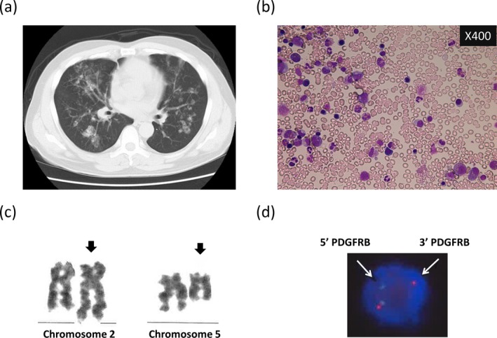
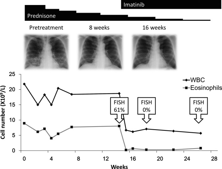
A 54‐year‐old man presenting with leukocytosis was referred to our hospital. Blood examination revealed eosinophilia—WBC 15.7 × 10⁹/L (neutrophils 28%, eosinophils 55%, basophils 1%, monocytes 3%, and lymphocytes 13%), Hb 13.0 g/dl, platelet count 339 × 109/L, and LDH 232 U/L (normal range: 100–220). Liver and renal functions were normal. Since no clinical symptom or organ damage was identified, a regular monthly follow‐up was advised. After 4 months, he developed respiratory symptoms including cough and dyspnea. Chest X‐ray and computed tomography (CT) scanning revealed bilateral lung infiltrates (Figure 1a). Bronchoalveolar lavage fluid obtained by bronchoscopy revealed increased probability of eosinophils (20.5% eosinophils, 78.0% macrophages, 1.0% lymphocytes, and 0.5% neutrophils). He was diagnosed with acute eosinophilic pneumonia and was given prednisone at a dose of 0.5 mg kg−1 day−1. The clinical course of the patient is shown in Figure 2. Although treatment with prednisone improved the shadow of infiltrates on the X‐ray and the respiratory symptoms, it did not reduce the increased number of eosinophils in circulation. Therefore, bone marrow examination was carried out and it revealed normocellularity with elevated eosinophils (22.1% of nuclear cells) without blastoid cell proliferation (0%) (Figure 1b). Cytogenetic analysis of the bone marrow showed 46, XY, t(2;5)(q37;q31) [16/20]/46, XY [4/20] (Figure 1c). FISH analysis in the peripheral blood leukocytes revealed the presence of a split signal at PDGFRB (Figure 1d). In addition, WT1 mRNA was positively expressed (1,200 copies/μg RNA) in the peripheral blood.
Figure 1.

Computed tomography scanning showing the development of acute eosinophilic pneumonia (a). Morphology of bone marrow examination before imatinib therapy stained with May‐Giemsa (b). Cytogenetic analysis of bone marrow sample showing translocation between chromosomes 2q37 and 5q31 (c). This abnormality was observed in 16 of 20 metaphases. Fluorescence in situ hybridization of peripheral blood showing the presence of a split signal on platelet‐derived growth factor receptor beta (PDGFRB) gene (d). This was observed in 61% of nucleated cells
Figure 2.

Clinical course of the patient after the incidence of acute eosinophilic leukemia. Low‐dose prednisone was initially administered, and it was effective for improving pneumonia but not in reducing the number of increased eosinophils. Imatinib administration rapidly reduced the number of eosinophils and the probability of cells harboring platelet‐derived growth factor receptor beta (PDGFRB) translocation in the peripheral blood. The probability of cells harboring PDGFRB translocation was evaluated by fluorescence in situ hybridization (FISH)
After the detection of PDGFRB rearrangement, imatinib was given at a dose of 400 mg/day, since previous studies have reported a positive outcome from this dose in patients with hematologic neoplasms with PDGFRB rearrangement (Cheah et al., 2014; Jawhar et al., 2017). Imatinib treatment was effective, with rapid regression of eosinophils in the peripheral blood and the pneumonia shadow on lung X‐rays. The eosinophil number was back to normal after a week of imatinib therapy and the pneumonia shadow disappeared in 6 weeks. Translocation analysis by FISH also revealed the absence of PDGFRB rearrangement in the peripheral blood leukocytes after a month of imatinib treatment. The disappearance of t(2;5)(q37;q31) and a normal eosinophil count in the bone marrow were confirmed after 3 months. WT1 mRNA expression was negative (<50 copies/μgRNA) in the peripheral blood at that time. The dose of imatinib was reduced to 200 mg/day after 12 months of treatment. No recurrence was observed under imatinib therapy for over 4 years. No severe adverse effects were recorded—a grade 1 liver dysfunction, increased CPK level, anemia, renal dysfunction, and edema according to the Common Terminology Criteria for Adverse Events ver.4.0 were the only adverse events that developed during the observation period. This study was approved by the research ethics board of Nihon University School of Medicine in accordance with the Declaration of Helsinki (identifier 150–0) and written informed consent was obtained from the patient before sample analysis.
3. MATERIALS AND METHODS
3.1. Whole‐genome sequencing
Whole‐genome sequencing (WGS) was conducted on DNA sample extracted from whole leukocytes of peripheral blood obtained from the patient before imatinib therapy. Genomic DNA was extracted from the whole blood using Maxwell® 16 LEV Blood DNA Kit (Promega, Fitchburg, WI), sheared into approximately 350 bp fragments, and used to make a library with TruSeq Nano DNA Sample Prep Kit (Illumina, San Diego, CA). Sequencing was performed on an Illumina HiSeq X platform in paired‐end 150 bp configuration.
3.2. Mapping and calling structural variations
Adapter sequences were removed by cutadapt (v1.2.1). After quality control, reads were mapped to the reference human genome (hg19) using BWA (ver.0.7.10). Mapping result was corrected using Picard (ver.1.73) for removing duplicates and GATK (ver.1.6‐13) for local alignment and quality score recalibration. Structural variation (SV) calls were performed using BreakDancer (ver.1.4.5). Annotations of SVs were based on RefSeq (UCSC Genome Browser, Feb 2017) and GENCODE (UCSC Genome Browser, ver. 19). SVs were further filtered according to the following criteria: (a) CTX (interchromosomal translocation), (b) the translocations between chromosomes 2 and 5, and (c) total number of supporting read pairs more than seven. The breakpoints of SVs were manually reviewed using Integrative Genomics Viewer (IGV).
3.3. PCR method
We designed oligonucleotide primers which were applicable to both amplification by PCR and direct sequencing by the dideoxy method. The forward primers were set 200 base‐pairs (bp) upstream from the breakpoints, and the reverse primers were set 200 bp downstream from the breakpoints. The following oligonucleotide primers were used for the detection of the fusion gene identified by WGS—forward 5′‐AAC AAC AAA CTA TGA TGT AGT TAG AG‐3′ and reverse 5′‐AGA GAA GGC AAG ACA CCA GCC CTA GGT‐3′.
The DNA used for WGS analysis was the same DNA that was used for direct sequencing. The concentration of genomic DNA was determined using a NanoDrop Onec Spectrophotometer (Thermo Fisher Scientific, Waltham, MA). The DNA was diluted to a final concentration of 100 ng/μL using nuclease‐free water. PCR was performed using a VeritiTM 200 thermal cycler (Thermo Fisher Scientific) with AmpliTaq Gold® 360 Master Mix (Thermo Fisher Scientific). Genomic DNA was used as the template with primers flanking the target gene. The PCR reaction conditions were 98°C for 3 m followed by 35 cycles of 98°C for 30 s, 60°C for 30 s, and 72°C for 30 s. Following PCR amplification, the amplification products were checked by agarose gel electrophoresis and purified using an ExoSAP‐IT purification kit for PCR products (Affymetrix/USB). The purification reaction conditions were 37°C for 15 m and 80°C for 15 m. Bidirectional sequencing was performed using forward and reverse primers. The reaction was carried out in a VeritiTM 200 thermal cycler using a BigDye® Terminator v1.1 Cycle Sequencing Kit (Thermo Fisher Scientific). The reaction conditions were 98°C for 1 m followed by 25 cycles of 98°C for 10 s, 50°C for 5 s, and 60°C for 2 m. The sequencing reaction products were purified using a BigDye XTerminatorTM Purification Kit (Thermo Fisher Scientific). Capillary electrophoresis was performed using an Applied BiosystemsTM 3,130 DNA Analyzer (Thermo Fisher Scientific) and the obtained nucleotide sequence data were compared against sequence data deposited in databases, such as GenBank at NCBI.
4. RESULTS
4.1. Whole‐genome analysis
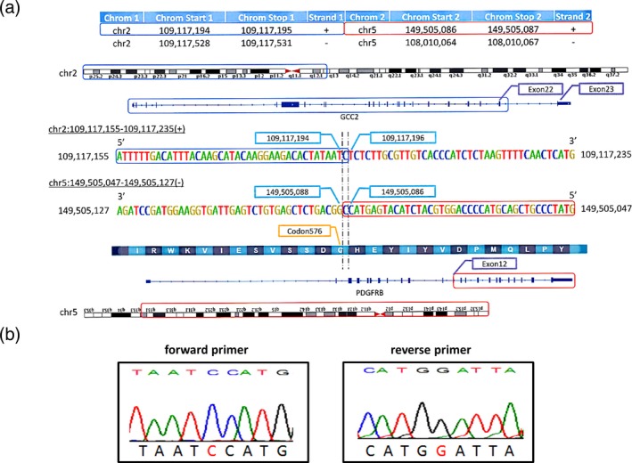
First, we detected the variant for each case by WGS analysis using a next‐generation sequencer in whole leukocytes obtained from the patient before imatinib therapy. The results from the WGS analysis showed the definitive nucleotide sequences including the breakpoints constituting the fusion gene—46, XY, t(2;5)(q37;q31)/46, XY. As shown in Figure 3a, we were able to detect a novel fusion gene between exon 22 of GCC2 and exon 12 of PDGFRB.
Figure 3.

Whole‐genome sequence identifying the breakpoint forming platelet‐derived growth factor receptor beta (PDGFRB) and GRIP and coiled‐coil domain containing 2 (GCC2) fusion gene (a). A novel fusion gene between exon 22 of GCC2 and exon 12 of PDGFRB was detected. Direct sequencing analysis confirming the presence of GCC2‐PDGFRB fusion gene (b)
4.2. PCR analysis
Next, we confirmed the nucleotide sequence for these breakpoint sites by direct sequencing analysis. The result of the direct sequencing analysis was in agreement with the data from the WGS analysis. As shown in Figure 3b, the presence of GCC2‐PDGFRB fusion gene was confirmed by direct sequencing analysis.
5. DISCUSSION
In this study, we were able to detect a novel fusion gene comprising of GCC2 (also called GCC185) and PDGFRB, involved in the pathogenesis of CEL. Results of G‐banding, FISH, WGS, and PCR analyses revealed the fusion of PDGFRB and GCC2 genes. In addition, blood examination revealed a positive expression for WT1 mRNA before imatinib therapy which turned negative after remission, suggesting that WT1 mRNA level is a possible marker that could distinguish CEL from HES. Interestingly, WT1 mRNA appeared to be unsuitable for detecting minimal residual disease, since it became negative at the achievement of complete cytogenetic remission (indicated by the absence of chromosomal abnormality). Furthermore, the efficacy of imatinib therapy was excellent and triggered the rapid regression of eosinophils and pneumonia shadow on X‐rays in this case. However, PCR detection of the fusion gene was not possible since the fusion gene was unknown. Imatinib therapy was well tolerated and the patient is still under the treatment.
CEL with PDGFRA or PDGFRB rearrangement is usually sensitive to imatinib monotherapy in the chronic phase, ensuring long‐lasting remission (Helbig et al., 2010; Jawhar et al., 2017). Although the characteristics of CEL with PDGFRA rearrangement and that with PDGFRB rearrangement are similar, some differences have been reported. Both CEL with PDGFRA rearrangement and that with PDGFRB rearrangement are predominant in adult males. Both are sensitive to imatinib, but maintenance therapy is required to retain remission of the disease (Cheah et al., 2014; Gotlib, 2017; Helbig et al., 2010; Jawhar et al., 2017). The optimal treatment doses for these diseases are considerably different. According to a study that tested kinase inhibitory profiles, the IC50 of imatinib was 3.2 nM for PDGFRA‐FIP1L1, 50 nM for ETV6‐PDGFRB, and 582 nM for BCR‐ABL1. This demonstrates the differences in sensitivity to imatinib between these conditions (Apperley et al., 2002; Chen et al., 2004). This suggests the requirement for the determination of the ideal daily dose of imatinib therapy for CEL. According to the report by Helbig et al. (2008), very low dose imatinib therapy (100 mg/week) for CEL with PDGFRA‐FIP1L1 fusion gene successfully maintained remission during the observation period. Patients with myeloid neoplasms with PDGFRB rearrangement have been treated with imatinib at a dose of 100–400 mg/day (Jawhar et al., 2017). Based on previous findings, we considered 100–200 mg/day to be the optimal therapeutic dose for CEL with PDGFRB rearrangement. Therefore, the dose of imatinib was reduced from 400 to 200 mg/day after cytogenetic remission (negative for PDGFRB rearrangement in the peripheral blood by FISH). After dose reduction, the patient sustained cytogenetic remission, suggesting that a dose of less than 200 mg/day is optimal for maintenance.
We found that the GCC2 gene was a fusion partner for PDGFRB in this patient. The protein translated from the GCC2 gene forms a part of the peripheral membrane protein localized to the trans‐Golgi networks (Luke, 2003). Although it is natural to consider that the GCC2 gene has a critical role in the constitutive activation of PDGFRB in this patient, the association of GCC2 has rarely been reported in human diseases. Indeed, GCC2 has never been described as a cause of a hematologic malignancy. However, in three patients with lung cancer, the GCC2 gene was shown to fuse with anaplastic lymphoma kinase (ALK; MIM #105590), which has a critical role in the pathogenesis of non‐small cell lung cancer, and targeting it with crizotinib was shown to be active against cancer (Jiang et al., 2018; Noh et al., 2017; Vendrell et al., 2017). The non‐small cell lung cancer harboring GCC2‐ALK fusion gene supports the hypothesis that the GCC2‐PDGFRB gene products could behave as an oncoprotein. The products translated from the fusion gene are considered to be activated constitutively, which result in the stimulation of downstream pathways for eosinophil differentiation.
The mechanism underlying the PDGFRB‐mediated accumulation of eosinophils is not well understood. Some evidence has shown the activation of the downstream pathways by transfecting PDGFRB fusion gene into cells. Ishibashi et al. (2016) showed that the transfection of ATF7IP‐PDGFRB gene to Ba/F3 conferred IL3‐independent cell growth accompanied by the activation of MAP kinase and AKT. In addition, STAT1, STAT3, and STAT5 are also reported to be activated by ETV6‐PDGFRB transfection (Montano‐Almendras et al., 2012; Wilbanks et al., 2000). Furthermore, the requirement of nuclear factor‐kappaB (NF‐κB) for eosinophil differentiation and cell growth with _ETV6‐PDGFRB_‐transfectant has been previously reported (Montano‐Almendras et al., 2012). With respect to the significance of IL5‐dependent STAT5 activation for differentiation toward eosinophils in normal hematopoiesis, we speculate that multiple pathways other than STAT5, including STAT1, STAT3, MAPK, AKT, and NF‐κB, are critical participants for CEL development and progression. The investigation for the direct effect of imatinib on eosinophils in patients with CEL harboring PDGFRA and PDGFRB rearrangements is currently underway.
In conclusion, we here reported the first case of CEL demonstrating GCC2 gene as a partner of PDGFRB. The functions of the fusion gene should be further investigated to clarify how it affects CEL pathogenesis.
CONFLICTS OF INTEREST
N.I. and Y.H. received honoraria and speaker fees from Novartis Pharma K.K. The remaining coauthors declare no competing financial interests.
ACKNOWLEDGMENTS
This work was supported by a grant from the Health Sciences Research Institute, Inc. (Yokohama, Japan) for Division of Companion Diagnostics, Department of Pathology of Microbiology, Nihon University School of Medicine, Tokyo, Japan. This study was also technically assisted by RIKEN GENESIS Co., Ltd. We also thank Miharu Watanabe and Eiko Ishizuka for supporting the study.
Iriyama N, Takahashi H, Naruse H, et al. A novel fusion gene involving PDGFRB and GCC2 in a chronic eosinophilic leukemia patient harboring t(2;5)(q37;q31). Mol Genet Genomic Med. 2019;7:e591 10.1002/mgg3.591
Noriyoshi Iriyama and Hiromichi Takahashi contributed equally to this work.
REFERENCES
- Apperley, J. F. , Gardembas, M. , Melo, J. V. , Russell‐Jones, R. , Bain, B. J. , Baxter, E. J. , … Goldman, J. M. (2002). Response to imatinib mesylate in patients with chronic myeloproliferative diseases with rearrangements of the platelet‐derived growth factor receptor beta. The New England Journal of Medicine, 347(7), 481–487. 10.1056/NEJMoa020150 [DOI] [PubMed] [Google Scholar]
- Cheah, C. Y. , Burbury, K. , Apperley, J. F. , Huguet, F. , Pitini, V. , Gardembas, M. , … Seymour, J. F. (2014). Patients with myeloid malignancies bearing PDGFRB fusion genes achieve durable long‐term remissions with imatinib. Blood, 123(23), 3574–3577. 10.1182/blood-2014-02-555607 [DOI] [PMC free article] [PubMed] [Google Scholar]
- Chen, J. , Wall, N. R. , Kocher, K. , Duclos, N. , Fabbro, D. , Neuberg, D. , … Gilliland, D. G. (2004). Stable expression of small interfering RNA sensitizes TEL‐PDGFbetaR to inhibition with imatinib or rapamycin. The Journal of Clinical Investigation, 113(12), 1784–1791. 10.1172/JCI20673 [DOI] [PMC free article] [PubMed] [Google Scholar]
- Cools, J. , DeAngelo, D. J. , Gotlib, J. , Stover, E. H. , Legare, R. D. , Cortes, J. , … Gilliland, D. G. (2003). A tyrosine kinase created by fusion of the PDGFRA and FIP1L1 genes as a therapeutic target of imatinib in idiopathic hypereosinophilic syndrome. The New England Journal of Medicine, 348(13), 1201–1214. 10.1056/NEJMoa025217 [DOI] [PubMed] [Google Scholar]
- Gong, S.‐L. , Guo, M.‐Q. , Tang, G.‐S. , Zhang, C.‐L. , Qiu, H.‐Y. , Hu, X.‐X. , & Yang, J.‐M. (2016). Fusion of platelet‐derived growth factor receptor β to CEV14 gene in chronic myelomonocytic leukemia: A case report and review of the literature. Oncology Letters, 11(1), 770–774. 10.3892/ol.2015.3949 [DOI] [PMC free article] [PubMed] [Google Scholar]
- Gotlib, J. (2017). World Health Organization‐defined eosinophilic disorders: 2017 update on diagnosis, risk stratification, and management. American Journal of Hematology, 92(11), 1243–1259. 10.1002/ajh.24880 [DOI] [PubMed] [Google Scholar]
- Helbig, G. , Moskwa, A. , Hus, M. , Piszcz, J. , Swiderska, A. , Urbanowicz, A. , … Krzemień, S. (2010). Clinical characteristics of patients with chronic eosinophilic leukaemia (CEL) harbouring FIP1L1‐PDGFRA fusion transcript–results of Polish multicentre study. Hematological Oncology, 28(2), 93–97. 10.1002/hon.919 [DOI] [PubMed] [Google Scholar]
- Helbig, G. , Stella‐Hołowiecka, B. , Majewski, M. , Całbecka, M. , Gajkowska, J. , Klimkiewicz, R. , … Hołowiecki, J. (2008). A single weekly dose of imatinib is sufficient to induce and maintain remission of chronic eosinophilic leukaemia in FIP1L1‐PDGFRA‐expressing patients. British Journal of Haematology, 141(2), 200–204. 10.1111/j.1365-2141.2008.07033.x [DOI] [PubMed] [Google Scholar]
- Hochhaus, A. , Larson, R. A. , Guilhot, F. , Radich, J. P. , Branford, S. , Hughes, T. P. , … Druker, B. J. (2017). Long‐term outcomes of imatinib treatment for chronic myeloid leukemia. The New England Journal of Medicine, 376(10), 917–927. 10.1056/NEJMoa1609324 [DOI] [PMC free article] [PubMed] [Google Scholar]
- Ishibashi, T. , Yaguchi, A. , Terada, K. , Ueno‐Yokohata, H. , Tomita, O. , Iijima, K. , … Kiyokawa, N. (2016). Ph‐like ALL‐related novel fusion kinase ATF7IP‐PDGFRB exhibits high sensitivity to tyrosine kinase inhibitors in murine cells. Experimental Hematology, 44(3), 177–88.e5. 10.1016/j.exphem.2015.11.009 [DOI] [PubMed] [Google Scholar]
- Jawhar, M. , Naumann, N. , Schwaab, J. , Baurmann, H. , Casper, J. , Dang, T.‐A. , … Metzgeroth, G. (2017). Imatinib in myeloid/lymphoid neoplasms with eosinophilia and rearrangement of PDGFRB in chronic or blast phase. Annals of Hematology, 96(9), 1463–1470. 10.1007/s00277-017-3067-x [DOI] [PubMed] [Google Scholar]
- Jiang, J. , Wu, X. , Tong, X. , Wei, W. , Chen, A. , Wang, X. , … Huang, J. (2018). GCC2‐ALK as a targetable fusion in lung adenocarcinoma and its enduring clinical responses to ALK inhibitors. Lung Cancer (Amsterdam, Netherlands), 115, 5–11. 10.1016/j.lungcan.2017.10.011 [DOI] [PubMed] [Google Scholar]
- Luke, M. R. , Kjer‐Nielsen, L. , Brown, D. L. , Stow, J. L. , & Gleeson, P. A. (2003). GRIP domain‐mediated targeting of two new coiled‐coil proteins, GCC88 and GCC185, to subcompartments of the trans‐Golgi network. The Journal of Biological Chemistry, 278(6), 4216–4226. 10.1074/jbc.M210387200 [DOI] [PubMed] [Google Scholar]
- Montano‐Almendras, C. P. , Essaghir, A. , Schoemans, H. , Varis, I. , Noël, L. A. , Velghe, A. I. , … Demoulin, J.‐B. (2012). ETV6‐PDGFRB and FIP1L1‐PDGFRA stimulate human hematopoietic progenitor cell proliferation and differentiation into eosinophils: The role of nuclear factor‐κB. Haematologica, 97(7), 1064–1072. 10.3324/haematol.2011.047530 [DOI] [PMC free article] [PubMed] [Google Scholar]
- Noh, K.‐W. , Lee, M.‐S. , Lee, S. E. , Song, J.‐Y. , Shin, H.‐T. , Kim, Y. J. , … Choi, Y.‐L. (2017). Molecular breakdown: A comprehensive view of anaplastic lymphoma kinase (ALK)‐rearranged non‐small cell lung cancer. The Journal of Pathology, 243(3), 307–319. 10.1002/path.4950 [DOI] [PubMed] [Google Scholar]
- Vendrell, J. A. , Taviaux, S. , Béganton, B. , Godreuil, S. , Audran, P. , Grand, D. , … Solassol, J. (2017). Detection of known and novel ALK fusion transcripts in lung cancer patients using next‐generation sequencing approaches. Scientific Reports, 7(1), 12510 10.1038/s41598-017-12679-8 [DOI] [PMC free article] [PubMed] [Google Scholar]
- Wilbanks, A. M. , Mahajan, S. , Frank, D. A. , Druker, B. J. , Gilliland, D. G. , & Carroll, M. (2000). TEL/PDGFbetaR fusion protein activates STAT1 and STAT5: A common mechanism for transformation by tyrosine kinase fusion proteins. Experimental Hematology, 28(5), 584–593. 10.1016/s0301-472x(00)00138-7 [DOI] [PubMed] [Google Scholar]
- Zhang, Y. , Qu, S. , Wang, Q. , Li, J. , Xu, Z. , Qin, T. , … Xiao, Z. (2018). A novel fusion of PDGFRB to TSC1, an intrinsic suppressor of mTOR‐signaling pathway, in a chronic eosinophilic leukemia patient with t(5;9)(q32;q34). Leukemia & Lymphoma, 59(10), 2506–2508. 10.1080/10428194.2018.1427855 [DOI] [PMC free article] [PubMed] [Google Scholar]
- Zou, Y. S. , Hoppman, N. L. , Singh, Z. N. , Sawhney, S. , Kotiah, S. D. , & Baer, M. R. (2017). Novel t(5;11)(q32;q13.4) with NUMA1‐PDGFRB fusion in a myeloid neoplasm with eosinophilia with response to imatinib mesylate. Cancer Genetics, 212–213, 38–44. 10.1016/j.cancergen.2017.03.004 [DOI] [PubMed] [Google Scholar]