The electrical response of turtle cones to flashes and steps of light (original) (raw)
Abstract
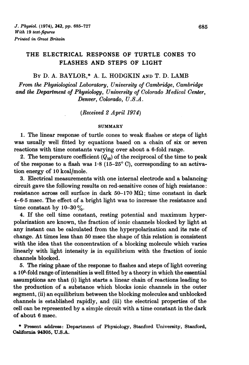
1. The linear response of turtle cones to weak flashes or steps of light was usually well fitted by equations based on a chain of six or seven reactions with time constants varying over about a 6-fold range.
2. The temperature coefficient (_Q_10) of the reciprocal of the time to peak of the response to a flash was 1·8 (15-25° C), corresponding to an activation energy of 10 kcal/mole.
3. Electrical measurements with one internal electrode and a balancing circuit gave the following results on red-sensitive cones of high resistance: resistance across cell surface in dark 50-170 MΩ; time constant in dark 4-6·5 msec. The effect of a bright light was to increase the resistance and time constant by 10-30%.
4. If the cell time constant, resting potential and maximum hyperpolarization are known, the fraction of ionic channels blocked by light at any instant can be calculated from the hyperpolarization and its rate of change. At times less than 50 msec the shape of this relation is consistent with the idea that the concentration of a blocking molecule which varies linearly with light intensity is in equilibrium with the fraction of ionic channels blocked.
5. The rising phase of the response to flashes and steps of light covering a 105-fold range of intensities is well fitted by a theory in which the essential assumptions are that (i) light starts a linear chain of reactions leading to the production of a substance which blocks ionic channels in the outer segment, (ii) an equilibrium between the blocking molecules and unblocked channels is established rapidly, and (iii) the electrical properties of the cell can be represented by a simple circuit with a time constant in the dark of about 6 msec.
6. Deviations from the simple theory which occur after 50 msec are attributed partly to a time-dependent desensitization mechanism and partly to a change in saturation potential resulting from a voltage-dependent change in conductance.
7. The existence of several components in the relaxation of the potential to its resting level can be explained by supposing that the `substance' which blocks light sensitive ionic channels is inactivated in a series of steps.

Selected References
These references are in PubMed. This may not be the complete list of references from this article.
- Baylor D. A., Fuortes M. G. Electrical responses of single cones in the retina of the turtle. J Physiol. 1970 Mar;207(1):77–92. doi: 10.1113/jphysiol.1970.sp009049. [DOI] [PMC free article] [PubMed] [Google Scholar]
- Baylor D. A., Fuortes M. G., O'Bryan P. M. Receptive fields of cones in the retina of the turtle. J Physiol. 1971 Apr;214(2):265–294. doi: 10.1113/jphysiol.1971.sp009432. [DOI] [PMC free article] [PubMed] [Google Scholar]
- Baylor D. A., Hodgkin A. L. Changes in time scale and sensitivity in turtle photoreceptors. J Physiol. 1974 Nov;242(3):729–758. doi: 10.1113/jphysiol.1974.sp010732. [DOI] [PMC free article] [PubMed] [Google Scholar]
- Baylor D. A., Hodgkin A. L. Detection and resolution of visual stimuli by turtle photoreceptors. J Physiol. 1973 Oct;234(1):163–198. doi: 10.1113/jphysiol.1973.sp010340. [DOI] [PMC free article] [PubMed] [Google Scholar]
- Baylor D. A., Hodgkin A. L., Lamb T. D. Reconstruction of the electrical responses of turtle cones to flashes and steps of light. J Physiol. 1974 Nov;242(3):759–791. doi: 10.1113/jphysiol.1974.sp010733. [DOI] [PMC free article] [PubMed] [Google Scholar]
- Borsellino A., Fuortes M. G., Smith T. G. Visual responses in Limulus. Cold Spring Harb Symp Quant Biol. 1965;30:429–443. doi: 10.1101/sqb.1965.030.01.042. [DOI] [PubMed] [Google Scholar]
- Briggs G. E., Haldane J. B. A Note on the Kinetics of Enzyme Action. Biochem J. 1925;19(2):338–339. doi: 10.1042/bj0190338. [DOI] [PMC free article] [PubMed] [Google Scholar]
- CONE R. A. THE RAT ELECTRORETINOGRAM. II. BLOCH'S LAW AND THE LATENCY MECHANISM OF THE B-WAVE. J Gen Physiol. 1964 Jul;47:1107–1116. doi: 10.1085/jgp.47.6.1107. [DOI] [PMC free article] [PubMed] [Google Scholar]
- FUORTES M. G., HODGKIN A. L. CHANGES IN TIME SCALE AND SENSITIVITY IN THE OMMATIDIA OF LIMULUS. J Physiol. 1964 Aug;172:239–263. doi: 10.1113/jphysiol.1964.sp007415. [DOI] [PMC free article] [PubMed] [Google Scholar]
- Fein H. Passing current through recording glass micro-pipette electrodes. IEEE Trans Biomed Eng. 1966 Oct;13(4):211–212. [PubMed] [Google Scholar]
- Hagins W. A., Penn R. D., Yoshikami S. Dark current and photocurrent in retinal rods. Biophys J. 1970 May;10(5):380–412. doi: 10.1016/S0006-3495(70)86308-1. [DOI] [PMC free article] [PubMed] [Google Scholar]
- Hagins W. A. The visual process: Excitatory mechanisms in the primary receptor cells. Annu Rev Biophys Bioeng. 1972;1:131–158. doi: 10.1146/annurev.bb.01.060172.001023. [DOI] [PubMed] [Google Scholar]
- Korenbrot J. I., Cone R. A. Dark ionic flux and the effects of light in isolated rod outer segments. J Gen Physiol. 1972 Jul;60(1):20–45. doi: 10.1085/jgp.60.1.20. [DOI] [PMC free article] [PubMed] [Google Scholar]
- Penn R. D., Hagins W. A. Kinetics of the photocurrent of retinal rods. Biophys J. 1972 Aug;12(8):1073–1094. doi: 10.1016/S0006-3495(72)86145-9. [DOI] [PMC free article] [PubMed] [Google Scholar]
- RUSHTON W. A. VISUAL ADAPTATION. Proc R Soc Lond B Biol Sci. 1965 Mar 16;162:20–46. doi: 10.1098/rspb.1965.0024. [DOI] [PubMed] [Google Scholar]
- Schwartz E. A. Responses of single rods in the retina of the turtle. J Physiol. 1973 Aug;232(3):503–514. doi: 10.1113/jphysiol.1973.sp010283. [DOI] [PMC free article] [PubMed] [Google Scholar]
- Tomita T. Electrical activity of vertebrate photoreceptors. Q Rev Biophys. 1970 May;3(2):179–222. doi: 10.1017/s0033583500004571. [DOI] [PubMed] [Google Scholar]
- Toyoda J., Nosaki H., Tomita T. Light-induced resistance changes in single photoreceptors of Necturus and Gekko. Vision Res. 1969 Apr;9(4):453–463. doi: 10.1016/0042-6989(69)90134-5. [DOI] [PubMed] [Google Scholar]