Obesity is associated with macrophage accumulation in adipose tissue - PubMed (original) (raw)
Obesity is associated with macrophage accumulation in adipose tissue
Stuart P Weisberg et al. J Clin Invest. 2003 Dec.
Abstract
Obesity alters adipose tissue metabolic and endocrine function and leads to an increased release of fatty acids, hormones, and proinflammatory molecules that contribute to obesity associated complications. To further characterize the changes that occur in adipose tissue with increasing adiposity, we profiled transcript expression in perigonadal adipose tissue from groups of mice in which adiposity varied due to sex, diet, and the obesity-related mutations agouti (Ay) and obese (Lepob). We found that the expression of 1,304 transcripts correlated significantly with body mass. Of the 100 most significantly correlated genes, 30% encoded proteins that are characteristic of macrophages and are positively correlated with body mass. Immunohistochemical analysis of perigonadal, perirenal, mesenteric, and subcutaneous adipose tissue revealed that the percentage of cells expressing the macrophage marker F4/80 (F4/80+) was significantly and positively correlated with both adipocyte size and body mass. Similar relationships were found in human subcutaneous adipose tissue stained for the macrophage antigen CD68. Bone marrow transplant studies and quantitation of macrophage number in adipose tissue from macrophage-deficient (Csf1op/op) mice suggest that these F4/80+ cells are CSF-1 dependent, bone marrow-derived adipose tissue macrophages. Expression analysis of macrophage and nonmacrophage cell populations isolated from adipose tissue demonstrates that adipose tissue macrophages are responsible for almost all adipose tissue TNF-alpha expression and significant amounts of iNOS and IL-6 expression. Adipose tissue macrophage numbers increase in obesity and participate in inflammatory pathways that are activated in adipose tissues of obese individuals.
Figures
Figure 1
Adipose tissue transcripts whose abundance was correlated with body mass in mice. The expression of more than 12,000 transcripts in parametrial and epididymal adipose tissue was monitored in C57BL/6J mice whose body mass varied secondary to sex, diet, or mutations in the agouti (Ay/+) or leptin (Lepob/ob) loci. Using Kendall’s τ, a nonparametric correlation metric, we identified 1,304 transcripts that correlated significantly with body mass when the false discovery rate was held to 0.03. Examples of adipose tissue transcripts whose expression correlated with body mass include (a) Csf1r and (b) CD68 antigen (Cd68), which correlated positively with body mass, (c) succinate dehydrogenase complex, subunit B, iron sulfur (Ip) (Sdhb), and (d) ubiquinol–cytochrome c reductase subunit (Uqcr), which correlated negatively with body mass. Gray and black symbols denote female and male mice, respectively. +, lean; triangles, Ay/+; squares, DIO; circles, Lepob/ob mice.
Figure 2
Adipose tissue macrophages in mice with varying degrees of adiposity. Immunohistochemical detection of the macrophage-specific antigen F4/80 (black arrows) in perigonadal adipose tissue from C57BL/6J mice: (a) lean female, (b) Ay/+ female, (c) Lepob/ob female, (d) lean male, (e) DIO male, and (f) Lepob/ob male. Macrophages are stained brown. In lean animals (a and d), F4/80-expressing cells were uniformly small, dispersed, and rarely seen in aggregates. The fraction of F4/80-expressing cells was greater in moderately obese mice (b and e) and greatest in the severely obese Lepob/ob mice (c and f). Depots from all animals contained small, isolated F4/80 expressing cells (black arrows). In addition, depots from obese animals contained aggregates of F4/80 expressing cells (large blue arrows). Some macrophage aggregates contained small lipid-like droplets (small thin black arrow). Calibration mark = 40 μm.
Figure 3
The relationship between adipocyte size and the percentage of macrophages in adipose tissue. Average adipocyte cross-sectional area and the percentage of F4/80+ cells (macrophages) in adipose tissue depots were determined for each mouse in this study. Average adipocyte cross-sectional area was a strong predictor of the percentage of F4/80+ cells in (a) perigonadal (_r_2 = 0.76, P < 10–4), (b) perirenal (_r_2 = 0.73, P < 10–4), (c) mesenteric (_r_2 = 0.9, P < 10–4), and (d) subcutaneous (_r_2 = 0.39, P < 0.01) adipose tissue depots. (e) Data collected from all mice and all depots were plotted together. Light gray and dark gray symbols denote female and male mice, respectively. +, lean; triangles, Ay/+; squares, DIO; circles, Lepob/ob mice.
Figure 4
Macrophages in the liver and muscle of lean and obese mice. Immunohistochemical detection of cells expressing the macrophage-specific antigen F4/80 (arrows) in extensor digitalis longus muscles from C57BL/6J (a and c) Lepob/ob female and (b and d) lean female mice. Macrophages were rarely detected in areas surrounding the myofibrils (c and d). However, muscle from both lean and obese animals was infiltrated and surrounded by adipose tissue that contained significant numbers of F4/80-positive macrophages (a and b). The percentage of F4/80-positive macrophages within this adipose tissue was markedly increased in obese compared with lean mice (e, P < 0.005). The percentage of F4/80-positive Kupffer cells within liver was not significantly altered in obesity. Calibration mark = 40 0m; white bars, lean mice; gray bars, Lepob/ob mice.
Figure 5
F4/80+ cells express macrophage markers. Perigonadal adipose tissue was collected from female B6.V Lepob/ob mice, digested, and centrifuged to yield a buoyant adipocyte-enriched cell population and a pellet of SVCs. The SVCs were separated into F4/80+ (black bars) and F4/80– (white bars) populations via FACS. Quantitative RT-PCR was used to measure the relative expression of macrophage markers (Emr1, Csf1r, Cd68) and an adipocyte-specific gene (Acrp30). Among the three isolated cell populations, the relative gene expression of macrophage markers was highest among the F4/80+ cells. *The F4/80– SVCs did not express detectable amounts (< 0.05 of mean of all populations) of the macrophage markers. The adipocyte-enriched population (gray bars) expressed small amounts of the macrophage markers (a), consistent with residual macrophage contamination seen by immunofluorescent staining of live cells (b). In the adipocyte-enriched fraction, large autofluorescent adipocyte cell membranes (green) are not recognized by fluorescently conjugated F4/80 antibody (red), but membrane staining of small nonautofluorescent cells is seen. Control fluorescently conjugated isotype antibody did not recognize these cells (c).
Figure 6
F4/80+ cells in adipose tissue are bone marrow–derived. Adipose tissue was collected and SVCs were isolated 6 weeks after lethal irradiation and bone marrow transplantation. The SVCs were incubated with APC-conjugated anti-F4/80 (F4/80-APC and either PE-conjugated anti-CD45.1 (CD45.1-PE) or PE-conjugated anti-CD45.2 (CD45.2-PE). Eighty-five percent of F4/80+ cells expressed the donor antigen, CD45.1 (right upper quadrant in a). Only 14% of the F4/80+ cells also expressed the recipient antigen, CD45.2 (right upper quadrant in b).
Figure 7
Macrophage-deficient FVB/NJ Csf1op/op mice are also deficient in F4/80+ cells in adipose tissue. SVCs were isolated from subcutaneous and perigonadal adipose tissue of macrophage-deficient (FVB/NJ Csf1op/op) and control (FVB/NJ Csf1+/+) mice. Flow cytometry of SVCs isolated from two perigondadal adipose tissue depot illustrates that tissue from macrophage-deficient mice (b) contains 34% the number of F4/80+ cells found in adipose tissue from control mice (a).
Figure 8
Adipose tissue macrophages express proinflammatory factors. Perigonadal adipose tissue was collected from female B6.V Lepob/ob mice, digested, and centrifuged to yield a buoyant adipocyte-enriched cell population (gray bars) and a pellet of SVCs. The SVCs were separated into F4/80+ macrophages (black bars) and F4/80– populations (white bars) via FACS. Quantitative RT-PCR was used to measure the relative expression of three proinflammatory genes (Tnfa, Nos2, and Il6). Expression of Tnfa was limited almost exclusively to adipose tissue macrophages. F4/80– SVCs did not express detectable amounts of F4/80 or Tnfa (as indicated by asterisks). The adipocyte-enriched fractions expressed Tnfa at levels commensurate F4/80 expression and consistent with macrophage contamination. Nos2 was expressed by both macrophages and F4/80– SVCs, and Il6 was detectably expressed by all three populations in adipose tissue.
Figure 9
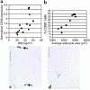
CD68 expression in human subcutaneous adipose tissue. Subcutaneous adipose tissue samples were aspirated from the subcutaneous abdominal region of human subjects whose BMIs ranged from 19.4 to 60.1 kg/m2. CD68 transcript expression was measured by quantitative real-time PCR. (a) BMI was a significant predictor of CD68 transcript expression (_r_2 = 0.43, P < 0.01). (b) Immunohistochemical detection and quantitation of CD68-expressing cells in subcutaneous adipose tissue from obese and lean subjects shows that the average adipocyte cross-sectional area was a strong predictor of the percentage of CD68-expressing cells (_r_2 = 0.86, P < 0.001). Typical micrographs from (c) an obese (BMI 50.8 kg/m2) female and (d) a lean (25.7 kg/m2) female subject are shown. Arrows in c indicate F4/80+ cells. Squares and diamonds denote female and male subjects, respectively. Calibration mark = 40 μm.
Comment in
- Obesity-induced inflammatory changes in adipose tissue.
Wellen KE, Hotamisligil GS. Wellen KE, et al. J Clin Invest. 2003 Dec;112(12):1785-8. doi: 10.1172/JCI20514. J Clin Invest. 2003. PMID: 14679172 Free PMC article. - Leptin Deficiency Shifts Mast Cells toward Anti-Inflammatory Actions and Protects Mice from Obesity and Diabetes by Polarizing M2 Macrophages.
Zhou Y, Yu X, Chen H, Sjöberg S, Roux J, Zhang L, Ivoulsou AH, Bensaid F, Liu CL, Liu J, Tordjman J, Clement K, Lee CH, Hotamisligil GS, Libby P, Shi GP. Zhou Y, et al. Cell Metab. 2015 Dec 1;22(6):1045-58. doi: 10.1016/j.cmet.2015.09.013. Epub 2015 Oct 17. Cell Metab. 2015. PMID: 26481668 Free PMC article.
Similar articles
- Mesenteric adipose tissue-derived monocyte chemoattractant protein-1 plays a crucial role in adipose tissue macrophage migration and activation in obese mice.
Yu R, Kim CS, Kwon BS, Kawada T. Yu R, et al. Obesity (Silver Spring). 2006 Aug;14(8):1353-62. doi: 10.1038/oby.2006.153. Obesity (Silver Spring). 2006. PMID: 16988077 - Telmisartan improves insulin resistance and modulates adipose tissue macrophage polarization in high-fat-fed mice.
Fujisaka S, Usui I, Kanatani Y, Ikutani M, Takasaki I, Tsuneyama K, Tabuchi Y, Bukhari A, Yamazaki Y, Suzuki H, Senda S, Aminuddin A, Nagai Y, Takatsu K, Kobayashi M, Tobe K. Fujisaka S, et al. Endocrinology. 2011 May;152(5):1789-99. doi: 10.1210/en.2010-1312. Epub 2011 Mar 22. Endocrinology. 2011. PMID: 21427223 - Adipose tissue collagen VI in obesity.
Pasarica M, Gowronska-Kozak B, Burk D, Remedios I, Hymel D, Gimble J, Ravussin E, Bray GA, Smith SR. Pasarica M, et al. J Clin Endocrinol Metab. 2009 Dec;94(12):5155-62. doi: 10.1210/jc.2009-0947. Epub 2009 Oct 16. J Clin Endocrinol Metab. 2009. PMID: 19837927 Free PMC article. - Adipocyte-Macrophage Cross-Talk in Obesity.
Engin AB. Engin AB. Adv Exp Med Biol. 2017;960:327-343. doi: 10.1007/978-3-319-48382-5_14. Adv Exp Med Biol. 2017. PMID: 28585206 Review. - Adipose tissue as an endocrine organ.
Galic S, Oakhill JS, Steinberg GR. Galic S, et al. Mol Cell Endocrinol. 2010 Mar 25;316(2):129-39. doi: 10.1016/j.mce.2009.08.018. Epub 2009 Aug 31. Mol Cell Endocrinol. 2010. PMID: 19723556 Review.
Cited by
- Unravelling monocyte functions: from the guardians of health to the regulators of disease.
Mildner A, Kim KW, Yona S. Mildner A, et al. Discov Immunol. 2024 Aug 30;3(1):kyae014. doi: 10.1093/discim/kyae014. eCollection 2024. Discov Immunol. 2024. PMID: 39430099 Free PMC article. Review. - Chemokine Expression in Inflamed Adipose Tissue Is Mainly Mediated by NF-κB.
Tourniaire F, Romier-Crouzet B, Lee JH, Marcotorchino J, Gouranton E, Salles J, Malezet C, Astier J, Darmon P, Blouin E, Walrand S, Ye J, Landrier JF. Tourniaire F, et al. PLoS One. 2013 Jun 18;8(6):e66515. doi: 10.1371/journal.pone.0066515. Print 2013. PLoS One. 2013. PMID: 23824685 Free PMC article. - Neonatal overfeeding attenuates acute central pro-inflammatory effects of short-term high fat diet.
Cai G, Dinan T, Barwood JM, De Luca SN, Soch A, Ziko I, Chan SM, Zeng XY, Li S, Molero J, Spencer SJ. Cai G, et al. Front Neurosci. 2015 Jan 13;8:446. doi: 10.3389/fnins.2014.00446. eCollection 2014. Front Neurosci. 2015. PMID: 25628527 Free PMC article. - Chronic tissue inflammation and metabolic disease.
Lee YS, Olefsky J. Lee YS, et al. Genes Dev. 2021 Mar 1;35(5-6):307-328. doi: 10.1101/gad.346312.120. Genes Dev. 2021. PMID: 33649162 Free PMC article. Review. - The Role of Leptin in the Association between Obesity and Psoriasis.
Hwang J, Yoo JA, Yoon H, Han T, Yoon J, An S, Cho JY, Lee J. Hwang J, et al. Biomol Ther (Seoul). 2021 Jan 1;29(1):11-21. doi: 10.4062/biomolther.2020.054. Biomol Ther (Seoul). 2021. PMID: 32690821 Free PMC article. Review.
References
- Tai ES, Lau TN, Ho SC, Fok AC, Tan CE. Body fat distribution and cardiovascular risk in normal weight women. Associations with insulin resistance, lipids and plasma leptin. Int. J. Obes. Relat. Metab. Disord. 2000;24:751–757. - PubMed
- Messerli FH, et al. Obesity and essential hypertension. Hemodynamics, intravascular volume, sodium excretion, and plasma renin activity. Arch. Intern. Med. 1981;141:81–85. - PubMed
- Liuzzi A, et al. Serum leptin concentration in moderate and severe obesity: relationship with clinical, anthropometric and metabolic factors. Int. J. Obes. Relat. Metab. Disord. 1999;23:1066–1073. - PubMed
- Janssen I, Katzmarzyk PT, Ross R. Body mass index, waist circumference, and health risk: evidence in support of current National Institutes of Health guidelines. Arch. Intern. Med. 2002;162:2074–2079. - PubMed
- Stolk RP, Meijer R, Mali WP, Grobbee DE, van der Graaf Y. Ultrasound measurements of intraabdominal fat estimate the metabolic syndrome better than do measurements of waist circumference. Am. J. Clin. Nutr. 2003;77:857–860. - PubMed
Publication types
MeSH terms
Substances
Grants and funding
- K08 DK-59960/DK/NIDDK NIH HHS/United States
- R01 DK066525/DK/NIDDK NIH HHS/United States
- R01 DK-66525/DK/NIDDK NIH HHS/United States
- R01 DK064773/DK/NIDDK NIH HHS/United States
- K08 DK059960/DK/NIDDK NIH HHS/United States
- R01 DK052431/DK/NIDDK NIH HHS/United States
- R01 DK-052431/DK/NIDDK NIH HHS/United States
LinkOut - more resources
Full Text Sources
Other Literature Sources
Medical
Research Materials
Miscellaneous


