Effects of B Cell Depletion on Early Mycobacterium tuberculosis Infection in Cynomolgus Macaques - PubMed (original) (raw)
Effects of B Cell Depletion on Early Mycobacterium tuberculosis Infection in Cynomolgus Macaques
Jiayao Phuah et al. Infect Immun. 2016.
Abstract
Although recent studies in mice have shown that components of B cell and humoral immunity can modulate the immune responses against Mycobacterium tuberculosis, the roles of these components in human and nonhuman primate infections are unknown. The cynomolgus macaque (Macaca fascicularis) model of M. tuberculosis infection closely mirrors the infection outcomes and pathology in human tuberculosis (TB). The present study used rituximab, an anti-CD20 antibody, to deplete B cells in M. tuberculosis-infected macaques to examine the contribution of B cells and humoral immunity to the control of TB in nonhuman primates during the acute phase of infection. While there was no difference in the overall pathology, disease profession, and clinical outcome between the rituximab-treated and untreated macaques in acute infection, analyzing individual granulomas revealed that B cell depletion resulted in altered local T cell and cytokine responses, increased bacterial burden, and lower levels of inflammation. There were elevated frequencies of T cells producing interleukin-2 (IL-2), IL-10, and IL-17 and decreased IL-6 and IL-10 levels within granulomas from B cell-depleted animals. The effects of B cell depletion varied among granulomas in an individual animal, as well as among animals, underscoring the previously reported heterogeneity of local immunologic characteristics of tuberculous granulomas in nonhuman primates. Taken together, our data clearly showed that B cells can modulate the local granulomatous response in M. tuberculosis-infected macaques during acute infection. The impact of these alterations on disease progression and outcome in the chronic phase remains to be determined.
Copyright © 2016, American Society for Microbiology. All Rights Reserved.
Figures
FIG 1
Confirmation of B cell depletion following rituximab treatment. Repeated measures of CD20 and CD79a by fluorescence-activated cell sorting (FACS) within PBMCs (A) and peripheral lymph node (pLN) biopsy specimens (B) to confirm B cell depletion within rituximab-treated animals (red) versus saline-treated controls (blue). Solid lines depict the group average, while dotted lines represent individual animals. Black arrows denote rituximab administration, the large red arrow denotes time of infection, and the red “X” denotes necropsy. n = 16 animals, 8 animals per group. An asterisk (*) represents statistical significance for values at the indicated time point with P < 0.05 using the Holm-Sidak method. (C) At necropsy, single cell suspensions obtained from granulomas were subjected to FACS staining for CD20 and CD79a quantification. Each point represents one granuloma (n = 16 animals, n = 100 granulomas, ∼6 granulomas per animal). (D) Similar staining to confirm B cell depletion was performed on thoracic LN samples obtained at necropsy. Each point represents one thoracic LN sample (n = 16 animas, n = 78 thoracic LN samples, ∼4 thoracic LN per animal). (E) Immunohistochemistry staining of paraffin-embedded granuloma (top four) and thoracic lymph node samples (bottom four panels) with CD20 (red), CD11b (green), and CD3 (blue) and H&E stains underneath each section confirmed the loss of B cell clusters within rituximab-treated animals. All statistical P values were obtained using the Mann-Whitney test unless otherwise stated.
FIG 2
Assessment of antibody profile of granuloma homogenates after rituximab treatment. (A) Granuloma homogenates obtained at necropsy were assayed for the amount of CFP-specific IgG, ESAT6-specific IgG, or total IgG present using ELISA. Antigen-specific IgG levels were significantly reduced after rituximab treatment compared to saline controls (the median granuloma antibody content was reduced ∼10-fold in the case of CFP and 5-fold for ESAT6). The total IgG content within rituximab-treated granulomas was also reduced by ∼10-fold. Each point represents one granuloma (n = 16 animals, n = 110 granulomas, ∼6 granulomas per animal). (B) Homogenates from lymph node samples were assayed for the amount of CFP-specific IgG and total IgG present. Both antigen specific IgG and total IgG were reduced with rituximab by approximately 2- to 5-fold. Each point represents one thoracic LN sample (n = 16 animals, n = 59 thoracic LN samples, ∼3 thoracic LN samples per animal). (C) The number of plasma cells generating IgG within the bone marrow, assayed by a plasma cell ELISPOT assay, were similar within both groups. Each point presents one animal (n = 16 animals, ∼8 animals per group). All statistical P values were obtained using the Mann-Whitney test unless otherwise stated.
FIG 3
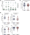
Clinical and pathology findings for animals at necropsy. (A) PET/CT scans were conducted prior to necropsy using [18F]FDG to identify and characterize granulomas. Within both groups, similar granuloma numbers were identified on scans, and the overall lung [18F]FDG avidities were comparable. Each symbol is an animal (n = 16 animals, 8 animals per group). The granuloma [18F]FDG avidity (standard uptake value [SUV]), although statistically significant, was only marginally lower in the rituximab-treated group. The sizes of granulomas were determined using CT. Each symbol is a granuloma (n = 319 granulomas, ∼19 granulomas per animal). (B) Gross pathology scores at necropsy. (C) Tissue sample homogenates obtained at necropsy were plated on 7H11 agar to enumerate the bacterial CFU. The total bacterial burden refers to the absolute number of CFU present within the thoracic cavity of each animal; each symbol is one animal (n = 16 animals). CFU counts from individual lung granulomas were then compared between both groups; each point represents one granuloma (n = 319 granulomas, ∼19 granulomas per animal). CFU counts from thoracic LN samples were comparable between both groups. Each point represents one thoracic LN sample (n = 138 samples, ∼9 lymph nodes per animal). (D) Granuloma CFU counts were further stratified according to individual animals. Each symbol is a single granuloma, and each individual monkey ID is on the x axis. Median CFU values per granuloma for the control group animals were ∼103 bacteria with some variability. However, variability within rituximab animals was much larger, with a higher distribution between animals within the group. A Brown-Forsythe test was used to establish that the observed difference in variability between both groups were statistically significant (P < 0.0001). (E) The capacity of granulomas and lymph nodes to sterilize bacteria was also assessed by comparing the number of granulomas with no recoverable CFU. Each symbol represents an animal (n = 16 animals, 8 animals per group). All statistical P values were obtained using the Mann-Whitney test unless otherwise stated.
FIG 4
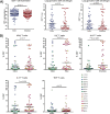
Effects of B cell depletion on T cell populations within the granuloma. (A) Single cell suspensions from granulomas were analyzed by FACS to identify T cells via CD3, CD4, and CD8 markers. Each symbol represents one granuloma. For CD3 enumeration, n = 16 animals and n = 191 granulomas, and thus there were ∼11 granulomas per animal. For CD4 and CD8 analysis, n = 16 animals and n = 93 granulomas, and thus there were ∼5 granulomas per animal. (B) The CD3 population were analyzed for cytokine production using intracellular cytokine staining via FACS, specifically IL-17, IL-2, IL-10, IFN-γ, and TNF. Each symbol represents a granuloma, and the colors differentiate granulomas from individual animals (n = 16 animals, n = 87 granulomas, ∼5 granulomas per animal for IL-17, IL-2, IFN-γ, and TNF). For IL-10, n = 10 animals and n = 62 granulomas, and thus there were ∼6 granulomas per animal. All statistical P values were obtained using the Mann-Whitney test.
FIG 5
Effects of B cell depletion on granuloma cytokine and neutrophil profiles. (A) Single cell suspensions from saline control granulomas were stained for B cell markers and cytokine antibodies to determine the cytokine secretion profile of B cells via intracellular FACS. Six cytokines were selected to be assayed: IL-2, IL-6, IL-10, IL-17, TNF, and IFN-γ. Each symbol is a granuloma, and colors denote granulomas from individual animals. For IL-6, IL-10, TNF, and IFN-γ, n = 8 animals and n = 27 granulomas, and thus there were ∼4 granulomas per animal. For IL-2 and IL-17, n = 5 animals and n = 22 granulomas, and thus there were ∼4 granulomas per animal. (B) ELISAs to quantify cytokine amounts were performed on granuloma homogenates for four cytokines, IL-6, IL-10, IL-17, and TNF to determine whether B cell depletion via rituximab perturbed the cytokine balance within the granulomas. Each symbol is a granuloma (n = 16 animals, n = 136 granulomas, ∼8 granulomas per animal). (C) Neutrophil amounts were quantified based on the amount of calprotectin present within granuloma homogenates. Each symbol is a granuloma (n = 16 animals, n = 155 granulomas, ∼10 granulomas per animal). All statistical P values were obtained using the Mann-Whitney test.
Similar articles
- Dynamics of Macrophage, T and B Cell Infiltration Within Pulmonary Granulomas Induced by Mycobacterium tuberculosis in Two Non-Human Primate Models of Aerosol Infection.
Hunter L, Hingley-Wilson S, Stewart GR, Sharpe SA, Salguero FJ. Hunter L, et al. Front Immunol. 2022 Jan 6;12:776913. doi: 10.3389/fimmu.2021.776913. eCollection 2021. Front Immunol. 2022. PMID: 35069548 Free PMC article. - Variability in tuberculosis granuloma T cell responses exists, but a balance of pro- and anti-inflammatory cytokines is associated with sterilization.
Gideon HP, Phuah J, Myers AJ, Bryson BD, Rodgers MA, Coleman MT, Maiello P, Rutledge T, Marino S, Fortune SM, Kirschner DE, Lin PL, Flynn JL. Gideon HP, et al. PLoS Pathog. 2015 Jan 22;11(1):e1004603. doi: 10.1371/journal.ppat.1004603. eCollection 2015 Jan. PLoS Pathog. 2015. PMID: 25611466 Free PMC article. - Activated B cells in the granulomas of nonhuman primates infected with Mycobacterium tuberculosis.
Phuah JY, Mattila JT, Lin PL, Flynn JL. Phuah JY, et al. Am J Pathol. 2012 Aug;181(2):508-14. doi: 10.1016/j.ajpath.2012.05.009. Epub 2012 Jun 19. Am J Pathol. 2012. PMID: 22721647 Free PMC article. - [Novel vaccines against M. tuberculosis].
Okada M. Okada M. Kekkaku. 2006 Dec;81(12):745-51. Kekkaku. 2006. PMID: 17240920 Review. Japanese. - Non-Human Primate Models of Tuberculosis.
Peña JC, Ho WZ. Peña JC, et al. Microbiol Spectr. 2016 Aug;4(4). doi: 10.1128/microbiolspec.TBTB2-0007-2016. Microbiol Spectr. 2016. PMID: 27726820 Review.
Cited by
- Markov Field network model of multi-modal data predicts effects of immune system perturbations on intravenous BCG vaccination in macaques.
Wang S, Myers AJ, Irvine EB, Wang C, Maiello P, Rodgers MA, Tomko J, Kracinovsky K, Borish HJ, Chao MC, Mugahid D, Darrah PA, Seder RA, Roederer M, Scanga CA, Lin PL, Alter G, Fortune SM, Flynn JL, Lauffenburger DA. Wang S, et al. bioRxiv [Preprint]. 2024 Oct 30:2024.04.13.589359. doi: 10.1101/2024.04.13.589359. bioRxiv. 2024. PMID: 39554028 Free PMC article. Updated. Preprint. - Spatial transcriptomic sequencing reveals immune microenvironment features of Mycobacterium tuberculosis granulomas in lung and omentum.
Qiu X, Zhong P, Yue L, Li C, Yun Z, Si G, Li M, Chen Z, Tan Y, Bao P. Qiu X, et al. Theranostics. 2024 Sep 23;14(16):6185-6201. doi: 10.7150/thno.99038. eCollection 2024. Theranostics. 2024. PMID: 39431015 Free PMC article. - Splenic marginal zone B cells restrict Mycobacterium tuberculosis infection by shaping the cytokine pattern and cell-mediated immunity.
Tsai CY, Oo M, Peh JH, Yeo BCM, Aptekmann A, Lee B, Liu JJJ, Tsao WS, Dick T, Fink K, Gengenbacher M. Tsai CY, et al. Cell Rep. 2024 Jul 23;43(7):114426. doi: 10.1016/j.celrep.2024.114426. Epub 2024 Jul 2. Cell Rep. 2024. PMID: 38959109 Free PMC article. - Potential biomarkers for evaluating the BCG vaccination response based on humoral immunity.
Chen YQ, Cao SH, Yang XY, Liu Y, Li CY. Chen YQ, et al. Heliyon. 2024 May 28;10(11):e32117. doi: 10.1016/j.heliyon.2024.e32117. eCollection 2024 Jun 15. Heliyon. 2024. PMID: 38947452 Free PMC article. - B cell heterogeneity in human tuberculosis highlights compartment-specific phenotype and functional roles.
Krause R, Ogongo P, Tezera L, Ahmed M, Mbano I, Chambers M, Ngoepe A, Magnoumba M, Muema D, Karim F, Khan K, Lumamba K, Nargan K, Madansein R, Steyn A, Shalek AK, Elkington P, Leslie A. Krause R, et al. Commun Biol. 2024 May 16;7(1):584. doi: 10.1038/s42003-024-06282-7. Commun Biol. 2024. PMID: 38755239 Free PMC article.
References
- Slight SR, Rangel-Moreno J, Gopal R, Lin Y, Fallert Junecko BA, Mehra S, Selman M, Becerril-Villanueva E, Baquera-Heredia J, Pavon L, Kaushal D, Reinhart TA, Randall TD, Khader SA. 2013. CXCR5+ T helper cells mediate protective immunity against tuberculosis. J Clin Invest 123:712–726. doi:10.1172/JCI65728. - DOI - PMC - PubMed
- Tsai MC, Chakravarty S, Zhu G, Xu J, Tanaka K, Koch C, Tufariello J, Flynn J, Chan J. 2006. Characterization of the tuberculous granuloma in murine and human lungs: cellular composition and relative tissue oxygen tension. Cell Microbiol 8:218–232. doi:10.1111/j.1462-5822.2005.00612.x. - DOI - PubMed
Publication types
MeSH terms
Substances
Grants and funding
- T32 AI060525/AI/NIAID NIH HHS/United States
- R01 HL075845/HL/NHLBI NIH HHS/United States
- R01 AI098925/AI/NIAID NIH HHS/United States
- P01 AI063537/AI/NIAID NIH HHS/United States
- R01 AI094745/AI/NIAID NIH HHS/United States
- R01 HL068526/HL/NHLBI NIH HHS/United States
LinkOut - more resources
Full Text Sources
Other Literature Sources
Medical


