Group 2 Innate Lymphoid Cells Promote an Early Antibody Response to a Respiratory Antigen in Mice - PubMed (original) (raw)
Group 2 Innate Lymphoid Cells Promote an Early Antibody Response to a Respiratory Antigen in Mice
Li Yin Drake et al. J Immunol. 2016.
Abstract
Innate lymphoid cells (ILCs) are a new family of immune cells that play important roles in innate immunity in mucosal tissues, and in the maintenance of tissue and metabolic homeostasis. Recently, group 2 ILCs (ILC2s) were found to promote the development and effector functions of Th2-type CD4(+) T cells by interacting directly with T cells or by activating dendritic cells, suggesting a role for ILC2s in regulating adaptive immunity. However, our current knowledge on the role of ILCs in humoral immunity is limited. In this study, we found that ILC2s isolated from the lungs of naive BALB/c mice enhanced the proliferation of B1- as well as B2-type B cells and promoted the production of IgM, IgG1, IgA, and IgE by these cells in vitro. Soluble factors secreted by ILC2s were sufficient to enhance B cell Ig production. By using blocking Abs and ILC2s isolated from IL-5-deficient mice, we found that ILC2-derived IL-5 is critically involved in the enhanced production of IgM. Furthermore, when adoptively transferred to Il7r(-/-) mice, which lack ILC2s and mature T cells, lung ILC2s promoted the production of IgM Abs to a polysaccharide Ag, 4-hydroxy-3-nitrophenylacetyl Ficoll, within 7 d of airway exposure in vivo. These findings add to the growing body of literature regarding the regulatory functions of ILCs in adaptive immunity, and suggest that lung ILC2s promote B cell production of early Abs to a respiratory Ag even in the absence of T cells.
Copyright © 2016 by The American Association of Immunologists, Inc.
Conflict of interest statement
Conflict of interest statement: All authors declare no conflict of interest.
Figures
FIGURE 1
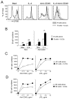
Lung ILC2s enhance the proliferation of B lymphocytes. Lung ILC2s and splenic or peritoneal B cells were isolated from naïve BALB/c mice. Panel A: CFSE-labeled splenic B cells were cultured at 2×10⁴ cells/well with or without ILC2s (104 cells/well) for 4 days. IL-4 (10 ng/ml) and/or anti-CD40 (1.5 μg/ml) were added to some cultures as indicated. The proliferation profiles of B cells were analyzed by FACS. Panel B: Splenic B cells were cultured with or without ILC2s for 4 days with medium (Med) alone or in the presence of IL-4 and/or anti-CD40. The numbers of CD19+ B cells were determined by FACS. Data (mean±SEM, n=3) are representative of two independent experiments. *, p<0.05; **, p<0.01 between the groups indicated by horizontal lines. Panel C and Panel D: Peritoneal B1 cells and B2 cells were cultured at 2×10⁴ cells/well with or without ILC2s (104 cells/well) in the presence of serial dilutions of anti-CD40 antibody or LPS plus IL-4 (10 ng/ml) for 4 days. The number of B220+ cells was determined by FACS. Each data point represents the cell numbers from one well. Data are representative of two independent experiments.
FIGURE 2
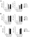
Lung ILC2s enhance Ig production by B lymphocytes. Lung ILC2s and splenic B cells were isolated from BALB/c mice. B cells were cultured at 2×10⁴ cells/well with or without ILC2s (104 cells/well) for 4 days. IL-4 (10 ng/ml), anti-CD40 (1.5 μg/ml) or LPS (5 μg/ml) were added to some cultures as indicated. Ig levels in the supernatants were analyzed by ELISA. Data (mean±SEM, n=3) are representative of two independent experiments. *, p<0.05; **, p<0.01 between the groups indicated by horizontal lines.
FIGURE 3
Lung ILC2s enhance Ig production by B1 and B2 cells. Lung ILC2s and peritoneal B1 and B2 cells were isolated from BALB/c mice. B1 cells (Panels A and C) and B2 cells (Panels B and D) were cultured at 2×10⁴ cells/well with or without ILC2s (104 cells/well) in the presence of serial dilutions of anti-CD40 or LPS plus IL-4 for 4 days. Ig levels in the supernatants were analyzed by ELISA. Each data point represents the Ig concentration from one well. Data are representative of three independent experiments.
FIGURE 4
Supernatants of ILC2s promote B cell Ig production. Lung ILC2s and splenic B cells were isolated from BALB/c mice. B cells were cultured with or without pooled supernatants (sup) of ILC2s in the presence or absence of anti-CD40 antibody (1.5 μg/ml) plus IL-4 (10 ng/ml) for 4 days. Ig concentrations in the culture supernatants were analyzed by ELISA. Data (mean±SEM, n=3) are representative of three independent experiments. *, p<0.05; **, p<0.01 compared to cells cultured in the absence of ILC2 supernatants.
FIGURE 5
IL-5 is involved in enhanced Ig production by B cells cultured with ILC2s. Lung ILC2s and splenic B cells were isolated from BALB/c mice. B cells (2×10⁴ cells/well) were cultured with or without ILC2s (104 cells/well) in the presence of anti-CD40 antibody (1.5 μg/ml) plus IL-4 for 4 days. Neutralizing antibodies to IL-5 or IL-13 (Panel A), to IL-6 or IL-9 (Panel B) or to GM-CSF (Panel C), or control antibody (Cont Ab, 2 μg/ml) were added to the culture. *, p<0.05 compared to no antibody. In Panel A, data (mean±SEM, n=3) are a pool of three independent experiments. In Panel B and Panel C, data (mean± SEM, n=3) are representative of two to three independent experiments.
FIGURE 6
Lung ILC2s from IL-5-deficient mice fail to promote IgM production by B cells. Splenic B cells were isolated from C57BL/6 mice. ILC2s were isolated from C57BL/6 or C57BL/6 Il5−/− mice. B cells (2×10⁴/well) were cultured with or without ILC2s (104/well) in the presence of anti-CD40 antibody (1.5 μg/ml, Panel A) or LPS (5 μg/ml, Panel B) plus IL-4 for 4 days. Ig concentrations in the culture supernatants were analyzed by ELISA. Data (mean±SEM, n=3) are representative of two independent experiments. *, p<0.05 between the groups indicated by horizontal lines.
FIGURE 7
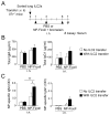
ILC2s promote antigen-specific IgM antibody production in IL-7R-deficient mice. (Panel A) Experimental protocol: Lung ILC2s were isolated from C57BL/6 mice and transferred into C57BL/6 _Il7r_−/− mice. Mice were then exposed i.n. to NP-Ficoll plus bromelain on days 1, 3, and 6, and sera were analyzed on day 7. The levels of total IgM or IgG1 (Panel B) or NP-Ficoll-specific IgM or IgG1 in sera were analyzed by ELISA. Data (mean±SEM, n=2 in PBS and n=6 in NP-Ficoll) are representative of two independent experiments. *, p<0.05; **, p<0.01 between the groups indicated by horizontal lines.
Similar articles
- Group 2 innate lymphoid cells and CD4+ T cells cooperate to mediate type 2 immune response in mice.
Drake LY, Iijima K, Kita H. Drake LY, et al. Allergy. 2014 Oct;69(10):1300-7. doi: 10.1111/all.12446. Epub 2014 Jun 17. Allergy. 2014. PMID: 24939388 Free PMC article. - T-bet inhibits innate lymphoid cell-mediated eosinophilic airway inflammation by suppressing IL-9 production.
Matsuki A, Takatori H, Makita S, Yokota M, Tamachi T, Suto A, Suzuki K, Hirose K, Nakajima H. Matsuki A, et al. J Allergy Clin Immunol. 2017 Apr;139(4):1355-1367.e6. doi: 10.1016/j.jaci.2016.08.022. Epub 2016 Sep 23. J Allergy Clin Immunol. 2017. PMID: 27670243 - Protein kinase Cθ controls type 2 innate lymphoid cell and TH2 responses to house dust mite allergen.
Madouri F, Chenuet P, Beuraud C, Fauconnier L, Marchiol T, Rouxel N, Ledru A, Gallerand M, Lombardi V, Mascarell L, Marquant Q, Apetoh L, Erard F, Le Bert M, Trovero F, Quesniaux VFJ, Ryffel B, Togbe D. Madouri F, et al. J Allergy Clin Immunol. 2017 May;139(5):1650-1666. doi: 10.1016/j.jaci.2016.08.044. Epub 2016 Oct 14. J Allergy Clin Immunol. 2017. PMID: 27746240 - Type 2 innate lymphoid cells: at the cross-roads in allergic asthma.
van Rijt L, von Richthofen H, van Ree R. van Rijt L, et al. Semin Immunopathol. 2016 Jul;38(4):483-96. doi: 10.1007/s00281-016-0556-2. Epub 2016 Mar 10. Semin Immunopathol. 2016. PMID: 26965110 Free PMC article. Review. - ILC2s and fungal allergy.
Kita H. Kita H. Allergol Int. 2015 Jul;64(3):219-26. doi: 10.1016/j.alit.2015.04.004. Epub 2015 May 18. Allergol Int. 2015. PMID: 26117252 Free PMC article. Review.
Cited by
- Guards at the gate: physiological and pathological roles of tissue-resident innate lymphoid cells in the lung.
Cheng H, Jin C, Wu J, Zhu S, Liu YJ, Chen J. Cheng H, et al. Protein Cell. 2017 Dec;8(12):878-895. doi: 10.1007/s13238-017-0379-5. Epub 2017 Mar 7. Protein Cell. 2017. PMID: 28271447 Free PMC article. Review. - Interactions between Innate Lymphoid Cells and Cells of the Innate and Adaptive Immune System.
Symowski C, Voehringer D. Symowski C, et al. Front Immunol. 2017 Oct 30;8:1422. doi: 10.3389/fimmu.2017.01422. eCollection 2017. Front Immunol. 2017. PMID: 29163497 Free PMC article. Review. - Exploring the Role of Innate Lymphocytes in the Immune System of Bats and Virus-Host Interactions.
Sia WR, Zheng Y, Han F, Chen S, Ma S, Wang LF, Leeansyah E. Sia WR, et al. Viruses. 2022 Jan 14;14(1):150. doi: 10.3390/v14010150. Viruses. 2022. PMID: 35062356 Free PMC article. Review. - Group 2 Innate Lymphoid Cells in Pulmonary Immunity and Tissue Homeostasis.
Mindt BC, Fritz JH, Duerr CU. Mindt BC, et al. Front Immunol. 2018 Apr 30;9:840. doi: 10.3389/fimmu.2018.00840. eCollection 2018. Front Immunol. 2018. PMID: 29760695 Free PMC article. Review. - Innate lymphoid cells and infectious diseases.
Yuan T, Zhou Q, Tian Y, Ou Y, Long Y, Tan Y. Yuan T, et al. Innate Immun. 2024 Aug;30(6-8):120-135. doi: 10.1177/17534259241287311. Epub 2024 Oct 3. Innate Immun. 2024. PMID: 39363687 Free PMC article. Review.
References
- Spits H, Artis D, Colonna M, Diefenbach A, Di Santo JP, Eberl G, Koyasu S, Locksley RM, McKenzie AN, Mebius RE, Powrie F, Vivier E. Innate lymphoid cells--a proposal for uniform nomenclature. Nat Rev Immunol. 2013;13:145–149. - PubMed
- Mirchandani AS, Besnard AG, Yip E, Scott C, Bain CC, Cerovic V, Salmond RJ, Liew FY. Type 2 innate lymphoid cells drive CD4+ Th2 cell responses. J Immunol. 2014;192:2442–2448. - PubMed
Publication types
MeSH terms
LinkOut - more resources
Full Text Sources
Other Literature Sources
Research Materials
Miscellaneous


